All chemicals and solvents were purchased from Sigma Aldrich (Chemie GmbH, Taufkirchen, Germany). The commercial pesticide Radiant SC 12% (Dow Agrosciences, Canada; CAS Number: 187166-40-1) was obtained from the Plant Protection Research Institute, Ministry of Agriculture, Egypt. Radiant’s active ingredient is spinetoram, a second generation of the spinosyns.
Endophytic actinobacteria and culture conditions
Seventy actinobacteria strains, previously isolated from six wild medicinal plant species (F. Compositae) in South Sinai, Egypt, were studied (El-Shatoury et al. 2013). The source-plant of the strains and their generic identity, based on the standard chemotaxonomy analysis is illustrated in Table 1. The strains were preserved as spore suspensions in 20% v/v glycerol (El Nasr Pharmaceutical Chemicals Co., ADWIC, Egypt) at − 15 °C, and were refreshed on starch casein agar (Sigma-Aldrich, Chemie GmbH, Taufkirchen, Germany), as described by Kieser et al. (2000).
Table 1 Numbers and generic identification of the actinobacteria isolated from six wild medicinal plant species (F. Compositae) in South Sinai, Egypt
Spodoptera littoralis larvae rearing
The laboratory Spodoptera littoralis larvae (L-larvae) was reared under constant laboratory conditions, at the Plant Protection Research Institute, Agricultural Research Center, Zagazig, Egypt. It was reared in an incubator at a temperature of 26 ± 2 °C, a relative humidity of 65 ± 10% RH, and 16: 8 h of light and dark, respectively, according to Mansour et al. (1966). The L-larvae were reared for thirty successive generations to guarantee that it is free from any resistance to pesticides. The field Spodoptera littoralis larvae (F-larvae) were collected from the local open field at Sharqia Governorate, Egypt (30°37'O6.6"N 31°32′54.8"E). They were transferred to the laboratory and reared for two successive generations, as described above.
Both L- and F- larval instars were fed on fresh castor bean leaves, Ricinus communis L., until reaching the accurate age of treating (fourth instar). The bioassay experiments were performed on the fourth instar larvae, because they represent the youth stage which causes massive crop damage and exhibits 72 resistance to the insecticides (El Sayed et al. 2022). During the bioassay experiments, the larvae were fed on castor bean leaves treated with the actinobacterial metabolic extracts for 48 h. Then replaced, with fresh castor bean leaves for the rest of their life until pupation. The commercial insecticide Radiant SC12%, was used as a positive control in the bioassay experiments. The detailed insect breeding protocol is shown in Additional file 1 (S1).
Actinobacteria fermentation and metabolites extraction
Two µl of spore suspension (2–8 × 10⁷ cfu/mL) of each actinobacterial strain was cultured in 50 mL sterilized starch casein broth (Sigma-Aldrich, Chemie GmbH, Taufkirchen, Germany) and incubated at 28 ± 2 °C for 21 days with continuous shaking at 100 rpm. The mycelia were separated by centrifugation at 5000 rpm. The filtrates were extracted three successive times using equal volumes of ethyl acetate (El Nasr Pharmaceutical Chemicals Co., ADWIC, Egypt). The solvent layers were combined, concentrated, and evaporated to dryness using a rotary evaporator (HS-2005S-N, HAHN SHIN Scientific Co., Korea) at 40 °C. The dried extracts were redissolved in ethyl acetate to prepare a stock concentration of 100 mg/mL and stored at 4 °C for bioactivity screening tests.
Screening effective metabolites for toxicity to fourth instar larvae of laboratory Spodoptera littoralis (L-larvae)
A series of four concentrations (0.6, 6, 60, 100 mg/mL) of crude metabolites, redissolved in ethyl acetate, from the seventy actinobacteria strains were prepared. Newly molted fourth instar laboratory, L-larvae, were starved for three-four hours prior to the treatment, to clear their alimentary canal and assure quick ingestion of treated leaves. Groups of larvae were transferred to 350 mL sterilized clean glass jars, and all jars were supplied with 7.0 cm filter paper to absorb any surplus moisture. Healthy, untreated leaves of castor, Ricinus communis L., were collected from the experimental field of the Plant Protection Research Institute. The leaves were washed, cut into equal discs using a cork borer, and impregnated with 50 µl of the corresponding metabolite concentration (i.e., equivalent to 0.03, 0.3, 3 and 5 µg/disc) using a leaf dipping technique. The toxicity was assessed in comparison to that of Radiant SC 12%, at LC50 0.5 mL/L (i.e., 0.05% concentration). All bioassay assessments were replicted (each replicate included four larvae), and were performed under constant laboratory conditions. The lethal effects (mortality %) were recorded, daily, and corrected according to Abbott's formula (Abbott 1925). The treated insects were followed up, until the pupation stage.
Screening effective metabolites for toxicity to fourth instar larvae of field Spodoptera littoralis (F-larvae)
Metabolic extracts (from 7 strains) that showed the highest activity against L-larvae were selected for investigation with the field F-larvae, at 100 mg/mL concentration, as detailed above. The most potent strain Streptomyces sp. ES2 was, then, selected for detailed characterization and toxicity investigations. The toxicity was assessed in comparison to that of Radiant SC 12%, at LC50 0.5 mL/L (i.e., 0.05% concentration). The experimental design included two control groups, fed on leaves treated with distilled water and ethyl acetate, respectively. All bioassay assessments were in triplicates (each replicate included four larvae), and were performed under constant laboratory conditions.
Identification of the most active strain Streptomyces sp. ES2
Streptomyces sp. ES2 was cultivated on ISP 4 medium, Inorganic Salt Starch Agar (HiMedia, India), (Shirling and Gottlieb 1966). The strain was incubated at 28 ± 2 °C for 14 days, and Scanning electron microscopy (SEM) images of the sporulated hyphae was investigated using LEO GEMINI-1530 high-resolution electron microscope (Carl Zeiss, SMT GmbH, Oberchoken, Germany). For the Detection of Diaminopimelic acid (DAP) isomers, ES2 strain was grown in Tryptone Soya broth (TSB) at 100 rpm, 28 °C for seven days. The mycelia were harvested by centrifugation at 12,000 × g, washed twice with sterile distilled water and dried. Two mg of the dried mycelia were hydrolyzed in 1 mL 6N HCl, as described by Staneck and Roberts (1974). The hydrolysate was filtered and run in parallel to DAP (DL-diamonipimelic acid) and LL-DAP standard on a thin layer chromatography plate. The bands were developed using ninhydrin solution and drying at 80 °C. The cultivation and phenotypic characterization of Streptomyces sp. ES2 was performed in triplicates, to guarantee the quality of the results.
For partial 16S rRNA gene sequence analysis, DNA was extracted using the salting-out method (Kieser et al. 2000), with an additional purification step using phenol/chloroform. The 16S rRNA gene of the strain was amplified using the universal primer set 27F (5´-AGA GTT TGA TCC TGG CTC AG-3´) and 1492R (5´-GGT TAC CTT GTT ACG ACT T-3´), (Metabion International AG, Planegg, Germany). Amplification conditions were according to Trujillo et al. (2010). Briefly: an initial denaturation step was performed for 9 min at 94 °C, followed by 30 cycles of denaturation for 1 min at 95 °C, annealing for 1 min at 55 °C and extension for 2 min at 72 °C. A final extension step was performed for 10 min at 72 °C.
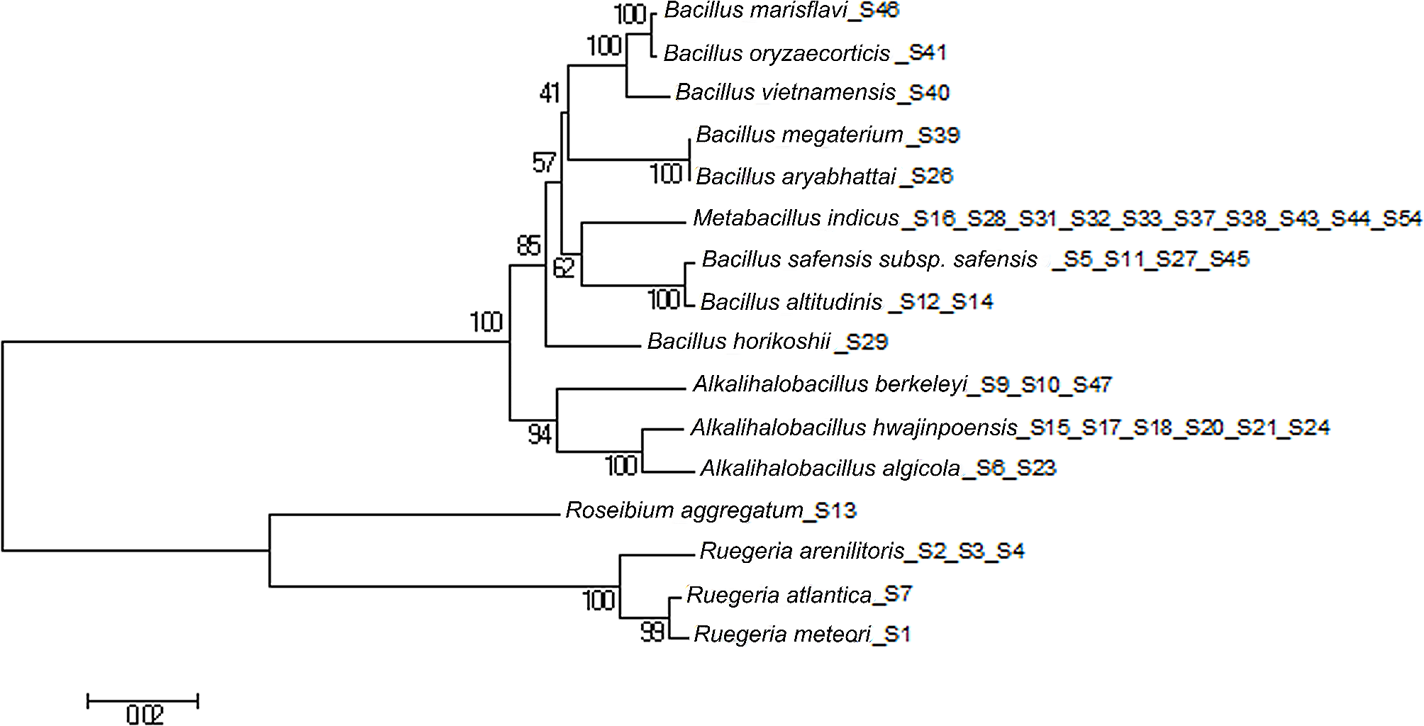
PCR product sequencing was performed at Macrogen Biotechnology, Ltd. (Korea) (https://dna.macrogen.com/eng/). The sequence obtained and those of its most closely related Streptomycetes spp., retrieved from GenBank, were aligned using BLASTN (Version: 2.9.0 +) (Zhang et al. 2000). The maximum identity score sequences were selected and aligned using the multiple alignment program ClustalW (Thompson et al. 1997). The phylogenetic tree was established by the maximum likelihood method, 1000 bootstrap, Tamura 3-parameter model; constructed using MEGA11 (Tamura et al. 2021).
Histopathological examinations of L-larvae treated with ES2 metabolic extract
The efficacy testing of natural products for toxicity and mortality should be documented 48 and/or 72 h after exposure, according to the WHO standards for laboratory and field testing of insecticidal activity (Yadav 2013). This is due to the possibility that natural products contain substances with fundamentally novel mechanisms of action on insects. Therefore, samples of the treated L-larvae and controls were collected at 48 and 72 h post treatment. They were preserved in 3 mL 10% formaldehyde (v/v), (El Nasr Pharmaceutical Chemicals Co., ADWIC, Egypt), in sterilized screw-capped tubes, dehydrated and embedded in paraffin wax. Serial longitudinal and transverse sections, at five microns thickness, were made with a microtome and mounted on clean slides using Mayer’s albumin (Stanbio laboratory, India). The sections were stained with Ehrlich’s hematoxylin–eosin (HE), (TissuePro Technology, Gainesville, FL, USA), (Ruiz et al. 2004). The histological sections were examined under a light binocular stereomicroscope (NOVEL; NLCD-120, China) at 100-X and 400-X magnifications.
Biochemical examinations of L-larvae treated with ES2 metabolic extract
Samples (groups of four) of the treated and control L-larvae were placed in clean screw-capped tubes and kept frozen overnight. The frozen samples were homogenized for three minutes in distilled water (50 mg/mL) using a chilled glass Teflon tissue homogenizer (ST–2 Mechanic-Preczyina, Poland) surrounded with a crushed ice jacket. Then, they were centrifuged at 8000 rpm for 15 min at 5 °C in a refrigerated microcentrifuge (Hettich, Kirchlengern, Germany). The supernatants, used as enzyme extracts, were stored at − 20 °C until use in biochemical assays. All biochemical measurements were performed in triplicates. A double beam UV spectrophotometer (Spectronic 1201, Milton Roy Co., Georgia, USA) was used to measure the absorbance of colored substances.
The total protein concentration was determined according to Bradford's method (Bradford 1976).
Acetylcholinesterase (AchE, EC 3.1.1.7) determination: acetylcholinesterase activity, a detoxification enzyme, was measured according to Simpson et al. (1964), using acetylcholine bromide (AchBr), (Sigma-Aldrich, Chemie GmbH, Taufkirchen, Germany), as a substrate. The samples were measured at 515 nm absorbance against a blank (ethanol in phosphate buffer, pH 8.0), (El Nasr Pharmaceutical Chemicals Co., ADWIC, Egypt). The activity was expressed as U/mg protein.
Protease (EC 3.4.21.112) determination: proteolytic activity was measured as described by Tatchell et al. (1972), with modifications, by measuring the increase in free amino acids split from a substrate protein (albumin) during one hour of incubation at 30 °C. Amino acids were colorimetrically assayed by ninhydrin reagent (Sigma-Aldrich, Chemie GmbH, Taufkirchen, Germany). The zero adjustment was performed at 570 nm against the reagent blank (100 µl distilled water). The amino acids were expressed as µg D, L-alanine/min/mg protein.
Lactate dehydrogenase (LDH, EC 1.1.1.27) determination: LDH activity was performed as described by Diamantino et al. (2001). The zero adjustment was performed against buffer without substrate. The activity was expressed as U/mg protein (1 U = 1 μmol substrate hydrolyzed per minute).
Non targeted metabolomics analysis
Liquid chromatography, combined with quadrupole-time-of-flight high-definition mass spectrometry, LC-Q-TOF-MS, was used to investigate the chemical constituents of the metabolites from Streptomyces sp. ES2 strain. This technique is a powerful tool for the characterization of microbial compounds with similar structures, particularly in the analysis of natural products (Liu et al. 2010). The analysis was performed using Triple TOF® 5600 + , Sciex system, Canada; pre-column (0.5 μm × 3.0 mm; Phenomenex Co., USA) and XBridge C18 column (3.5 μm, 2.1 × 50 mm; Waters Co., USA) with two LC columns, in-line filter discs, at 40 °C. Detailed preparation and processing of the sample is provided in the Additional file 1: (S2). Based on their fragments, MasterView was used to define peaks using Build-in databases (Data acquisition Analyst TF 1.7.1 software, Sciex). Reaxys ChemDraw software, version 18.0.0.20 (https://www.reaxys.com) was used to the compounds that can effectively target lethality to the larvae (Table 4).
Molecular docking simulation
Molecular docking aimed to illustrate the virtual mechanism of binding of selected compounds which towards acetylcholinesterase (AchE, PDB = 4EY5), lactate dehydrogenase (LDH, PDB = 1LDG), and protease (SREBPs, PDB = 5GPD) target proteins. The data were freely accessible through the protein data bank. Both proteins and ligands were optimized, and the molecular docking study was carried out using AutoDock Vina as the computational software (Trott and Olson 2010). Each complex was analyzed for 3D interaction images taken by Chimera (UCSF) (Pettersen et al. 2004).
Statistical analysis
All data were formulated as means ± standard error of the mean (SEM). The data wee subjected to normality testing using Kolmogorov–Smirnov at 0.05 level. Accordingly, LDH, protease and AchE were parametric and parametric data analysis applied. One-way ANOVA was applied to assess the difference between treatment groups, ANOVA was followed by Duncan’s Multiple Range tests (DMRTs) as a post-hoc test at 0.05 level.


留言 (0)