USE OF OPTICAL COHERENCE TOMOGRAPHY IN DETECTING RETINAL TEARS IN ACUTE, SYMPTOMATIC POSTERIOR VITREOUS DETACHMENT
Purpose:
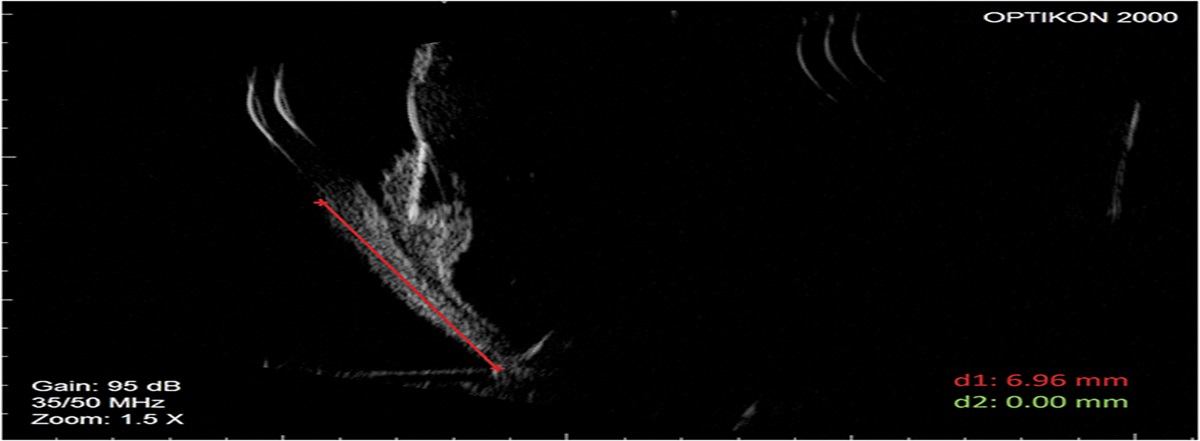
To evaluate the association of posterior vitreous opacities (PVOs) on optical coherence tomography with retinal tears identified on examination in patients with acute, symptomatic posterior vitreous detachment (PVD).
Methods:
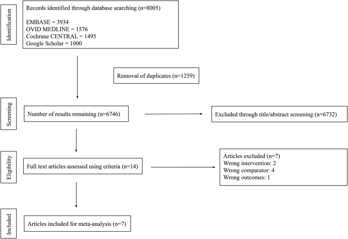
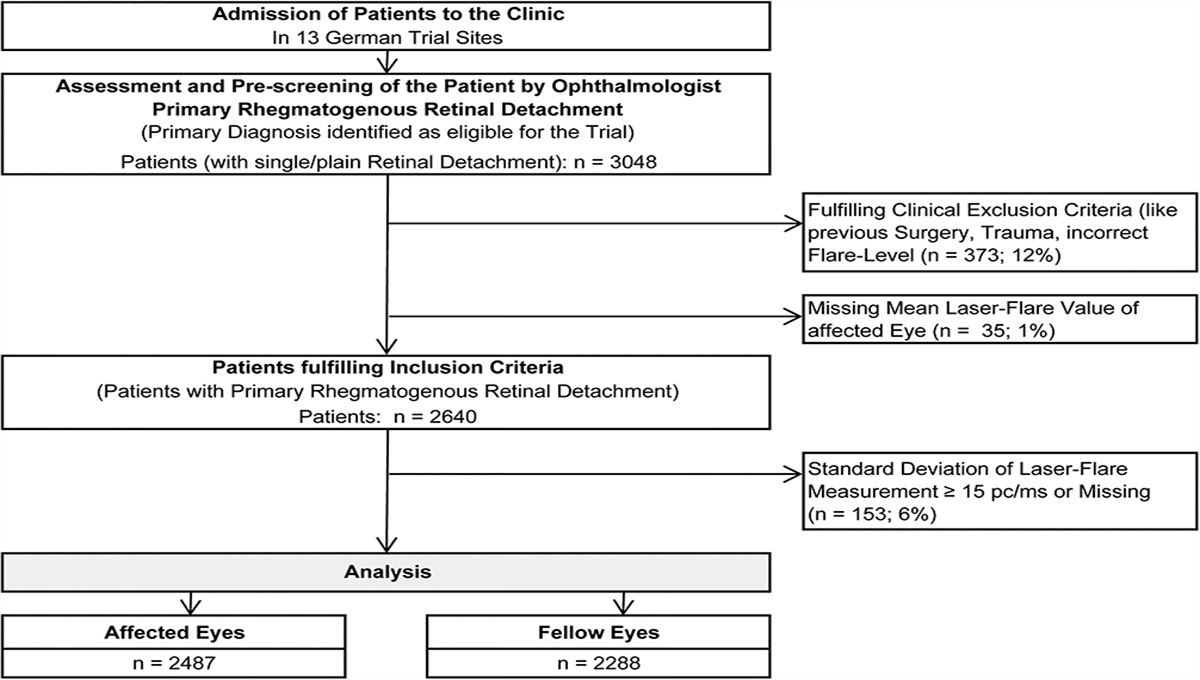
Data were retrospectively collected from the medical records of 388 patients with acute, symptomatic PVD between January 1, 2021, and June 30, 2021. Included patients had received a primary diagnosis of PVD and presented with flashes and/or floaters. Optical coherence tomography scans were reviewed by two separate readers for the presence of PVOs. The primary outcome was the presence of retinal tear on fundus photograph and on examination.
Results:
Of 388 patients who presented with acute PVD symptoms, 90 (23.2%) were found to have a retinal tear on dilated fundus examination. Among these patients, 78 (86.7%) were found to have PVOs on optical coherence tomography. Statistical analysis demonstrated a significant relationship between the presence of PVOs and retinal tear (P < 0.01). The sensitivity and specificity of this finding was 86.7% and 72.5%, respectively. Further analysis included area under the curve from receiver operating characteristic curve which was found to be 0.80.
Conclusion:
The presence of PVOs on optical coherence tomography is suggestive of a retinal tear in patients with acute, symptomatic PVD.


留言 (0)