Our study confirmed that high proportion of patients have persisting or new pulmonary symptoms following COVID-19 infection. HRCT changes were present in 13.8% of patients, who need follow-up of lung abnormalities.
The proportion of patients with ILD was lower than in a UK study (Myall et al. 2021), in which 24% of patients were suspected of having ILD. In that study, CT abnormalities were identified in 76.6% of patients referred for MDD, with the highest rates of organizing pneumonia and GGO. Based on the MDD diagnosis, ILD was diagnosed in 4.8% of the total patient population (837 patients). Patients with ILD were started on steroid therapy, and a subsequent follow-up study assessed the reduction of previous symptoms and described the regression of CT abnormalities; highlighting that CT lesions did not progress to fibrosis in patients treated with steroids (Myall et al. 2021). In our patients steroids were used during the hospital stay in high proportion as standard of care treatment, non-treated patients were mainly from the group of referrals who had their infection at home (Polivka et al. 2022). This might have contributed to the lower number of pulmonary changes on LDCT.
In our study, the most frequently observed symptom was fatigue, which persisted in one third of patients. Fatigue was also the leading symptom in all previous studies. The second most common symptom, affecting about a quarter of patients, was respiratory in origin. The most common were dyspnoea (25.2% as a persistent symptom); and cough (22.6% as a persistent symptom), and the third most common complaint was sleep disturbance (insomnia 13.2% and sleepiness 8.2% as a persistent symptom). ILD suspected group reported more often cough and sleep disorders.
Several other studies described the prevalence of post-COVID symptoms. A Chinese study described fatigue in 63% of patients 6 months after COVID-19; sleep disturbance in 26%; and anxiety and/or depression in 23% (Huang et al. 2021). Also in an Austrian study, 41% of patients experienced symptoms after the infection had passed; the most common symptoms being dyspnoea (36%); night sweats (24%) and sleep disturbance (22%). In this study, at a later follow-up visit (100 days after the onset of infection), patients had a reduced rate of symptoms, indicating that post-COVID syndrome is a dynamically changing and generally improving condition (Sonnweber et al. 2021).
Our study also compares the symptoms of patients with suspected ILD with those of controls (non-ILD patients) who have undergone COVID-19. The difference between the two groups was significant for new-onset symptoms, with cough and sleepiness being more frequent in patients with suspected ILD. This may suggest that the persistence of these two symptoms and a history of COVID-19 disease may raise the clinicians’ attention for the possibility of lung abnormalities, including ILD.
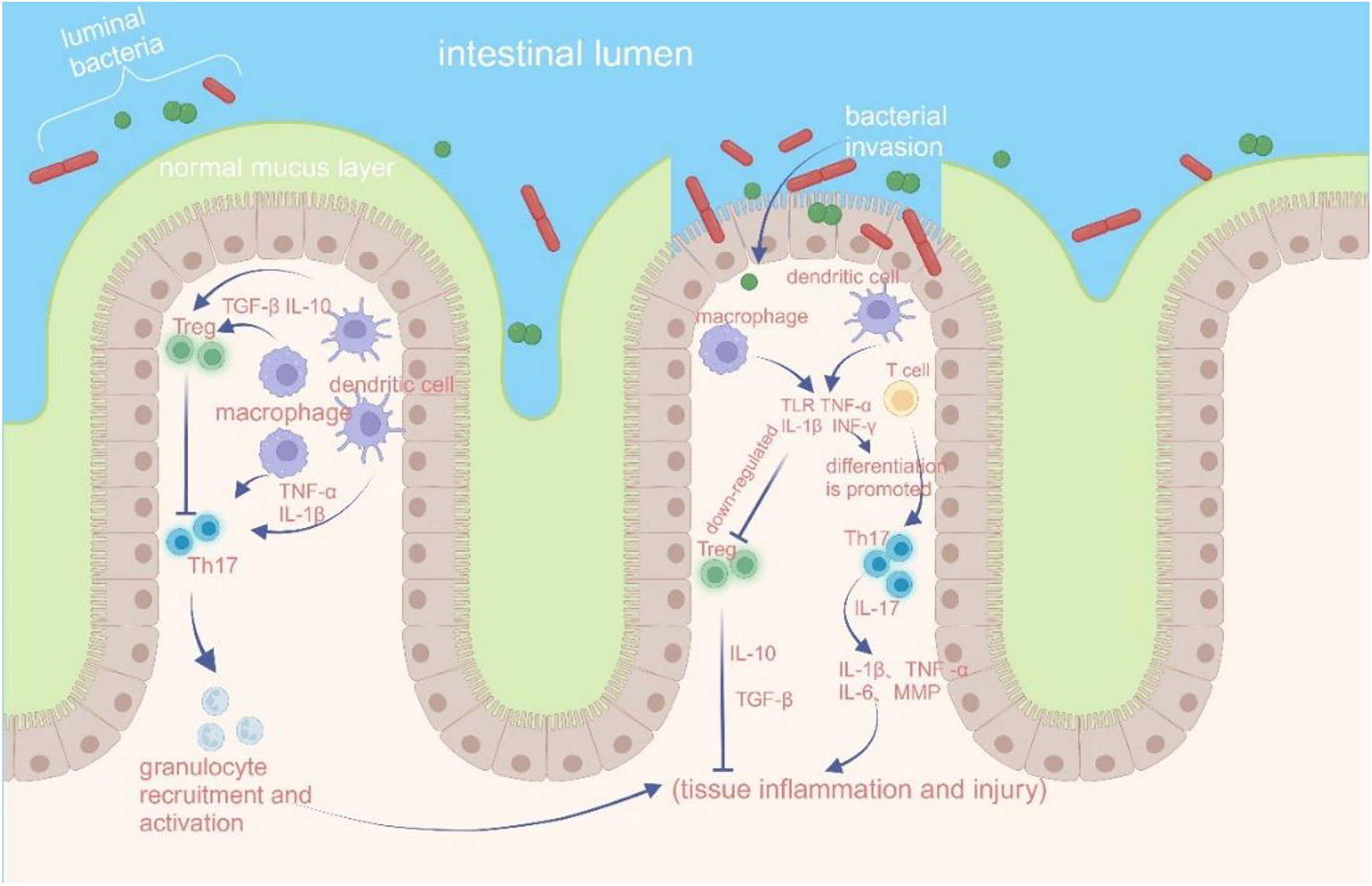
Lesions with characteristics of ILD after COVID-19 disease can be identified in varying degrees and patterns in affected individuals. The most typical patterns are GGO, reticulation and consolidation (Sonnweber et al. 2021; Lerum et al. 2021; McGroder et al. 2021; Besutti et al. 2022; Liu et al. 2021) which are rarely associated with fibrosis immediately after the infection and often show regression (Mandal et al. 2021; Sonnweber et al. 2021; Myall et al. 2021). Two international studies highlight organizing pneumonia (Myall et al. 2021) and usual interstitial pneumonia (UIP) (Konopka et al. 2021) as the most common patterns. Many cases with CT lesions are not associated with respiratory symptoms or impairment of lung function or 6MWT; therefore, Sonnweber et al. propose the use of CT scans for diagnosis or evaluation of post-COVID ILD instead of functional measurements (Sonnweber et al. 2021). Abnormal patterns in the lung parenchyma have been associated with the length of invasive ventilation, which may damage lung tissue due to a possible barotrauma; or could also be associated with the more severe form of COVID-19 in patients receiving invasive ventilatory support, which has been correlated with CT abnormalities in several studies (Huang et al. 2021)(Guler et al. 2021).
Regarding respiratory function results, the currently available literature is consistent in describing a pattern of restrictive ventilatory dysfunction (reduced TLC, FVC, RV) and diffusion reduction (TLCO; KLCO) (Huang et al. 2021; Lerum et al. 2021; Sonnweber et al. 2021; Guler et al. 2021; Myall et al. 2021; Stockley et al. 2021). Several studies have highlighted the impairment in diffusion parameters, which may be related to the severity of COVID-19 disease (Huang et al. 2021; Guler et al. 2021), the presence of CT lesions (McGroder et al. 2021), and the use of mechanical ventilation (Guler et al. 2021). In the present study, impairment in respiratory function and decreased diffusion capacity were also measured in the ILD suspect group, with a significant difference compared to the control group, where the decrease in these parameters was not significant. Regarding performance, 6MWT parameters, including desaturation and reduced distance parameters are characteristics of patients with suspected ILD, similar to previous observations (McGroder et al. 2021; Guler et al. 2021).
The exact prevalence of post-COVID disease is not yet known, and many studies have aimed to identify risk factors and predisposing factors. The international literature associates female sex (Huang et al. 2021), older age (Huang et al. 2021), obesity (Stockley et al. 2021), invasive ventilatory support (Lerum et al. 2021) and more severe COVID-19 disease (Huang et al. 2021) with the disease, while steroid therapy is considered as a beneficial factor (Myall et al. 2021). In contrast, in the present study, male sex is more prevalent in both groups, with no significant difference between the two groups. The ILD suspected patient group did not have a higher prevalence of invasive ventilatory support; and there was no difference in obesity as a risk factor compared to the control group. Regarding age, in accordance with the literature, the mean age was higher in the ILD suspect group, which might have contributed to the observed differences. Steroid therapy, oxygen supplementation need, and anticoagulant therapy were more frequent in the ILD suspect group, which can be associated with the fact that this patient group was more frequently admitted to hospital for COVID-19 infection, where these therapies are widely used according to guidelines.
Limitation of our study is the short follow-up for a limited number of patients and the lack of re-evaluation of LDCT scans and function.

留言 (0)