It has been suggested that stage 4 IMHs seem to be associated with less satisfactory surgical outcomes comparing to stage 3 IMHs [9, 11]. This may be explained by the histological findings that the percentage of internal limiting membrane with vitreous remnant is higher in stage 4 holes against in stage 3 holes, so that stage 4 holes may be more difficult to be sealed if the extent of internal limiting membrane peeling is not sufficient [14, 15]. However, in the report of Brockmann and colleagues’, stage 4 IMHs exhibited a closure rate without statistical significance comparing to that of stage 3, though lower in value [10]. Meanwhile, according to the comparison of the initially closed cases and unclosed cases by Hasegawa and colleagues, there was no significant difference in primary closure rate between stage 3 and stage 4 IMHs [12]. Actually, surgical outcomes include not only the closure rate, but also the visual function and other morphological parameters. Whether IMHs of the two stages differ in surgical outcomes still needs direct evidence for further elucidation, but few targeted studies have discussed about it. The present study focused on IMHs of stage 3 and stage 4 with a relatively large sample, and for the first time compared their visual function and morphological manifestations after vitrectomy. The results demonstrate that IMHs of the two stages are identical in anatomical and visual outcomes.
As is shown in our previous report [8] and confirmed in this study, IMHs of stage 3 and stage 4 exhibited no significant differences in preoperative clinical features and morphological characteristics. According to Gass’s classification applied in this study, the overwhelming majority of stage 3 and stage 4 IMHs were larger than 400 μm, so the size of the hole tended to be approximate between stages. Moreover, with posterior vitreous detachment accomplished in the fovea, the adhesion at the optic disc seems no longer to affect the evolution of the hole, so that IMHs of the two stages demonstrate identical characteristics.
Vitrectomy with internal limiting membrane peeling is considered as an efficient surgical treatment for IMHs with a primary closure rate of more than 90% according to previous reports [16]. Conventionally, gas tamponade like sulfur hexafluoride and octafluoropropane is applied. Recent years, internal limiting membrane peeling with air tamponade also exhibited satisfactory outcomes [12, 17]. In this study, gas and air tamponade were respectively applied in two time periods because gas were not available after July 2016 in mainland China. Different tamponade may affect the outcomes of surgery [18], but the two stages in this study exhibited comparable ratio of gas/air tamponade, suggesting that types of tamponade may not make a difference in comparison of the outcomes. In fact, in eyes with neither gas nor air tamponade did the primary closure rate exhibit a difference between stage 3 and stage 4. Since the age of patients and the preoperative BCVA were comparable between the two stages, it is not surprising that the rate of combined cataract surgery exhibited no difference.
Postoperatively, the primary closure rate exhibited no significant differences between the two stages. Eleven eyes with stage 3 IMHs and three eyes with stage 4 IMHs underwent a second operation and obtained final closure; five eyes with stage 3 IMHs and three eyes with stage 4 IMHs did not take a second operation. There seems to be no evident difference in the secondary closure rate between stages, but statistical analysis was not carried out considering the relatively small sample. The postoperative BCVA is considered to be associated with not only the preoperative minimum diameter [19], base diameter [19, 20], but also the primary closure [16]. Therefore, within a comparable duration of follow-up, it is understandable that the BCVA at last visit appeared identical between stage 3 and stage 4, since IMHs of the two stage exhibited comparable size and primary closure rate.
Besides closure rate and postoperative vision, the morphology of the fovea after surgery is also noteworthy. In a prospective interventional series by Apostolopoulos et al., the thickness of the fovea was manually measured and exhibited significant association with the BCVA 1 year postoperatively [21]. Takamura et al. also found the average central retinal thickness 1 month after surgery, which was computed automatically within 1 mm diameter central area, was correlated with the macular hole diameter and the BCVA 1 year after surgery [22]. Given different methods and point-in-time of the measurements, the relationship among MLD, postoperative FRT, and postoperative BCVA needs to be further discussed. In the present study, the manually measured FRT exhibited no significant difference between stages at the end of the comparable follow-up; for IMHs of either stage, FRT was indeed negatively correlated with MLD and positively correlated with the BCVA at the same point-in-time. However, whether the FRT at this moment has influence on long-term vision recovery still needs to be elucidated by further studies.
The ORD in this study, which is also referred as foveolar lucency, is a common cystic space underlying the fovea with continuous inner retina in the surgically sealed macular hole [23]. Kang et al. reviewed 96 eyes with IMH (mean MLD around 425 μm) and considered that the foveolar lucency was detected predominantly in smaller macular holes; [24] on the contrary, Grewal et al.’s report of 45 eyes (mean MLD of IMH around 227 μm) suggested that MLD ≥ 330 mm was predictive of foveolar lucency development [25]. Meanwhile, it still remains controversial whether this sign is correlated with the visual outcomes [23, 25]. In the present study, prevalence of ORD showed no significant differences between not only stage 3 and stage 4, but also smaller than 650 μm and larger. Stage 2 IMHs were not enrolled here and the mean MLD was larger than those of Kang et al’s [24] and Grewal et al’s [25], and that may be why we did not found the relationship between hole size and the prevalence of ORD. Accordingly, it could be inferred that ORD prevalence may be not correlated to stage of the hole, but to what extent ORD is influenced by hole size and whether its presence makes sense to postoperative vision still need further elucidation. That’s what we have been working on.
Considering all above, IMHs of stage 3 and stage 4 exhibited identical anatomical and visual outcomes based on comparable baseline characteristics and surgical interventions. It could be inferred that IMHs of the two stages may have an identity of the intrinsic nature in wound healing.
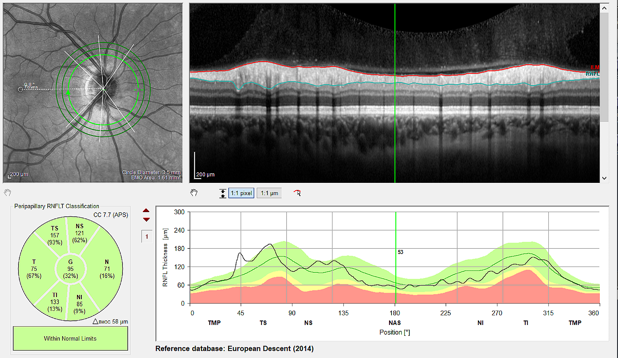
There are quantities of parameters provided by OCT to depict the postoperative morphology of IMH, among which the integrity of the ellipsoid zone, once referred as the IS/OS junction, is frequently applied and widely considered to be associated with the vision recovery [26, 27]. However, the definition of its abnormality varies a lot among different studies, such as defect, discontinuity, and disruption. Therefore, in the present study, two more visible anatomical parameters ——FRT and ORD, were applied to depict the fovea postoperatively.
A previous report involving 2456 IMH eyes indicated that duration > 9 months reduced the MH closure rate [28]. In this study, the proportion of duration > 9 months showed no significant difference between stage 3 and stage 4. Thus, duration of symptoms may not affect the comparison of closure rate.
This study staged IMHs according to Gass’s classification in 1995 [6] and all the IMHs enrolled were larger than 400 μm except ten of stage 4, so that the overwhelming majority here is considered as large IMHs [7]. Recently, it has been suggested that large IMHs demonstrated different success rates by a diameter around 650 μm [29, 30]. Thus, we divided all the cases into two groups by 650 μm and found in neither group did IMHs exhibit significant differences in outcomes between the two stages. However, when comparing IMHs of different sizes, those which were smaller than 650 μm exhibited significantly better outcomes than larger ones. This indicated that in large IMHs (> 400 μm), the size seems to play a more crucial role in surgical outcomes than the stage, and is a more noteworthy factor for choice of surgical intervention and prediction of prognosis.
This study of stage 3 and stage 4 IMHs has a limitation of its retrospective nature. With a relatively large sample and strict inclusion criteria, the results are considerably reliable, but further studies, especially prospective ones and randomized controlled trials are still in need. Meanwhile, the mean follow-up of about 6 months seems to be relatively short, because the visual outcomes could continue to improve in the long run [31]. However, the duration of follow-up was comparable between the two stages, so this limitation may not affect the comparison. Studies of long-term outcomes are meaningful, and that’s what we are working on.
In conclusion, this study with a relatively large sample compared stage 3 and stage 4 IMHs based on the Gass’s classification in 1995, and found IMHs of the two stages exhibited considerable identity not only in preoperative characteristics, but also in anatomical and visual outcomes after vitrectomy, internal limiting membrane and other comparable intraoperative interventions. For IMHs larger than 400 μm, the size, instead of the stage, may be more valuable for outcome prediction and decision-making in proper technique for hole closure.


留言 (0)