Large assemblies of purified enzymes from across intermediary metabolism have been documented since their initial purifications in the 1960s. Early examples of enzymes that reversibly assemble in large filaments include glutamine synthetase [1], acetyl CoA carboxylase [2,3], glutaminase [4], and phosphoribosyl pyrophosphate synthetase [5,6] (for a comprehensive review, see Park & Horton, 2019 [7]). In each case, assembly was modulated by the presence of specific ligands known to affect enzyme activity, suggesting that assembly might play a role in regulation. These early examples of enzyme filaments were characterized using analytical centrifugation, size exclusion chromatography, and transmission electron microscopy, and in some cases changes in activity could be correlated with assembly states. Due to the technical challenges associated with studying large, dynamic polymers at the time, however, the structural underpinnings of the enzyme assemblies and the biochemical and physiological effects of assembly remained uncharacterized.
Advances in light microscopy and subsequent identification of cellular protein assemblies containing enzymes from intermediary metabolism have supported physiological roles for the large polymers. Screens using fluorescently labeled proteins have identified numerous enzyme assemblies in budding yeast, with some paralogs or enzymes within the same pathways co-localizing within the cell [8, 9, 10, 11]. Recent studies have begun to define the conditions under which the punctate and filamentous assemblies appear in cells, demonstrating that the macrostructures are dynamic and assemble reversibly [12, 13, 14, 15, 16, 17∗∗, 18, 19]. The assemblies often appear at times of cell stress, including starvation, cell division, and proliferation. Many of the assemblies tend to contain proteins that sit at regulatory nodes within metabolic networks, suggesting that assembly provides another layer of enzyme regulation within the cell and driving the exploration of the molecular mechanisms and functional consequences of polymerization [10].
The uncovering of physiological roles for large-scale assemblies prompted the exploration of their structural organization. Cryo-electron microscopy, which can provide high-resolution structures of polymers spanning tens of nanometers in length, has greatly expanded our understanding of the molecular underpinnings of the protein assemblies observed in cells. This technique, paired with other structural and biochemical tools, has made it possible to dissect the functional consequences of polymerization [20]. Mutagenesis studies targeted to filament interfaces show that filament assembly serves as a platform for allosteric regulation [21∗, 22∗, 23∗, 24], where mutations lead to changes in catalytic activity. Here, we reflect on recent work, describing patterns in the filaments assembled from enzymes in intermediary metabolism. We discuss the allosteric regulatory mechanisms made possible by filament formation, and we contemplate the next steps in the field.
Filaments from intermediary metabolism share some traits with the better-understood dynamic cytoskeletal polymers. For example, actin filaments and microtubules are extremely well conserved and found in all eukaryotes, and both have homologues in prokaryotes. Both are dynamic and change assembly states in response to changes in cellular conditions. However, both actin filaments and microtubules are directional (polar) and assemble from monomers (actin) (Figure 1a) or asymmetric dimers (tubulin) into helical or pseudo-helical filaments [25].
Unlike actin and tubulin, metabolic enzymes that form polymers generally assemble into helical filaments that lack directionality (i.e. apolar filaments) (Figure 1b–e), even when the assembling protomers are monomers. For example, glucokinase 1 from Saccharomyces cerevisiae, a distant actin homolog that catalyzes the first step in glycolysis, assembles from monomers into filaments [26]. The repeating unit in the filament of glucokinase 1 (black outline in Figure 1b, bottom row) contains twofold symmetry, which adds a symmetry axis perpendicular to the helical axis. This axial symmetry coupled with the helical symmetry places the filament in dihedral filament symmetry group D1 [27] (Figure 1b), where a 180° rotation around the axis perpendicular to the helical axis will superimpose the filament on itself.
Most other metabolic enzyme filaments assemble from protomers that are themselves oligomers with closed point group symmetry. In most cases, the defined oligomerization is required for activity or allosteric regulation. Panels C through H in Figure 1 illustrate several examples of nondirectional filaments formed from oligomers of increasing symmetry. Acetyl CoA-carboxylase forms a symmetric dimer in solution, and these dimers stack to form filaments with D1 symmetry (Figure 1c) [28]. Higher dihedral symmetries are also observed (Figure 1d–h) [16,21∗, 22∗, 23∗,29, 30, 31, 32], and oligomers with D3 symmetry or greater tend to stack with their highest symmetry axes coincident with the helical axis of the filament. Oligomers composed of multiple proteins or protein isoforms with pseudo dihedral filament symmetry also have been identified and characterized [33,34].
A filament interface containing dihedral symmetry becomes much more sensitive to a single amino acid change at the protomer level and can more rapidly evolve towards or away from filament assembly (Figure 2) [35]. When a mutated amino acid is located on the outer surface of the oligomer, the change can result in a “self-interacting patch” on the protein, triggering formation of filaments [36]. The propensity for assembly into filaments can be calculated [36] and increases with higher-order symmetries due to avidity effects. Negative selection also occurs at potential assembly interfaces on dihedral oligomers, with specific surface residues preventing filament formation [37]. The degree to which the filament interfaces are evolutionarily constrained varies by protein and suggests polymerization can evolve. For example, there is evidence for widespread conservation of PRPP synthetase filament interfaces [16,23], but CTP synthase forms different interfaces in animals, fungi, and prokaryotes, implying that polymerization has arisen multiple times in evolution [22,29].
Individual filaments and their larger organization within the cell can be described in terms of their quinary structures, or their quasi-regular assembly into agglomerates via weak interactions [38,39]. Individual filaments like those in Figure 1 can be defined as a level of quinary structure, as they are not bound by a specific protomer number; they are theoretically infinite, and their assembly is dependent on their environment. Quinary structural assemblies build upon the characteristics generated by quaternary assembly by reversibly tuning protein behavior in a variety of ways [40, 41, 42, 43]. Quinary assembly can stabilize or destabilize protein structure (Figure 3a, PRPP synthetase [23]), modulate protein activity (Figure 3b, IMP dehydrogenase [30]), and localize proteins to specific regions within a cell [37], all in response to changes in the cellular environment.
Many filaments are hypothesized to form larger lateral arrays in cells, with notable examples in the literature [9,22,44,45]. While some of these assemblies may consist of quasi-ordered filament arrays without a strictly ordered lattice, some filaments have been shown to assemble with defined geometry. Both CTP synthase isoforms from S. cerevisiae assemble reversibly into multi-filament bundles with well-defined inter-filament assembly contacts [22]. Bundles of IMP dehydrogenase filaments have been observed in vivo via ultrathin section EM [9] and bundles can be induced by crowding in vitro [45,46].
Enzyme filament structures are intimately linked to their effects on biochemical function, which are surprisingly varied among different enzymes. Filament assembly can act as a mechanism of allosteric control, either by directly stabilizing active or inactive states of an enzyme or by influencing the affinity for other allosteric effectors (Figure 3). Some filaments accommodate conformational changes between different functional states, influencing the equilibria between active and inhibited conformations. In other cases, filaments act as an integration node, where signals from binding partners and phosphorylation can be merged [28]. One advantage of such assembly-based regulation is that it can happen rapidly in response to sudden changes in metabolic demand, while other forms of control like transcriptional reprogramming or degradation require longer response times. Below, we discuss several recent examples of assembly-based control of enzyme activity (Figure 3, Figure 4) and suggest other possible mechanisms.
Filaments can act as a mechanism of inhibition and “protein buffering” within a cell. For example, glucokinase 1 (Glk1), which regulates entry into glycolysis, is active as a monomer. Glk1 has a high affinity for glucose, and is typically expressed when glucose levels are low, where it remains monomeric and active. However, when glucose levels spike Glk1 assembles into filaments in which it is prevented from turning over substrate, so that only the small pool of unassembled protein at the critical concentration remains active (Figure 4a) [26]. Assembly-based inactivation of Glk1 prevents toxic over commitment to glycolysis, acting as a “molecular surge protector” to prevent overactivity due to sudden sharp changes in metabolic state.
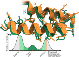
Enzyme filaments, however, are not all inactive. For example, assembly into filaments stabilizes the active conformation of human CTP synthase 1, which upregulates its activity (Figure 4b) [47]. CTP synthase 2 from humans assembles into filaments that can dynamically shift from the inhibited state to the active state and vice versa [29]. Physical coupling of CTP synthase 2 protomers within a filament results in highly cooperative transitions between low- and high-activity conformations via propagation along the filament (Figure 4c). The cooperativity of CTP synthase 2 filaments makes for a conformational switch that is exquisitely sensitive to the balance of substrates and allosteric inhibitor, allowing cells to quickly adapt to changes in nucleotide demand.
Assembly of filaments can also act to stabilize ligand binding sites, promoting activity, inhibition, or both. For example, human PRPP synthetase 1 assembles into filaments that stabilize a critical allosteric site that binds either the activator phosphate or the inhibitor ADP [23]. In free hexamers, the short helix that forms the allosteric site is disordered, making PRPP synthetase 1 insensitive to activation by phosphate. But in the filament, assembly contacts constrain the short helix and pre-patterns the allosteric pocket (Figure 3a). This stabilizes binding of either phosphate or ADP, and the filaments then adopt either the active or inhibited conformation of the enzyme (Figure 4d). Similar filaments and regulatory mechanisms have also been observed in the Escherichia coli PRPP synthetase [16], suggesting this mode of regulation is broadly conserved.
Filaments can also tune the response of enzymes to allosteric effectors. Assembly of IMP dehydrogenase 2 into filaments decreases its response to the allosteric inhibitor GTP. Binding of GTP compresses and allosterically inhibits free octamers of IMP dehydrogenase 2, but filament contacts stabilize a conformation of the catalytic core that resists this GTP-induced compression, making the filaments less sensitive to GTP inhibition (Figure 3, Figure 4e) [30]. Filaments formed by IMP dehydrogenase 1 also modulate the enzyme's response to GTP, and inhibition is further tuned by N- and C-terminal extensions that are a part of the retinal splice variants of the protein [21]. This ability to remain functional at high GTP levels is especially important in cells that have a high nucleotide demand, like those in the retina, where the enzyme must maintain activity in the presence of large pools of a feedback inhibitor.
Many other regulatory mechanisms have been associated with filament formation in enzymes. Filament assembly can occur as a direct response to environmental cues, such as divalent cations, as is the case in glutamine synthetase [32,48], or pH, as for CTP synthase from S. cerevisiae [22] and arginine and lysine decarboxylases in bacteria [31,49]. Assembly can provide a platform for integration of regulatory signals; filament formation of acetyl-CoA carboxylase allows for the integration of signals from binding partners and phosphorylation [28]. Filament formation can stabilize the domains located at the filament interface, inhibiting the activity of those domains, as happens in methylcrotonyl-CoA carboxylase [50]. Filaments can create a cofactor “nanowire” required for the transfer of electrons [44,51]. Recent work on aldehyde-alcohol dehydrogenase has demonstrated that “spirosome” filament assembly is not only critical for its activity, but that changes in the filament architecture facilitate substrate channeling between different active sites in the enzyme (Figure 4f) [52, 53, 54].
We can postulate additional mechanisms of assembly-based regulation, beyond those observed to date. Like the allosteric site stabilization in filaments of PRPP synthetase, assembly of filaments could form emergent allosteric sites not found in the enzyme protomers, making filaments required for activity or inhibition. Filament formation might also accomplish the opposite affect; by assembling into a filament, the active or allosteric sites of an enzyme could be occluded, preventing activity or regulation. Filaments might impose hysteresis on an enzyme; once the filament has assembled in either the active or inactive form, a higher effector concentration might be required to switch the enzyme into the opposing conformation than is required when the enzyme is in its monomeric or stoichiometric oligomer form. This modality would buffer enzyme activity or inhibition against short or shallow spikes in ligand concentration, preventing the cell from shifting into a new regime prematurely. Whether any such mechanisms exist in nature remains to be seen, and considering these alternatives may be useful for efforts to design novel filamentous assemblies to control biochemical activities [55, 56, 57].


留言 (0)