
記住我


Primary and secondary syphilis are periods defined by protean clinical manifestations that last for several months [8]. Transmission to others occurs through direct contact with the lesions of primary syphilis (chancres), or the mucocutaneous lesions of secondary syphilis (“syphilids”). Public health efforts focus on identifying and treating disease during these stages because they are infectious and the clinical manifestations, while varied, are more readily apparent. This contrasts with the non-infectious latent stages where the clinical manifestations take years to develop and are more systemically devastating to the patient. Adults acquire syphilis most commonly through sexual transmission during the infectious primary and secondary stages, while congenital syphilis (infection of the fetus in utero) may be transmitted to the fetus at any stage, even latent [9].
CDC Case DefinitionsThe most up-to-date CDC staging nomenclature was published in 2018 [10]. Classification guides management, including treatment decisions and partner notification systems. Primary syphilis is defined by the presence of one or more ulcerative lesions (chancres) at the inoculation site days to weeks following exposure. Secondary syphilis is defined by polymorphic mucocutaneous lesions most often accompanied by generalized lymphadenopathy (among other clinical manifestations) presenting concurrently or weeks following the primary chancre. Early non-primary non-secondary syphilis is defined by serologic confirmation of infection without signs or symptoms of primary or secondary syphilis acquired within the past 12 months. Latent syphilis is defined by serologic confirmation of infection without signs or symptoms of primary or secondary syphilis acquired greater than 12 months prior; it may be further differentiated as “late” or of “unknown duration”. Late clinical manifestations, neurologic or otherwise, may be present for months or years following initial infection [10].
Primary SyphilisChancres are the hallmark manifestation of primary syphilis. These lesions are classically ulcerative, indurated, and painless with raised borders (Fig. 1). The ulcers are clean based, may be pink, red, or grayish, and range in size from 0.5 to 3 cm [11]. Chancres form at the site of inoculation 3–90 days (average 3 weeks) following exposure and will heal without scarring in 3–6 weeks if the infection is untreated [8, 9, 12]. Single or multiple lesions may occur and are most commonly found in the genital area; however, chancres are not limited to this location as inoculation can occur at any exposed site [8]. Extragenital chancres may be found on the mucosal or keratinized surfaces of the mouth or anogenital area, nipples, and fingers, and are more likely to have an atypical appearance [8, 11, 13,14,15,16,17]. Up to 80% of patients will have accompanying painless regional lymphadenopathy, particularly if the chancre is located in the genital area [8, 14, 18, 19].
Fig. 1
Primary syphilis (chancre) of the urethral meatus
Genital ulcers prompt a broad differential diagnosis, but certain features are sensitive and/or specific to syphilitic chancres. A painless ulcerated lesion with a clean base is a combination of features that has a sensitivity of 31% and specificity of 98% [19]. A lack of purulence (involving less than 30% of the base) has been reported to be the single most sensitive finding [19]. The single most specific sign is induration of the ulcer, occurring in 47–92% of patients [18, 19]. Despite these distinguishing features, the presentation of chancres is variable and other causes of genital ulcers should be ruled out.
Secondary SyphilisClinical manifestations of secondary syphilis are caused by hematogenous and lymphatic dissemination of spirochetes. Although distinctive signs and symptoms exist, secondary syphilis is infamously protean, presenting with a wide array of subtle signs and symptoms that mimic other diseases [20].
Secondary syphilis is characterized by localized or diffuse mucocutaneous lesions often accompanied by generalized lymphadenopathy [10, 14, 20, 21]. Common regions of lymphadenopathy are suboccipital, cervical, postauricular, epitrochlear, and inguinal; nodes are discrete, rubbery, and typically non-tender and non-suppurative [20, 21]. Clinical manifestations typically present 3–12 weeks following resolution of the primary chancre, as many as one-third of patients with secondary syphilis have a concurrent chancre [12, 14, 20]. Other less common systemic signs include malaise, a sore throat, myalgias, weight loss, and low-grade fevers [8, 12, 14, 20]. Untreated secondary syphilis manifestations take weeks to months to resolve [9].
The mucocutaneous manifestations of syphilis, other than the chancre of primary syphilis, are termed “syphilids”. Distribution and character of syphilids vary. Syphilids have a generalized and symmetric distribution although localization to the palms and soles or genitals is common [20]. Pruritus may or may not be present [8, 20].
A mucocutaneous eruption is the most common syphilid of secondary syphilis. It classically presents in a diffuse and symmetric pattern involving the trunk and extremities, including the palms and soles, with discrete 0.5–2 cm macules or papules that are red-brown (“copper-colored” or “ham-colored”) and scaly (Figs. 2 and 3). Consistent with “the Great Imitator” moniker of syphilis, the mucocutaneous eruption of secondary syphilis frequently deviates from this classic morphology. Psoriasiform, follicular, pustular, lichenoid, nodular, or annular morphologies have all been reported [8, 11]. Furthermore, the mucocutaneous eruption may be inconspicuous and thus overlooked by patients and physicians alike [8].
Fig. 2
Secondary syphilis of the plantar surface
Fig. 3
Secondary syphilis of the palmar surfaces
Mucous patches and condyloma lata are two syphilids that are highly infectious. Mucous patches are primarily found on the tongue, buccal mucosa, and lips; several subtypes exist. One subtype is characterized by well-defined, slightly elevated oval plaques (at times ulcerated) with an overlying gray or white pseudomembrane. Multiple mucous patches may also coalesce, forming serpiginous “snail track ulcers.” A final common subtype is “leukoplakia-like” mucous patches that have a verrucous surface [22]. Specifically, on the dorsum of the tongue, erythema, grooves, fissures, or patches of depapillation may occur. A painful, unilateral, and fissured papule of the labial commissure, “false cheilitis,” may be misdiagnosed as angular cheilitis; however, these “split papules” are associated with additional oral lesions and submandibular lymphadenopathies, thus suggestive of secondary syphilis [23]. As expected, atypical oral presentations of secondary syphilis are not uncommon, and a high index of suspicion is warranted [22].
Condyloma lata are papules or nodules found in moist warm areas of skin apposition (anogenital area, medial thighs, inframammary creases, periumbilical area, and pedal interdigital spaces), and multiple areas may be affected at once (Fig. 4) [11, 24,25,26]. Condyloma lata also commonly exist adjacent to the location of the primary chancre likely due to the direct spread of treponemes [8]. The surface may be covered with an exudate, smooth, hypertrophic, or verrucous, and lesions may be painful [11, 27]. Because of similar morphology, condyloma acuminata and carcinoma are included in the differential diagnosis [28]. The unique histopathology of syphilis stages can be found in Table 1 [29,30,31,32,33,34,35].
Fig. 4
Condyloma lata of the anus
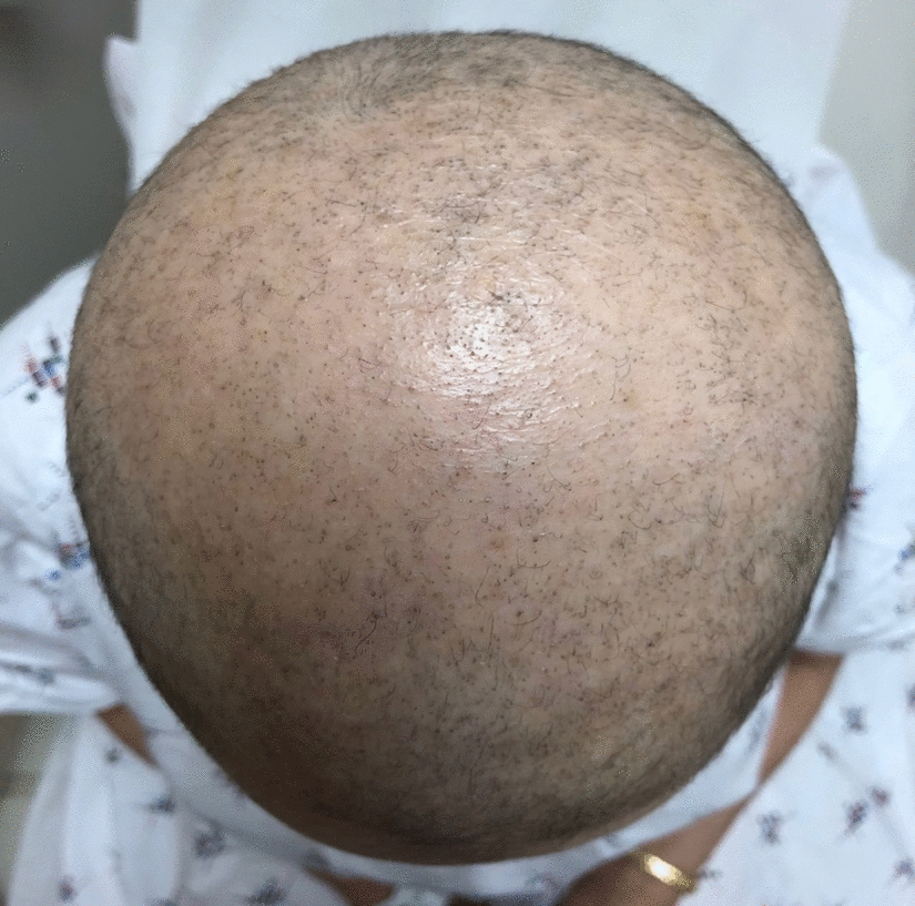
Table 1 Histopathologic features of syphilis [29,30,31,32,33,34,35]Alopecia is an uncommon manifestation of secondary syphilis, occurring in less than 10% of patients. Alopecia syphilitica (AS) is nonscarring with non-inflammatory hair loss, and hair regrowth will be achieved following 5–12 weeks of treatment. Alopecia syphilitica may be accompanied by syphilitic lesions on the scalp (symptomatic AS) or alone (essential AS). Subclassifications of essential AS are moth-eaten, diffuse, or mixed pattern, with the moth-eaten pattern being the most common manifestation. Moth-eaten AS presents as a patchy pattern of alopecic areas throughout the scalp and mimics other causes of alopecia, including alopecia areata, tinea capitis, and trichotillomania [36]. The diffuse subtype may mimic telogen effluvium. Alopecia syphilitica is usually accompanied by other secondary syphilis manifestations, but in some cases may be the only clinical sign of disease [37].
Lues maligna, also called malignant or ulceronodular syphilis, is a rare and aggressive manifestation of secondary syphilis that is found most often in immunocompromised individuals, such as those with HIV [38]. Lesions of lues maligna are oval, ulcerative, or necrotic papules and plaques with a thick scale or crust that mainly affects the trunk and extremities. Lesions on the face, scalp, palms, soles, and mucous membranes may also occur. Lues maligna is associated with high treponemal titers, systemic symptoms, and severe Jarisch–Herxheimer reaction. Associated comorbidities include HIV infection, diabetes mellitus, alcoholism, drug abuse, psoriasis, and hepatitis [39]. Other notable dermatologic manifestations of secondary syphilis can be observed in the nails (onycholysis, onychomadesis, brittleness, splitting, paronychia) and through pigmentation changes (“leucoderma syphiliticum,” scattered hypomelanotic macules at sites of previous inflammation or within an area of hyperpigmentation on the neck) [11, 40].
Early Non-primary Non-secondaryThe diagnosis of early non-primary non-secondary syphilis is made through positive serologic studies combined with an absence of signs or symptoms of primary or secondary syphilis and evidence that initial infection was acquired within the past 12 months [10]. Previously referred to as “early latent” syphilis, this stage is still considered infectious because of the 25% risk of relapse to secondary syphilis, most often occurring within the first year [41].
Unknown Duration or Late SyphilisThe stage classified as late syphilis or unknown duration differs from early non-primary non-secondary syphilis through evidence that infection occurred > 12 months previously or a lack of evidence to support infection occurred < 12 months previously, respectively. This stage of infection is supported by positive serologic studies and clinical evidence of late manifestations and/or neurologic syphilis; as before, signs and symptoms of primary or secondary syphilis are absent.
Late clinical manifestations are likely to present in this stage as these inflammatory processes take years to develop. “Tertiary syphilis” is no longer considered a stage with the updated classification system. Research on the natural history of untreated syphilis estimates that one-third of patients will develop late manifestations, including cardiovascular syphilis, late benign syphilis (“gummatous syphilis,” involving the skin, mucous membranes, bones, and joints), and neurosyphilis [41]. Cardiovascular and late benign syphilis are rare in the modern era of antibiotics, while neurosyphilis is still prevalent. Although included as one of several probable criteria in the revised case definition of unknown duration of late syphilis, neurologic syphilis (including otic and ocular syphilis) may occur at any stage of infection.
Cardiovascular syphilis predominantly affects the heart and great vessels. It may present as an aneurysm of the ascending aorta, aortic valve insufficiency, aortitis, myocarditis, and coronary vessel disease [9, 42].
Late benign syphilis is perhaps most closely associated with lesions of the skin, but virtually any tissue may be affected. Lesions of the skin may be of two forms: gummatous or noduloulcerative. Arising from a reactive granulomatous process, gummatous lesions of the skin are painless and rubbery nodules that are usually grouped and few in number. Gummas typically perforate the skin, leaving a distinctive punched-out ulcer from which necrotic material drains. Large lesions are destructive, heal with retracted and hyperpigmented scars, and may invade deeper tissues or arise from another tissue entirely. The noduloulcerative form appears as indurated and elevated papules or nodules that are red-brown in color. They expand to form serpiginous or arciform plaques, often healing centrally and occasionally eroding or ulcerating with thick crusts [43].
Neurosyphilis, Ocular Syphilis, and Otic SyphilisInfection of the nervous system by T. pallidum results in neurosyphilis, ocular syphilis, or otic syphilis. Each is considered a separate disease state but may occur concomitantly. Within days of the primary infection, treponemes invade the nervous system, thus neurologic involvement may occur at any stage of infection.
Late Benign (Tertiary) SyphilisEarly tertiary lesions may lack granulomata. Nodular lesions will show granulomatous inflammation (multinucleated giant cells, lymphocytes, plasma cells, and scattered epithelioid cells), inconspicuous necrosis, and possible endothelial swelling of vessels; at times, nodular lesions will lack granulomata. Gummatous lesions will also show granulomatous inflammation in addition to foci of acellular necrosis, and endarteritis obliterans of the dermal and subcutaneous vessels [29].
Congenital SyphilisClinical signs of congenital syphilis appear within the first 4–8 weeks of life. Cutaneous manifestations of early congenital syphilis include persistent rhinitis (“snuffles”), a vesiculobullous or maculopapular rash, and generalized nontender lymphadenopathy [44]. Condylomata, mucous patches, and fissures may also be observed at mucous membrane sites similar to cutaneous lesions found in infected adults [45].
留言 (0)