1. Tumor location: Geographic epicenter of the tumor or the location of the largest part of the tumor. It can be a single or more than one lobe. Location includes the following: Frontal, Parietal, Temporal, Occipital, Insular, Deep gray matter and Brainstem and cerebellum.
2. Tumor laterality: The side at which the tumor is epicentered, namely
-Right, Left and Central or bilateral
3. Enhancement pattern: It is calculated based on subjective qualitative assessment of the degree of contrast enhancement of the entire or part of the tumor on post contrast T1w sequences comparing to pre contrast T1w sequence.
It is further subdivided into
3. Homogenous/heterogenous: Whether the tumor is enhancing homogenously or heterogeneously in its entire portion.
4. Mild/Moderate and severe: Qualitative visual assessment of the degree of contrast enhancement of the entire tumor or part of the tumor.
5. Rim/Nodular/Solid/Patchy: Rim enhancement is provisionally defined as peripheral rim enhancement with central non-enhancing areas. Nodular enhancement is provisionally defined as enhancing nodule with remaining non-enhancing areas. Solid enhancement is provisionally defined as near homogenous or heterogenous enhancement of the entire tumor. Patchy enhancement is provisionally defined as non-uniform distribution of enhancing and non-enhancing areas within the tumor.
6. Proportion enhancing: It is a subjective qualitative assessment of the proportion of the enhancing tumor and is provisionally grouped into four categories namely
-No enhancing part, <25% of tumor, 25–50% of tumor and >50% of tumor.
7. Eloquent brain involvement: Involvement of the eloquent cortex or involvement of the underlying subcortical white matter of the eloquent cortex.
Following eloquent cortex were included in our study,
-Wernicke’s, Broca’s, Motor and Vision.
8. Necrosis: It is the portion of the tumor which is not enhancing on post contrast sequence and is T1 hypointense and T2 hyperintense with irregular borders. It is provisionally grouped into four subjective categories, namely
-No necrosis, <25% of tumor, 25–50% of tumor and >50% of tumor.
9. Edema: It is defined as the T2 and FLAIR hyperintense areas/ region surrounding the tumor. It is provisionally grouped into four subjective categories, namely
-No edema, less than tumor volume, equal to tumor volume and greater than tumor volume.
10. Hemorrhage: It is defined as the area of GRE/SWI blooming within the tumor or any area of T1 hyperintensity or T2 hypointensity.
11. Cysts: It is defined as relatively well-defined round or oval areas of T2 hyperintensity and T1 hypointensity, paralleling the CSF intensity in all sequences with a smooth, thin and regular enhancing or non-enhancing rim. Internal septations can be present or absent.
12. Multifocal/multicentric:
-Focal: It is defined as single tumor localized to one region of the brain parenchyma.
-Multifocal: It is defined as at least one not contiguous lesion with the dominant tumor and is outside the region of edema surrounding the dominant tumor. It can be either enhancing or non-enhancing.
This can be defined as those resulting from dissemination or growth or spread via commissural or other pathways, or via CSF channels or local metastases.
-Multicentric: Two or more widely separated lesions in different lobes or hemispheres that are not attributed to one of the above-mentioned pathways of spread.
-Gliomatosis: It refers to generalized neoplastic transformation of the white matter of most of a hemisphere.
13. Tumor size: It is defined as the maximum longest diameter of the tumor on axial plane (anteroposterior and transverse dimensions) and the craniocaudal dimension measured on coronal or sagittal plane. However, the single largest dimension was only taken in to account. It was further subdivided into
-Maximum dimension less than 2 cm, between 2–5 cm and more than 5 cm.
14. Satellite lesions: It is defined as an area of enhancement within the region of edema surrounding the index lesion and is not contiguous in any part with the major tumor mass.
15. Leptomeningeal spread: It is defined as the enhancement of the overlying pia/arachnoid layer either in continuity or not in continuity with enhancing or non-enhancing tumor.
16. Midline shift: It is defined as the mass effect with resulting midline shift of the brain to contralateral side. It was further subdivided into
-No midline shift, mild midline shift of less than 5 mm, moderate midline shift between 5–10 mm and severe midline shift of more than 10 mm.
17. Tumor crossing midline: It is defined as either the enhancing or non- enhancing tumor crosses into contralateral hemisphere through white matter commissures (not including the herniated ipsilateral parenchyma).
18. Calvarial remodelling: It is defined as erosion of inner table of skull either directly by the tumor or indirectly by the mass effect.
19. Diffusion restriction: It is defined as either presence of restricted diffusion or absence of restriction or facilitated diffusion.
-Restricted diffusion: Hyperintense on diffusion sequence and hypointense on ADC maps.
-No Restriction: Hypointense on diffusion sequence and hyperintense on ADC maps.
-Facilitated diffusion: Hyperintense on diffusion sequence and hyperintense on ADC maps.
20. Ependymal invasion: It is defined as invasion of adjacent ependymal surface by the tumor in continuity with it.
21. Epicenter: It is defined as the epicenter of the tumor which can either be cortical/ deep white matter.
22. Subcortical involvement: It is defined as the presence or absence of the subcortical region by the cortical or deep white matter epicenteredtumor.
23. Tumor margins: The margin characteristics of the tumor are provisionally grouped into three subjective categories, namely
-Well defined: All the margins were smooth and well defined.
-Ill defined: Most of the margins were ill-defined and irregular.
-Well defined with areas of focal infiltration: Most of the margins were smooth and well defined with few areas of ill-defined and irregular margins.
24. FLAIR/T2 mismatch: Homogeneous or heterogeneous signal intensity on T2WI and relative hypointensity on FLAIR with a peripheral hyper intense rim (T2- FLAIR mismatch).
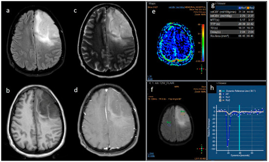
25. rCBV value: The value of relative cerebral blood volume is calculated based on the dynamic susceptibility contrast MRI.
26. Closeness to subventricular zone: The distance of the closest margin of the tumor from the ventricular margin. It is subdivided into
-Distance of the tumor less than 5 mm and more than 5 mm.
27. Dural enhancement: It is defined as the enhancement along the dural lining in case of a cortical based tumor.
28. Presence of vessel on T2w images: Presence or absence of vessels within the tumor on T2 weighted images.


留言 (0)