1. IntroductionObstructive sleep apnea (OSA) is the most common type of sleep-disordered breathing. This condition is characterized by recurrent episodes of partial or complete airway obstruction during sleep leading to repetitive apneas or hypopneas. Airway obstruction results from upper airway collapse or anatomic airway obstruction, even though respiratory effort is still present [
1]. The clinical signs and symptoms include sleep interruption, snoring, and daytime sleepiness, which can lead to significant impairment in the quality of life. In order to assess the severity of sleep apnea, the apnea–hypopnea index (AHI) is used. The AHI is based on the total number of apneas and hypopneas occurring per hour of sleep; an AHI
2].OSA represents a major public health issue: In fact, nearly one billion adults aged 30–69 years suffer from OSA worldwide [
3]. Population-based epidemiologic studies have uncovered the prevalence of OSA ranging between 4% and 24% in middle-aged people [
4]. In addition, the prevalence of sleep apnea has been reported to be 49.7% in men and 23.4% in women aged 40 years or older in a large population-based sample in Western Europe [
5]. Further data reported that 88% of men aged 65–69 years had five or more apneic or hypoxic events per hour, increasing to 90% in men aged 70–85 years [
6]. The variation in the estimated prevalence is likely to reflect the different health status of the older populations studied and the definition of the disease [
7]. For instance, Peppard et al. were among the first authors to show that the apparent increase in AHI with age was strongly correlated with concomitant weight gain [
8]. Notably, being overweight, and in particular having an elevated body mass index (BMI) is the strongest risk factor for developing OSA [
9]. Conversely, not all obese individuals have OSA. Furthermore, Hoch et al. found that AHI only increased with age among patients with moderate to severe OSA [
10]. Contrarily, a recent meta-analysis showed no statistical difference regarding baseline AHI between individuals younger or older than 65 years [
11]. In addition, the role of comorbidities in OSA patients has emerged over the years and the association between OSA and medical conditions such as hypertension, diabetes mellitus, cardiovascular disease, pulmonary disease or chronic mental health disorders is a cause for concern, since a bidirectional relationship is suspected [
12]. However, it remains controversial whether increasing age alone is associated with an increase in OSA severity, or whether this is due to individual biological changes or the result of more age-related comorbidities.
Therefore, the aim of the present study was to investigate and compare polysomnographic parameters of younger and older individuals in a retrospective analysis of all sleep laboratory polysomnographic recordings of one year, taking baseline study population characteristics, especially OSA-related comorbidities, sex and BMI into consideration.
3. Results 3.1. Combined (Male and Female) Study Population
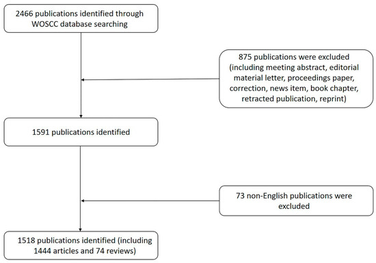
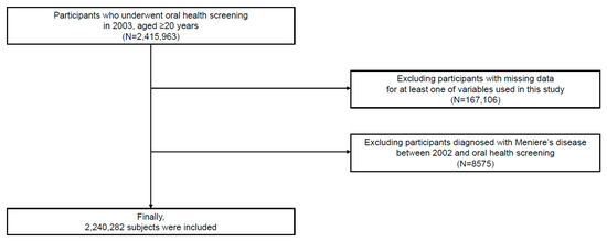
From 1 January 2020 to 31 December 2020 a total of 197 individuals underwent first-time full-night polysomnography in our sleep laboratory. Eleven individuals were excluded from the study due to age under 18 years, medical history of unstable cardiac diseases (e.g., history of heart attack), medical history of severe chronic mental health disorders (e.g., schizophrenia, and psychosis), or forms of sleep-disordered breathing other than OSA (e.g., central apnea). A total of 186 individuals met inclusion criteria and were included in the study.
63 male individuals (63.6%) and 36 female individuals (36.4%) were assigned to group “m+f < 55 yrs”. Age distribution in this group was 22.6–54.9 years (median 44.6 (37.1–51.7) years) and median BMI was 28 (25–32) kg/m². The review of all medical records of group “m+f < 55 yrs” revealed arterial hypertension, diabetes mellitus, cardiovascular diseases, pulmonary diseases and chronic mental health disorders to be present in 27 (27.3%), 3 (3%), 6 (6.1%), 4 (4%), and 11 (11.1%) individuals, respectively. The number of permanent medication taken daily by individuals assigned to group “m+f < 55 yrs” was 1 (0–2). At the time of the medical evaluation, 53 (53.5%) individuals assigned to group “m+f < 55 yrs” were non-smokers, 28 (28.3%) were current smokers, 4 (4%) were ex-smokers and 14 (14.1%) individuals lacked information about their smoking status. A comparison of different smoking statuses did not reveal statistical significance in this group.
Accordingly, 55 male individuals (63.2%) and 32 female individuals (36.8%) were assigned to group “m+f > 55 yrs”. Age distribution in this group was 55.1–85.1 years (median 62.8 (58.6–68.8) years) and median BMI was 30 (26–34) kg/m². The review of all medical records of group “m+f > 55 yrs” revealed arterial hypertension, diabetes mellitus, cardiovascular diseases, pulmonary diseases and chronic mental health disorders to be present in 51 (58.6%), 13 (14.9%), 16 (18.4%), 16 (18.4%), and 18 (20.7%) individuals, respectively. The number of permanent medication taken daily by individuals assigned to group “m+f > 55 yrs” was 3 (2–5). At the time of the medical evaluation, 58 (66.7%) individuals assigned to group “m+f > 55 yrs” were non-smokers, 14 (16.1%) were current smokers, 2 (2.3%) were ex-smokers and 13 (14.9%) individuals lacked information about their smoking status. A comparison of different smoking statuses did not reveal statistical significance in this group. All baseline characteristics of the combined (male and female) study population are shown in
Table 1. 3.2. Comparison of Sleep Parameters of the Combined (Male and Female) Study PopulationThe primary goal of this study was to compare sleep parameters in individuals less than 55 years of age (m+f 55 yrs). In this initial analysis, we compared mixed-gender study population groups. All respiratory parameters, such as AHI, AI, and HI, but not SI, were significantly higher in group “m+f > 55 yrs” (p = 0.0001, p = 0.0011, p = 0.0015, and p = 0.0739, respectively). An evaluation of pulse oximetry measurements found ODI and t90 to be significantly higher, and mOS significantly lower in older individuals (p = 0.0015, ppp = 0.0521). The distribution of sleep stages, more precisely percentages of N3 and REM sleep, did not differ significantly between groups (p = 0.0812, and p = 0.1677, respectively). Most of the young and aged study population showed supine positional OSA, but no age-related statistical difference was found (p = 0.2405). All the above-mentioned sleep parameters of the mixed-gender study population are shown in
Table 2 and presented in
Figure 1. Given the tested sample size, all significant group differences achieved a statistical power > 0.8 at an alpha = 0.05 following log-normalization of polysomnographic data to account for unequal variance between groups. 3.3. Male Study Population
A total of 63 male individuals were assigned to group “m < 55 yrs”. Age distribution in this group was 22.6–54.9 years (median 44.2 (38–51) years) and median BMI was 29 (26–33) kg/m². The review of all medical records of group “m < 55 yrs” revealed arterial hypertension, diabetes mellitus, cardiovascular diseases, pulmonary diseases, and chronic mental health disorders to be present in 16 (25.4%), 1 (1.6%), 3 (4.8%), 3 (4.8%), and 4 (6.3%) individuals, respectively. Regarding pulmonary diseases, bronchial asthma (BA) was found in two cases and chronic obstructive pulmonary disease (COPD) in one case. The number of permanent medication taken daily by individuals assigned to group “m < 55 yrs” was 0 (0–2). At the time of the medical evaluation, 30 (47.6%) individuals assigned to group “m < 55 yrs” were non-smokers, 19 (30.2%) were current smokers, 3 (4.8%) were ex-smokers and 11 (17.5%) individuals lacked information about their smoking status. A comparison of different smoking statuses did not reveal statistical significance in this group.
Accordingly, 55 male individuals were assigned to group “m > 55 yrs”. Age distribution in this group was 55.3–85.1 years (median 61.5 (58.4–68.7) years) and median BMI was 30 (26–34) kg/m². The review of all medical records of group “m > 55 yrs” revealed arterial hypertension, diabetes mellitus, cardiovascular diseases, pulmonary diseases, and chronic mental health disorders to be present in 34 (61.8%), 10 (18.2%), 10 (18.2%), 7 (12.7%), and 10 (18.2%) individuals, respectively. Regarding pulmonary diseases, BA was found in five cases and COPD in two cases. The number of permanent medication taken daily by individuals assigned to group “m > 55 yrs” was 3 (2–5). At the time of the medical evaluation, 36 (65.5%) individuals assigned to group “m > 55 yrs” were non-smokers, 11 (20%) were current smokers, 2 (3.6%) were ex-smokers and 6 (10.9%) individuals lacked information about their smoking status. Comparison of different smoking status did not reveal statistical significance in this group. All baseline characteristics of the male study population are shown in
Table 3. 3.4. Comparison of Sleep Parameters of the Male Study PopulationAfter comparing sleep parameters of the combined (male and female) study population, we aimed to compare sleep parameters of only male individuals less than 55 years of age (m 55 yrs). In contrast to the combined (male and female) analysis, AHI and AI were the only respiratory parameters being significantly higher in group “m > 55 yrs” compared to group “m p = 0.0067, and p = 0.0135, respectively) while HI and SI did not differ significantly between groups (p = 0.1044, and 0.3396, respectively). Pulse oximetry measurements of ODI and t90 were found to be significantly higher in older individuals (p = 0.0248, and p = 0.0002, respectively), while mOS was found to be significantly lower (p = 0.0007) in older individuals. Also, TST was significantly lower in older individuals (p = 0.0141). The distribution of sleep stages, more precisely percentages of N3 and REM sleep, did not differ significantly between groups (p = 0.3459, and p = 0.1870, respectively). Supine positional OSA was significantly more prevalent in younger males compared to older individuals (p = 0.0048). All the above-mentioned sleep parameters of the male study population are shown in
Table 4 and presented in
Figure 1. It is noteworthy that, of the polysomnographic data reported as significant for the male collective, only AHI was found to have a statistical power of > 0.8, mainly because of the very high interindividual variance. 3.5. Female Study Population
A total of 36 female individuals were assigned to group “f < 55 yrs”. Age distribution in this group was 22.9–54.8 (median 49.7 (36.7–52.8) years and median BMI was 27.5 (23–30) kg/m². The review of all medical records of group “f < 55 yrs” revealed arterial hypertension, diabetes mellitus, cardiovascular diseases, pulmonary diseases and chronic mental health disorders to be present in 11 (30.6%), 2 (5.6%), 3 (8.3%), 1 (2.8%), and 7 (19.4%) individuals, respectively. Regarding pulmonary diseases, BA was reported. The number of permanent medication taken daily by individuals assigned to group “f < 55 yrs” was 1 (0–4). At the time of the medical evaluation, 23 (63.9%) individuals assigned to group “f < 55 yrs” were non-smokers, 9 (25%) were current smokers, 1 (2.8%) were ex-smokers and 3 (8.3%) individuals lacked information about their smoking status. Comparison of different smoking statuses did not reveal statistical significance in this group.
Accordingly, 32 female individuals were assigned to group “f > 55 yrs”. Age distribution in this group was 55.1–83.8 years (median 66.1 (61–70.1) years) and median BMI was 28.5 (24–34.5 kg/m². The review of all medical records of group “f > 55 yrs” revealed arterial hypertension, diabetes mellitus, cardiovascular diseases, pulmonary diseases, and chronic mental health disorders to be present in 17 (53.1%), 3 (9.4%), 6 (18.8%), 9 (28.1%), and 8 (25%) individuals, respectively. Regarding pulmonary diseases, BA was found in six cases and COPD in three cases. In addition, none of the female study participants suffered from anemia in need of specific medical therapy (e.g., iron substitution), the same applies to all male study participants. The number of permanent medication taken daily by individuals assigned to group “f > 55 yrs” was 4 (2–7). At the time of the medical evaluation, 22 (68.8%) individuals assigned to group “f > 55 yrs” were non-smokers, 3 (9.4%) were current smokers, 7 (21.8%) were ex-smokers and zero individuals lacked information about their smoking status. Comparison of different smoking status did not reveal statistical significance in this group. All baseline characteristics of the female study population are shown in
Table 5. 3.6. Comparison of Sleep Parameters of the Female Study PopulationAfter comparing sleep parameters of the combined (male and female) and the male study population, we finally aimed to compare sleep parameters of only female individuals less than 55 years of age (f 55 yrs). Matching the combined study population, all respiratory parameters, such as AHI, AI, and HI, but not SI, were significantly higher in group “f > 55 yrs”, compared to group “f p = 0.0005, p = 0.0027, p = 0.001, and p = 0.0947, respectively). Evaluation of pulse oximetry measurements found ODI and t90 to be significantly higher, and mOS significantly lower in older individuals (p = 0.0031, p = 0.003, and p = 0.0002, respectively). TST did not differ significantly between groups (p = 0.8106). The percentage of N3 sleep was significantly higher in younger women (p = 0.0224), unlike the percentage of REM sleep, which did not differ significantly between groups (p = 0.3846). Most of the young and aged female subgroups showed supine position-related OSA, but no age-related statistical difference was found (p = 0.2405). All the above-mentioned sleep parameters of the female study population are shown in
Table 6 and presented in
Figure 1. In contrast to males, all significant polysomnographic data are reported with a statistical power > 0.8, except for percentage of N3 sleep. 4. Discussion
In this study, we showed that several important sleep parameters differ significantly when younger and older individuals were compared: First, the core parameter for determining the severity of OSA-AHI was significantly higher in older than in younger participants, when considering the combined (male and female) study population, as well as the male and female cohorts. This holds also true for AI, which was significantly higher in all older cohorts. Second, HI was higher in all older cohorts, with statistical significance found only in the combined and female subpopulation. Third, pulse oximetry measurements of ODI and t90 were found to be significantly higher in all older cohorts, while mOS was found to be significantly lower in these subpopulations. Interestingly, SI was not significantly different in any subpopulation analysis.
The aforementioned results are in line with recently published studies: Kopel et al. demonstrated in a large retrospective analysis of 3993 diagnostic sleep test results that AHI less likely is normal as individuals get older [
18]. In addition, Ernst et al. proved OSA prevalence to be higher among elderly individuals (>65 years) compared to younger cohorts (18–45 years and 46–65 years) in a retrospective study of 2491 respiratory polygraph recordings [
19]. Likewise, Leppänen et al. proved that AHI and duration of apneas, hypopneas, and desaturations increase with increasing age in a retrospective analysis of 1090 individuals with AHI ≥5 [
20]. Moreover, Gabbay et al. found that OSA increased with age in both men and women, but men had consistently higher AHIs for each age group [
21]. Interestingly, a meta-analysis by Iannella et al. indeed proved a direct correlation between aging and AHI values but found no significant differences in baseline AHI in comparison of individuals younger or older than 65 years [
11].The relationship between OSA severity and age, and also with BMI and medical conditions has recently been of increased interest [
9,
12]. In fact, the strongest risk factor for developing OSA is being overweight, and in particular an elevated BMI [
9]. Obesity increases the risk for OSA by 10–14 times and weight loss reduces the risk for this condition [
22]. Notably, neither the combined (male and female) population, nor the male or female subpopulation analyzed in the present study showed significant differences in BMI when younger cohorts were compared with older ones, therefore attenuating a major confounder in the consideration of age-related OSA severity.Besides BMI, several authors suggested that the age-related increase in OSA severity is due to concomitant age-related increase in OSA-enhancing comorbidities such as chronic heart failure, diabetes mellitus, and renal failure [
7,
12,
23]. This is also the case in the present study: The review of all medical records revealed arterial hypertension, diabetes mellitus, cardiovascular diseases, pulmonary diseases, and chronic mental health disorders to be significantly more prevalent in the combined (male and female) population, as well as the male subpopulation (except for pulmonary diseases). This fact should be taken into account when considering age-related increase in OSA severity.Interestingly, the data of the present study showed that within the female subpopulation all investigated comorbidities (except for pulmonary diseases) did not differ significantly between the young and old cohort. In summary, within the older cohort of the female subpopulation, all investigated sleep parameters were significantly increased (AHI, AI, HI, ODI, and t90) or decreased (mOS), although there were no significant differences neither in comorbidities nor in BMI compared to the younger cohort. Our results are consistent with a study by Fietze et al. that found a positive linear association between AHI severity and prevalence with age for both men and women. However, OSA onset was later in women [
24]. Given that BMI did not differ significantly within all investigated subpopulations, we suggest that OSA severity may increase with age due to the increasing accumulation of comorbidities in males, but not in females.This finding may be due to the effect of menopause on sleep-disordered breathing in females or may be due to the existing differential systemic pro-inflammatory (or other) effects of intermittent hypoxia between females and males [
25,
26]. Therefore, these findings provide new evidence suggesting a differential association of sex with the aging process that promotes OSA severity.Another possible explanation for the increased prevalence of sleep apnea amongst the elderly could be age-related changes in breathing control at sleep onset increasing apneic or hypoxic events during sleep. However, Browne et al. proved stable central breathing control in older people, indicating that aging per se does not promote central sleep apnea [
27]. Alternatively, different authors suggested the higher prevalence of OSA in older people may be associated with age-dependent mass reduction or lengthening of pharyngeal skeletal muscles, leading to increase in pharyngeal resistance [
28,
29]. This thesis might be supported by the fact that the majority of sleep-disordered events are predominantly obstructive.
Due to the cross-sectional study design, the effect of aging on the increase in OSA severity could not be investigated in each individual patient; this limitation may be addressed in future prospective studies. It is acknowledged that future studies are needed to investigate the effects of age on severity of individual AHI and other basic sleep parameters. Notably, the number of permanent medications was significantly higher in all older cohorts, which may also have influenced OSA parameters. Another potential source of error could be that all baseline evaluation of comorbidities as well as the intake of permanent medication was based on self-reporting, although all information was confirmed throughout an in-person interview. Although an attempt was made to compare comorbidities influencing OSA severity or BMI in the different groups, it cannot be excluded that individual diseases or past OSA-specific surgeries were selectively causative for the expression of sleep apnea. Furthermore, it must be acknowledged that all patients examined in our sleep laboratory presented with complaints of clinically relevant sleep apnea syndrome. Thus, our study population does not include asymptomatic OSA patients. Moreover, due to the retrospective study design it was not possible to access the number of women who were menopausal at the time of polysomnography. This limitation makes it impossible to correlate menopause with OSA severity, especially among older women in the study population.
However, to the best of our knowledge, this is the first study to compare different sleep parameters in young versus old mixed-gender cohorts, as well as only male or female subpopulations taking OSA-related baseline characteristics into consideration. This evidence should be considered in the context of an aging global population to prevent the health impacts of OSA and its associated comorbidities, especially in older patients. In addition, it seems that a different strategy of prevention should be followed between females and males.


留言 (0)