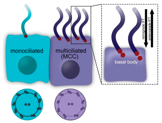
Several of the aforementioned monogenic neurodevelopmental disorders exhibit common neuropsychiatric manifestations including early onset epilepsy, intellectual disability, and ASD. In addition, the majority of these disorders exhibit similar neuronal primary cilia defects, namely fewer and shorter cilia (
Table 1). Given the critical role of primary cilia in the brain, it is possible that defects in these organelles contribute to the neuropsychiatric manifestations. It is worth noting however, that primary cilia dysfunction might cause different changes on the molecular level depending on the cell type and the specific genetic perturbation. Hence, while the phenotypic manifestations are shared, different mechanisms could be responsible in each disorder. Therefore, it is important to examine (1) whether and how the genetic perturbations underlying these disorders lead to abnormal cilia structure and function, and (2) if and to what extent, primary cilia dysfunction is involved in this wide range of neurologic abnormalities in each of these disorders. Such studies could open the road for discovery of novel therapeutic targets and development of new treatment strategies. Crosstalk between Signaling Pathways in Monogenic Neurodevelopmental Disorders Most studies identifying deficits in primary cilia in monogenic neurodevelopmental disorders have not characterized the mechanisms involved. Some of these studies, however, have suggested that alterations in major cellular functions, such as autophagy, are involved [
44,
75]. It is unclear whether there is a convergent mechanism or independent mechanisms underlying cilia deficits in each of these diseases. While each disorder likely has unique features, several lines of evidence support bidirectional interactions among key components of the molecular cascades involved in these disorders. Interestingly, many potential shared mechanisms center around direct or indirect mTOR dysregulation [
76].Genetic mutations underlying FCD and TSC affect mTOR signaling directly by altering the balance between activation and inhibition [
40,
43]. However, there are also proposed interactions between the RTT related protein, MeCP2, and mTOR protein. Specifically, aberrant mTOR signaling has been shown in patients with RTT syndrome [
77]. In addition, Mecp2 mutations in mouse models of RTT lead to downregulation of mTOR signaling activity and reduced neuronal size [
78], a known phenotype controlled by the mTOR pathway. Interestingly, in Mecp2 null or heterozygous mice, downregulation of the phosphorylated form of ribosomal protein S6 (p-rpS6), a well-established mTOR target, is detectable prior to the appearance of obvious RTT-related neurologic manifestations [
79]. mTOR signaling was also found to be altered in FXS [
75,
80]. For example, Sharma et al. showed increased mTORC1 activity in the hippocampal region of a mouse model of fragile X [
80]. Additionally, Yan et al., showed that mTOR-dependent decreased autophagy is responsible for several of the phenotypes observed in Fmr1-KO mice, including spine and synaptic plasticity defects as well as impaired cognition [
75]. MeCP2 was also shown to interact with the FMRP. Specifically, a study showed reciprocal regulation between the expression levels of these two proteins both in vitro and in vivo [
81].CDKL5 has also been shown to affect the mTOR pathway [
82,
83,
84]. Studies in Cdkl5 mutant mouse models reveal downregulation of Akt and mTORC1 activity, hence disruption of the Akt/mTOR signaling cascade [
83,
84]. Notably, one of these studies showed that by boosting phosphorylation of GSK-3b, an Akt downstream target, in Cdkl5 null neuronal precursor cells, several developmental alterations including neuronal survival and maturation were rescued [
84]. Another study examined how loss of Cdkl5 affected the mTOR signaling cascade by examining components of the mTOR pathway in different neuronal types. The authors examined cortical excitatory and inhibitory neurons, as well as striatal inhibitory neurons, and observed differential perturbation of the mTOR signaling cascade, suggesting that Cdkl5 affects mTOR in a cell type-dependent manner [
82].The mTOR dysregulation seen in FCD, TSC, RTT, FXS and CDD is noteworthy given that several lines of evidence propose that that mTOR and primary cilia regulate each other [
85]. Specifically, primary cilia inhibit mTORC1 activity via several proposed mechanisms involving proteins such as Lkb1, Folliculin, AMPK and polycystin-1 [
86,
87,
88]. Reciprocally, several studies have shown that mTORC1 activity affects cilia formation and length [
44,
89,
90]. Outside of mTOR, MeCP2 and CDKL5 have been shown to interact in various systems. Specifically, it has been shown that MeCP2 can be phosphorylated in a Cdkl5-dependent manner [
91,
92] and that Cdkl5 is a MeCP2-repressed target gene in the rat brain [
93]. In addition, patient stem cells that express mutated MeCP2 or CDKL5 exhibit common phenotypes such as upregulation of glutamate D1 receptor (GluD1) [
94].Taken together these data suggest that there is some crosstalk between components of these signaling pathways and support the hypothesis of a convergent mechanism that could act independently or synergistically with other mechanisms to underlie cilia defects. One of the most intriguing signaling cascades that appears dysregulated in all these disorders is the Akt/mTOR signaling pathway. In FCD, TSC and FXS models Akt/mTOR appears upregulated and the number of primary neuronal cilia is reduced [
37,
39,
40,
43,
44,
75,
80]. Additionally, in FCD and FXS, remaining primary cilia are also shorter in length [
37,
44]. On the other hand, a few studies have shown that in CDD, Akt/mTOR is downregulated and primary neuronal cilia length is increased [
45,
82,
83,
84]. Taken together these data suggest that Akt/mTOR activity can bidirectionally affect the number and length of primary neuronal cilia. However, RTT syndrome appears to be an exception to this Akt/mTOR activity–cilia phenotype pattern as mTOR in RTT is downregulated and primary cilia are reduced both in number and length [
48,
78,
79]. One explanation could be that different mechanisms in each genetic perturbations could underlie and/or contribute to the primary cilia phenotypes. Further studies are warranted to elucidate the molecular mechanisms leading to impaired ciliation in these disorders.


留言 (0)