
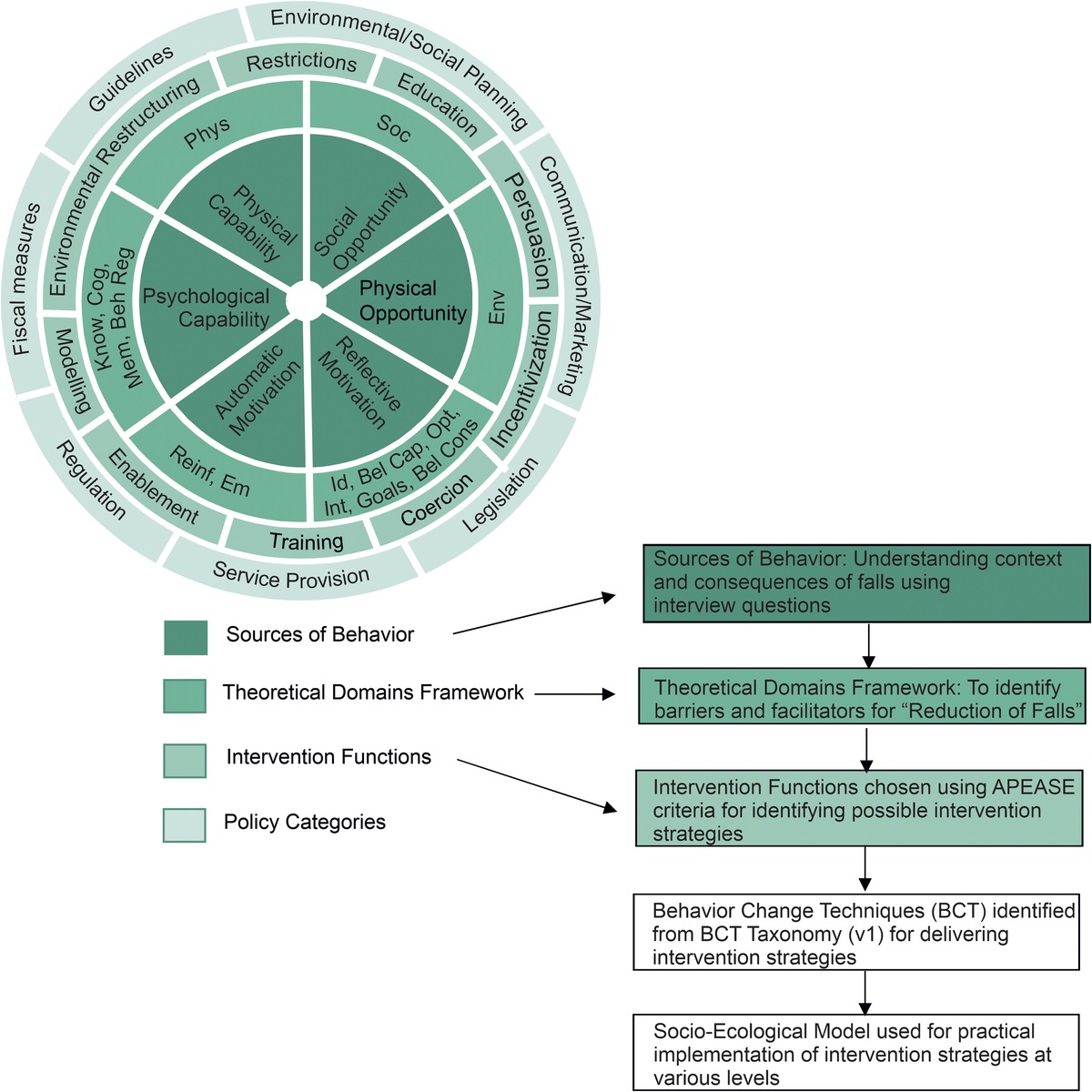
記住我


People with impaired vision can face increased challenges when performing many activities, leading to reduced quality of life.1–9 When visual impairment cannot be corrected with medical treatment or conventional refractive optics, low-vision care and rehabilitation services are an important option. These services involve providing information, training, and devices that aim to help individuals to maximize their residual vision and to participate in desired activities.10 Basic rehabilitation is tailored toward an individual's needs and goals but can also be informed by empirical research on common challenges faced by people with impaired vision and on the efficacy of compensatory strategies and devices.11–15
To better understand the needs of people seeking low-vision care, as well as the services provided to them at clinics, several prospective and retrospective clinical studies have been conducted.5,16–21 These prior clinical studies identified activities such as reading, driving, and mobility as being among the most common goals for patients with low vision, which is consistent with laboratory research on the impact of visual impairment on performance of these tasks.22,23 Whereas some studies have investigated differences in needs associated with different demographics (e.g., age and sex),5,13 none have examined how the goals of patients with low vision are changing over time. Examining longitudinal trends can play an important role in keeping up with potential shifts in the needs and priorities for services. Other studies have characterized patterns and longitudinal trends in prescribing rehabilitative devices from the 1970s to the early 2000s, but information about potential shifts in practice patterns in recent years is lacking.24–27 Here, we used a retrospective study design to characterize trends over the past decade in the topics discussed during low-vision patient case histories and the types of rehabilitative devices assessed in low-vision examinations at two clinics.
METHODS Data SetA retrospective study was conducted on deidentified electronic health record data from the Low Vision Clinic at the Meredith W. Morgan University Eye Center in Berkeley, California, and LightHouse for the Blind and Visually Impaired in San Francisco, California, between June 1, 2009, and June 1, 2019. This research was reviewed by an independent ethical review board and conforms with the principles and applicable guidelines for the protection of human subjects in biomedical research.
The two sites have a shared electronic health record system. No major version updates were made during the period under study, and most of the care providers worked at both sites. The care providers were optometry interns and residents under the supervision of faculty. The electronic health record fields analyzed include examination year, patient age at examination, patient sex, diagnosis codes, complaint notes, and refraction fields. Complaint notes contain free-form text with clinician notes that are recorded during the case history, in which patients are typically asked about their vision goals, current devices and adaptations, mobility, light sensitivity, and activities of daily living. The refraction fields contain information about trial frame refraction and the different rehabilitative devices that were assessed during the examination. At both sites, rehabilitative specialists are available to make additional assistive technology recommendations, but these encounters are not recorded in the electronic health record.
Only the first examination from each patient who visited the clinics during this period was analyzed. Thus, if a patient returned for multiple visits, only the earliest visit was analyzed. Patients may have also visited the clinics before the start date of our analysis (i.e., they may already be established patients). However, it was standard practice to take a detailed case history at each visit, regardless of whether the patient was established or new. The analysis was restricted to patients who were 18 years or older at the time of the examination. A total of 3638 examinations that met these initial criteria were retrieved. Examinations were excluded under the following conditions: no diagnosis code or refraction information present, incorrect examination format used (i.e., not a low-vision examination), and low-vision demonstration only (i.e., not a comprehensive low-vision examination). In total, 168 examinations were removed based on these exclusions, leaving 3470 examinations for analysis. The numbers of examinations per calendar year were 329 (2009), 446 (2010), 414 (2011), 353 (2012), 349 (2013), 292 (2014), 325 (2015), 275 (2016), 264 (2017), 283 (2018), and 140 (2019).
Of the patients included in this study, 2585 (74%) had International Classification of Diseases (ICD) codes for level of visual impairment recorded, with either ICD-9 or ICD-10 codes used. These codes were used in the analysis as the metric of visual ability because of variability in recording practices for other metrics, such as visual acuity, visual field size, and contrast sensitivity. We dichotomized patients into two groups: moderate impairment (group 1) and severe impairment to total blindness (group 2). The levels of visual impairment associated with these two groups are summarized in Table 1.28,29 Note that the transition from ICD-9 to ICD-10 in October 2015 included a shift in the acuity ranges included in each ICD category. Therefore, group 1 includes moderate low vision (ICD-9) and category 1 (ICD-10); group 2 includes all other categories in ICD-9 and ICD-10. For analyses of overall frequency, we consider all 3470 records; for regression analyses, we consider only the 2585 records with ICD codes so that impairment level can be used as a regressor. Missing ICD codes likely reflect cases in which the clinician determined that the visual acuity or visual fields did not meet the ICD requirements for visual impairment.
TABLE 1 - Categories used for level of visual impairment and associated descriptions (ICD-9 and ICD-10) Visit date International classification of disease category Corrected visual acuity or visual field of better-seeing eye Group 1: moderate impairment Before October 2015 ICD-9: moderate low vision 20/70 to 20/160 October 2015 and after ICD-10: category 1 20/80 to 20/200 Group 2: severe impairment to total blindness Before October 2015 ICD-9: Severe low vision to total blindness 20/200 or worse October 2015 and after ICD-10: categories 2–5 20/250 to NLP or VF less than 10° in diameter For ICD-10 codes, the first value in the official ranges are 20/70 and 20/200 noninclusive. Here, we indicate the next possible line on the visual acuity chart for consistency with ICD-9 codes such that all ranges in the table are inclusive. Descriptions are based on published ICD guidelines.28,29 NLP = no light perception; VF = visual field.All statistical analyses were conducted in MATLAB (The MathWorks, Inc., Natick, MA), and a P value of less than .05 was deemed as statistically significant. Because multiple comparisons were conducted in each analysis, the Benjamini-Hochberg procedure was used to control the false discovery rate at 5%.30
Topics Discussed during Case HistoryThe frequency with which different topics were discussed during the case history was quantified using an automated search for a set of keywords in the complaint notes. The keywords were classified into predetermined topic categories modeled after the Low Vision Rehabilitation Outcomes Study, in which a specialist generated a standard set of query terms related to a set of functional complaints (Table 2).5 We omitted the hobby category because of the challenge of generating a comprehensive hobby list, and we omitted the assistive device category because we instead include an in-depth analysis of rehabilitative devices that were assessed with each patient. We combined the out-of-home activities and walking categories into a single mobility category, as was done for some analyses in the previous study.5 Lastly, additional words were added to some categories to be more comprehensive. In particular, we added more technology words to allow for a more focused analysis of technology-related trends over time. To expand the technology word list, common technology words, such as those in the original list, were input in the web-based tool relatedwords.org. Patients for whom the complaint notes were empty (n = 408) or for whom a standard template with some keywords included was copied into the record (n = 22) were omitted from this analysis. These omissions resulted in 3040 patients in the overall frequency analysis and 2267 patients in the regression analysis (because of the additional exclusion of empty ICD codes for regression), which represents 88% of the possible data.
TABLE 2 - Topics and associated keywords Topic Keywords Reading Read, print, reading Television Television, TV Driving Driv*, drove, road, street Social interaction Face, kids, people, son, daughter, facial, church, faces, husband, wife, spouse, parent, friend, friends Light related Glare, dim, dark, night, light, photosen* Employment and school School, blackboard, job, courses, employ*, occupation, work, chalkboard, whiteboard, class, classroom Technology Computer, type, typing, mouse, palm pilot, kindle, laptop, iPod, iPad, tablet, Galaxy, Android, Mac, phone, application, apps, Bluetooth, touchscreen, WiFi, wireless, cellular, Blackberry, Motorola, email, texting, messaging, camera, browser, ebook, internet, Google, chat, Skype, stream, streaming, video, Microsoft Home activities Writ*, cook, cooking, mail, ingredients, recipes, clothes, packag*, clean*, paperwork, bill, check, yard, microwave, bath, label, oven, eat* Mobility Getting around, getting places, shop, transportation, go out, going out, travel, leav* home, store, grocery, supermarket, bump, curb, ran into, step, walk, navigat*, balance, trip, fall, stair, ground, moving around, o&m, orientation and mobility, terrain, maneuvering *Indicates that we allowed for multiple permutations of a word (e.g., drive, driving, driver). Boldface indicates words that we added to the original source word list. Both the topics and keywords were modeled after Table 1 in the Low Vision Rehabilitation Outcomes Study with additional keywords added for specific topics.5The frequency, number, and type of rehabilitative devices that were demonstrated to each patient were determined using an automated search of free-form text for each patient. Many different types of devices were demonstrated over the decade, so we generated a set of categories after consultation with low-vision specialists and included both “low-tech” (optical) and “high-tech” (electronic) tools (Table 3). Both clinical sites offer separate electronic device evaluations with a low-vision rehabilitation specialist, which are not recorded in the electronic health records. Information about which demonstrated devices, if any, were ultimately prescribed to the patient is also not recorded. Thus, although electronic tools are demonstrated in the examination room, the tools included here are only a partial list. Nonetheless, these data provide an important snapshot of the primary tools that are being demonstrated to patients during their examination. Patients for whom there were no demonstrations (n = 227) were omitted from this analysis. These omissions resulted in 3243 patients in the overall frequency analysis and 2465 patients in the regression analysis (~95% of the possible data).
TABLE 3 - Categorization and description of rehabilitative devices Device Type Definition Tinted lenses Optical Colored lenses and filters (e.g., to reduce glare and ocular discomfort) Refractive spectacles Optical Ophthalmic glasses that require a refraction (e.g., progressive addition lenses, bifocals, single-vision lenses) Handheld magnifier Optical Handheld optical devices that magnify nearby objects Handheld telescope Optical Handheld optical devices that spot or magnify distant objects, or expand visual field when used inversely Other head-borne optical devices Optical Prefabricated (i.e., “over the counter”) or customized low vision tools worn like spectacles (e.g., prism readers, OptiVisor, microscopics, bioptics, prism fitting for hemianopic field loss) Stand magnifier Optical Optical devices mounted into a base, magnifying objects when rested on the surface underneath Electronic magnifier Electronic Desktop and portable digital magnifiers Other electronics Electronic Built-in accessibility features on consumer computers, tablets, and phones as well as advanced low-vision aids (e.g., NuEyes, OrCAM, IrisVision)We used multivariate logistic regression to model the probability of each topic being discussed and the probability of each device being demonstrated at an examination as a function of the year of examination, as well as the patient age (in years), patient sex, and impairment level (group 1 vs. group 2). Individual patients were modeled as random effects. For the topics analysis, we found that the verbosity of the records increased over the decade (e.g., the average number of words recorded per patient was 151 in 2009 and 237 in 2019), so we included a variable in the model controlling for overall word count. In addition to the main effects listed previously, we included one interaction term between patient age and year of examination, because we hypothesized that different age groups may have experienced changes in needs differently over the decade. Models were fit to binary data. Models fit using the raw values of continuous predictors did not converge, so the continuous predictors (patient age, year of examination, and word count) were z scored before fitting to facilitate finding an accurate model. In the results text and all tables, odds ratios for main effects of year of examination and age at examination are reported for rescaled coefficients to reflect the change in units of 10 years. Models were fit to a random sample of 80% of the data (1814 for the analysis of topics discussed, 1972 for the device demonstrations), and accuracy for predicting the remaining 20% was assessed by calculating the area under the receiver operating characteristic curve, which represents the trade-off between false-positive and true-positive classifications for a range of thresholds on the response probabilities estimated by the regression (ranging from 0 to 1).31
To visualize differences in trends for patients of different ages, we grouped patients into five age groups. Because there is no uniform standard for age groups in low vision, we base these cohorts on the Medical Subject Heading dictionary (https://www.ncbi.nlm.nih.gov/mesh/68009273). The five groups correspond to young adult (18 to 24 years), adult (25 to 44 years), middle aged (45 to 64 years), aged (65 to 79 years), and 80 years and older (≥80 years). For the word frequency regression analysis, the numbers of patients in each age group were 134, 312, 482, 336, and 550, respectively. For the device demonstration regression analysis, the numbers were 133, 326, 510, 382, and 621, respectively. We also used these age groups to conduct follow-up stratified analyses in the case of interactions between patient age and examination year. Specifically, when a significant (or marginal) interaction was observed, we calculated simple effects coefficients for examination year in each of the age groups and examined them separately. Other than the removal of the coefficient for age, these models were the same as the main models.
RESULTS Patient DemographicsCollapsing across all years, there was a relatively balanced sex distribution with 44% males and 56% females. The mean age was 62 years (range: 18 to 103 years), and the average female was older than the average male (female mean age = 65 years; male mean age = 58 years). The four most prevalent causes of visual impairment were age-related macular degeneration (29%), glaucoma (16%), retinitis pigmentosa (7%), and diabetic retinopathy (7%). Of the patients with ICD codes, the majority (65%) were in group 1 rather than group 2 (the group 1 percentage was 63% during the period in which ICD-9 codes were used, and 69% in the period when ICD-10 codes were used). These demographic trends were relatively consistent over time, as shown in Fig. 1, and consistent with prior studies reporting characteristics of individuals seeking low-vision care in the United States.16,17
 FIGURE 1: Patient demographics over the decade. (A) Percentage of patients with female (green) and male (gray) sex for each year. (B) For each year, violin plots illustrate the distribution of patient ages for females (left/green) and males (right/gray). The mean for each year is indicated with a circle/square for females/males, respectively. (C) Percentage of patients with each of the top four most common diagnoses for each year. Note that some patients had multiple diagnoses. (D) Percentage of patients in each visual impairment group for each year (Table 1). These percentages are relative to the total number of patients with ICD vision impairment codes filled in. AMD = age-related macular degeneration; DR = diabetic retinopathy; GL = glaucoma; RP = retinitis pigmentosa.Topics Discussed during Case History
Overall Frequency of Topics Discussed
FIGURE 1: Patient demographics over the decade. (A) Percentage of patients with female (green) and male (gray) sex for each year. (B) For each year, violin plots illustrate the distribution of patient ages for females (left/green) and males (right/gray). The mean for each year is indicated with a circle/square for females/males, respectively. (C) Percentage of patients with each of the top four most common diagnoses for each year. Note that some patients had multiple diagnoses. (D) Percentage of patients in each visual impairment group for each year (Table 1). These percentages are relative to the total number of patients with ICD vision impairment codes filled in. AMD = age-related macular degeneration; DR = diabetic retinopathy; GL = glaucoma; RP = retinitis pigmentosa.Topics Discussed during Case History
Overall Frequency of Topics Discussed
Collapsing across all years, the most common topic that appeared in the case history notes was reading (78%, n = 2372) (Fig. 2). Light-related, technology, social interaction, and employment/school words were also noted in more than half of the case histories. However, the number of keywords in each topic category was not equal, so in subsequent analyses we focus on changes in these topics over time, rather than overall frequency. To examine whether any topics had a consistent tendency to co-occur, we conducted an exploratory analysis in which we examined the pairwise joint probability distribution of these topics. When compared with the expected joint probabilities if all topics were independent, we did not see strong evidence for correlations, so in subsequent analyses, we continue to treat these topics as separate variables.
 FIGURE 2:
 Percentage of patients for whom at least one word in each topic category was noted in the case history, collapsed across all years. Error bars represent 95% binomial confidence intervals.
Regression Model of Topics DiscussedWe first focus on the main effects of the examination year, along with interactions between examination year and patient age (Fig. 3; Tables 4, 5). We found that more recent years were associated with greater odds of discussing technology, social interaction, mobility, and driving (Figs. 3C, D, G, H). Odds ratios for these topics, reflecting the increase in probability that a topic was discussed from the beginning to the end of the decade, ranged from 1.9 (driving) to 4.5 (technology). For mobility, we observed a significant interaction between examination year and patient age, suggesting that the increase in discussing mobility over time was steeper for younger patients than for older patients (Fig. 3G, Table 5). For social interaction, we observed an age-related interaction in the opposite direction: the upward trend over time was steeper for older patients (Fig. 3D, Table 5). Only one topic, reading, was associated with a significant decrease over time, with an odds ratio of 0.57 over the decade (Fig. 3A). Whereas this decrease was descriptively steeper in younger ages, the interaction with age was not statistically significant.
 FIGURE 3:
 Raw data and logistic regression model for topics discussed in the case history as a function of examination year and patient age. Each panel plots the percentage of patients with at least one word in each topic noted in the case history as circles, binned in increments of 2 years for examination year and into the age cohorts for patient age in years: 18 to 24 (red), 25 to 44 (blue), 45 to 64 (gold), 65 to 79 (green), and ≥80 (black). Data represent a random sample of 80% of patients to which the model was fit. Lines show the average fitted probability for these data in each year and age bin. Note that the data points and fits both reflect additional variation because of different proportions of patient sex and visual impairment level in each bin.
TABLE 4 - Logistic regression models for each topic Reading Light Technology Social Employment/school In home Mobility Driving TV Exam year Odds ratio* 0.57 1.47 4.53 3.31 1.33 1.46 2.71 1.95 1.55 95% CI 0.35–0.94 0.97–2.23 3.07–6.69 2.24–4.90 0.91–1.96 1.02–2.11 1.88–3.88 1.35–2.80 0.99–2.45 t Stat −2.22 1.81 7.63 5.99 1.46 2.05 5.40 3.60 1.90 P .03 .07 <.001 <.001 .14 .04 <.001 <.001 .06 Age at exam Odds ratio* 1.39 1.00 0.92 1.38 0.66 1.23 0.93 1.02 1.51 95% CI 1.31–1.47 0.95–1.05 0.88–0.96 1.31–1.44 0.63–0.69 1.17–1.28 0.89–0.98 0.98–1.07 1.42–1.61 t Stat 11.53 −0.09 −3.58 12.90 −16.05 8.81 −3.04 1.11 13.06 P <.001 .93 <.001 <.001 <.001 <.001 .002 .27 <.001 Year × age Ratio of odds ratio 1.08 0.98 0.93 1.19 0.94 0.95 0.86 1.07 0.93 95% CI 0.95–1.23 0.88–1.10 0.84–1.04 1.06–1.33 0.83–1.05 0.85–1.05 0.78–0.96 0.97–1.19 0.81–1.07 t Stat 1.14 −0.3 −1.23 2.99 −1.12 −0.99 −2.80 1.3 −1.0 P .25 .75 .22 .003 .26 .32 .005 .19 .30 Sex (F) Odds ratio 1.44 0.88 0.7 1.36 0.77 1.37 1.00 0.86 0.96 95% CI 1.13–1.85 0.70–1.10 0.61–0.93 1.10–1.67 0.63–0.95 1.12–1.67 0.82–1.22 0.70–1.05 0.76–1.21 t Stat 2.91 −1.13 −2.6 2.85 −2.41 3.06 −0.01 −1.47 −0.36 P .004 .26 .008 .004 .02 .002 .99 .14 .73 Impairment level (group 2) Odds ratio 0.49 0.85 0.75 1.45 0.91 1.34 1.29 0.38 1.33 95% CI 0.38–0.63 0.68–1.07 0.61–0.93 1.16–1.80 0.73–1.13 1.09–1.65 1.05–1.59 0.31–0.47 1.05–1.69 t Stat −5.69 −1.4 −2.60 3.30 −0.84 2.75 2.46 −8.8 2.33 P <.001 .16 .009 .001 .40 .006 .01 <.001 .02 Word count Odds ratio 1.94 2.33 2.08 1.60
留言 (0)