
記住我


This manuscript was written according to CONSORT statement. The study was approved by an independent Ethics Committee (Fundació Unio Catalana Hospitals) on 27 January 2015 (CEIC 15/03). All patients signed an informed consent to participate. The study was conducted according to the Declaration of Helsinki and all local legal and regulatory requirements. Trial registration: NCT02479321 (24/06/2015).
Study designThis is a single-centre, non-randomized, hospital-based intervention study with a historical control group (CG) and 12-month follow-up after hospital discharge.
Inclusion/exclusion criteriaPatients over 64 years with hip fracture within an ERP who underwent surgical treatment were included.
Exclusion criteria were as follows: patients with pathological fractures, traffic-related fractures and refractures; patients with known contraindication or limitations to advanced haemodynamic monitoring with ClearSight® system and EV1000 platform (Edwards Lifesciences, Irvine, USA) (Saugel et al. 2015); patients with Raynaud disease, with aortic valve prosthesis, proximal aortic aneurysm, known intra-cardiac shunts; moderate to severe mitral or aortic regurgitation; moderate to severe aortic or mitral stenosis; patients with poor-quality arterial waveform signal (see below) and patients with significant preoperative psychomotor agitation.
Conduct of the studyPerioperative management common to both groupsBoth groups were treated during the perioperative period in a multidisciplinary ERP unit created in 2010 exclusively dedicated to patients undergoing hip fracture repair (Reguant et al. 2019).
This unit’s objectives were to optimize patient health status before surgery, minimize preoperative stress, prevent and/or treat electrolyte imbalance, prevent and/or treat cardiovascular, respiratory, infectious and cognitive disorders, improve nutritional status and reduce surgical delay. The team comprised orthopaedic surgeons, anaesthesiologists, internists, a nurse case manager, a social worker, a physiotherapist and a nutritionist.
The main interventions of this multidisciplinary ERP unit for patients with hip fracture are shown in Table 1.
Table 1 Main interventions of enhanced recovery pathway unit for hip fracture patients Intraoperative periodAll subjects received standard of care with a 3-lead electrocardiogram, pulse oximetry and two peripheral intravenous lines. Patients in both groups received standard measures to maintain oxygen saturation by pulse oximetry >94% and heart rate <100 beats/min. Anaesthetic technique was at the discretion of the anaesthetist.
Post-anaesthetic care unit (PACU)After surgery, patients were treated in the PACU. The attendant physician determined discharge from this unit according to the local protocol.
Study arms Control groupData from patients who underwent surgery for hip fracture between October 2010 and November 2011 with follow-up to December 2012 were used for the CG (Reguant et al. 2019).
Haemodynamic management was at the discretion of the attending anaesthetist, using fluid therapy with crystalloids (0.9% saline, lactated Ringer or Isofundin®), colloids (Voluven®, Gelaspan ®) and/or cardiovascular drugs (in bolus—ephedrine, or continuous infusion—noradrenaline, dobutamine).
Non-invasive, intermittent arterial pressure measurement was obtained at least every 5 min using a cuff (Dahtex Ohmeda-GE S/5 Aespire ®).
Intervention groupData from patients who underwent surgery for hip fracture between June 2015 and February 2018 with follow-up to March 2019 were used as the IG.
Pre- and intraoperative non-invasive haemodynamic monitoring was conducted using ClearSight® monitor (Edwards Lifesciences, Irvine, USA). This monitoring system is based on the volume clamp method to continuously measure arterial pressure and the Physiocal method that periodically recalibrates the system (Saugel et al. 2015). Baseline haemodynamic measurements were taken when the Physiocal value exceeded 30 (Wesseling et al. 1995). If a Physiocal value over 30 was not obtained after 7 min monitoring, the patient was excluded due to a poor-quality arterial waveform signal (Wesseling et al. 1995).
Haemodynamic optimisation was performed according to the following GDHT protocol.
GDHT protocol (Fig. 1)
Fig. 1
Algorithms for goal-directed haemodynamic therapy phases
Three groups of cardiac index (CI) goals were formed according to age and prior functional capacity expressed in metabolic equivalents (METS) (Montenij et al. 2014) Additional file 1.
Fluids were given based on a protocolized haemodynamic algorithm to achieve and maintain an adequate indexed stroke volume (SVI) using crystalloids (0.9% Saline, Lactated Ringer or Isofundin ®) or colloids (if preoperative glomerular filtration rate was above 60 mL/min using Modification of Diet in Renal Disease (MDRD) equation (Ishihara 2014)- Voluven ®, Gelaspan ®). Choice of fluid type was based on anaesthesiologist criteria.
If a fluid bolus (FB) was not indicated and/or the target perfusion pressure was not been achieved with its infusion, vasopressor was administered to maintain systolic arterial pressure (SAP) above 90 mmHg (in bolus—ephedrine, phenylephrine, or continuous infusion—noradrenaline) or continuous infusion of dobutamine was added to achieve in addition, the individualized CI goal.
Phase 1: Preoperative resuscitation
On arrival in the surgical area, patients received a FB of 250 ml of 5 min. If SVI increased by 10% or more (First Fluid Bolus Responder), the fluid bolus was repeated (Cecconi et al. 2011). Fluid boluses of 250 ml were repeated until the SVI failed to increase by 10%.
Once preoperative resuscitation was completed, prophylactic antibiotic was infused (Table 1). This fluid contribution covered the estimated insensible losses during surgery (Jacob et al. 2007).
Phase 2: Post-incision optimisation
Post-incision optimisation began 15 min after the surgical incision, if the haemodynamic stabilization was achieved (SAP and heart rate variation < 10% for 3 min); meanwhile, the haemodynamic priority was the maintenance of arterial pressure above goal set (Tassoudis et al. 2011).
Haemodynamic optimisation consisted of a 100-ml fluid bolus administered of less than 3 min (Guinot et al. 2015; Mallat et al. 2015; Marik 2015; Muller et al. 2011). If SVI rose >10%, the 100 ml fluid bolus was repeated. The trigger SVI during surgery was calculated by subtracting 10% from the SVI obtained from the last positive 100 ml fluid bolus (Muñoz et al. 2016).
Phase 3: Maintenance during surgery
If at least one of the following objectives, SVI>SVI trigger and /or SAP>90mmHg, were not achieved, SVI was analysed:
If it was lower than the trigger SVI, a 100 ml FB was administered.
If SVI was higher than trigger SVI, we look at the CI.
If its value was under goal level, dobutamine was added.
When CI was above goal level, a vasopressor was chosen.
After each therapy, we re-evaluated the achievement of SAP and SVI goals.
Measurements and data handlingProcedureIntraoperative haemodynamic parameters (arterial pressure, heart rate, SpO2 in CG and also CI and SVI in IG) were registered at 15-min intervals. Haemodynamic instability, between intervals, was registered as an event in the next record. Fluids and cardiovascular drugs used from the patient’s arrival in the surgical area to their admission to the PACU were collected. In both groups, the evaluation of intraoperative complications was based on the intraoperative anaesthesia charts, whereas the postoperative complications were documented in the clinical course and hospital discharge report.
Post-discharge follow-up consisted of a structured telephone interview at 1, 3, 6 and 12 months after surgery. When the information could not directly be obtained from the patients (including deceased patients), the interview was done with next of kin or carer.
Assessment of outcomes Primary outcome measuresThe primary outcome was the percentage of patients who developed intraoperative haemodynamic instability, defined as one measurement of SAP < 90 mmHg in the CG and for at least 1 min in the IG and/or the need for a bolus of vasoconstrictor.
Secondary outcome measuresIntraoperative arrhythmias: defined as electrocardiographic evidence of cardiac rhythm disturbance.
Postoperative complications, grouped as follows:
Major cardiovascular complications: acute myocardial infarction, acute pulmonary oedema, ischemic stroke, pulmonary thromboembolism and cardiorespiratory arrest.
Minor cardiovascular complications: haemodynamic instability, defined as one measurement of SAP < 90 and arrhythmias.
Respiratory: hypoxia, defined as oxygen saturation <92%. Other respiratory complications: decompensation of chronic obstructive pulmonary disease, acute respiratory infection (clinical and radiological diagnosis and antibiotic treatment) and others.
Renal: presence of at least one of the following: oligoanuria, defined as urine output under 0.5ml/kg per hour, including absence of urine output. Acute renal failure, defined as an increase in urea > 50 mg/dl and creatinine levels > 1.09 mg/dl in any analysis during admission.
Infections: surgical wound (infection within 30 days after surgery that involves only skin and subcutaneous tissue of the incision), urinary (positive urine culture causing patient’s symptoms and which were not present on admission to hospital), systemic (fever >38 °C and positive blood antigen test with appropriate antimicrobial therapy instituted by a physician).
Surgical reintervention during hospital stay.
Total intraoperative volume and type of administered fluids, doses of cardiovascular drugs used, perioperative packed red blood cell transfusion, length of hospital stay, readmission within 30 days of surgery, and survival within 12 months after surgery.
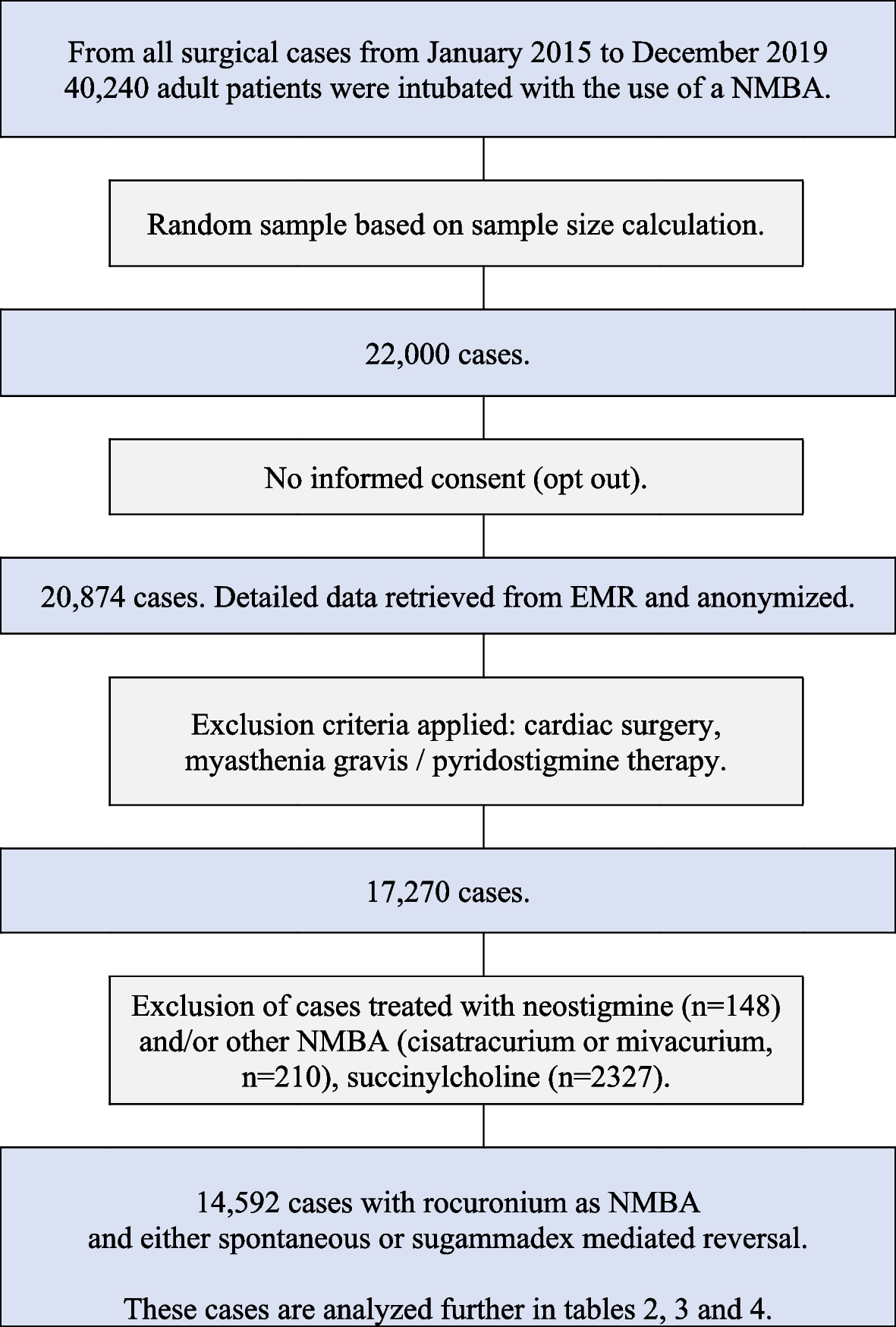
Statistical analysisSample sizeThe rate of intraoperative haemodynamic instability described with standard of care was 37.5% (Reguant et al. 2019). We planned a relative risk reduction of 30% in IG.
To achieve a power of 80% using a bilateral χ2 test for two independent samples with a level of significance of 0.05, 538 patients had to be included (269 patients in each group). With a potential dropout of 5%, 568 patients were included.
The percentage of patients who developed one or more postoperative complications in CG was 45.2%. A meta-analysis by Grocott and colleagues suggested a RR reduction of 0.68 for complications in patients undergoing major surgery (Grocott et al. 2013). A sample size of 568 patients, 284 in each group, would have 80% power to detect a reduction of at least 22% in the number of IG patients presenting one or more postoperative complications, using a bilateral χ2 test for two independent samples.
Statistical analysisCategorical variables were presented as absolute values and relative frequencies. Continuous variables are summarized as means and standard deviation for normal distribution and by the median and interquartile range (IQR) (25th to 75th percentiles) for non-normal distributions.
In the bivariate analysis, we used the Student’s t-test or the non-parametric Mann–Whitney U test for continuous variables. We used the χ2 test for categorical variables, and Fisher’s exact test or bilateral exact p-values in contingency tables when the expected frequencies were less than five.
One-year survival Kaplan–Meier curves were constructed, and the log-rank test was used to compare them. Crude and adjusted hazard ratios and confidence intervals (CI 95%) were calculated using Cox proportional regression models. The proportionality of hazards was verified by examining Schoenfeld residual plots.
Outcomes were analysed on an intention-to-treat basis. The level of statistical significance was two-sided 5% (p < 0.05). The IBM SPSS Statistics v.26 (IBM Corporation®, Armonk, New York) and Stata v.14 (StataCorp LP®, College Station, Texas) programmes were used for statistical analysis.
留言 (0)