This case report presents our management of a patient with bilateral, mobile, and pedunculated masses on both aryepiglottic folds. From the collaborative preoperative evaluation and planning with the ENT surgeon, the main focus of airway management was to minimize the risk of damage, bleeding, or displacement of the masses while maintaining airway patency. These risks would have increased if the process of awake intubation, which includes topical anesthesia and flexible bronchoscopic intubation, did not proceed smoothly. Therefore, our airway management of this patient may seem to have strayed from the conventional gold standard for difficult airway management. Because the concept of a perceived difficult airway encompasses a wide range of circumstances, an individual approach may be more appropriate to ensure optimal decisions rather than a strict adherence to a uniform difficult airway algorithm (Apfelbaum et al. 2022).
First, awake intubation was not attempted, although difficult ventilation and intubation were predicted. Instead, tracheal intubation was attempted after induction of general anesthesia, including neuromuscular blockade. Although awake intubation has been cited as the gold standard for management of anticipated difficult airways, it may be prudent to choose between awake and post-induction airway management on a case-by-case basis (Apfelbaum et al. 2022). In addition, the literature is insufficient to evaluate the benefits or risks of maintenance versus ablation of spontaneous ventilation and the use of neuromuscular blockade to improve mask ventilation (Apfelbaum et al. 2022). Our assessment was that manual mask ventilation would be maintained after ablation of spontaneous ventilation and administration of neuromuscular blockade for this patient, as there were no signs of stridor or dyspnea in any position including supine position. Instead, concerns were raised about the risk of the patient’s hanging masses being dislodged due to upper airway reflexes, such as coughing and laryngospasm. Despite the relatively low likelihood of such an event occurring due to the patient’s own upper airway reflexes, we could not completely rule out the risk. This was particularly due to the fragility of the masses’ stalks, as confirmed by the experienced ENT surgeon’s preoperative examination. This could occur during the application of topical anesthesia for awake intubation or the process of endotracheal intubation using a flexible bronchoscope. It could even occur during post-induction intubation if the depth of general anesthesia was insufficient to suppress upper airway reflexes. To address this, adequate doses of propofol, remifentanil, and rocuronium were sequentially administered while ensuring that mask ventilation was maintained with ease. When the attending anesthesiologist determined that the patient was sufficiently anesthetized to suppress upper airway reflexes, a gentle videolaryngoscopy was performed to evaluate the feasibility of intubation.
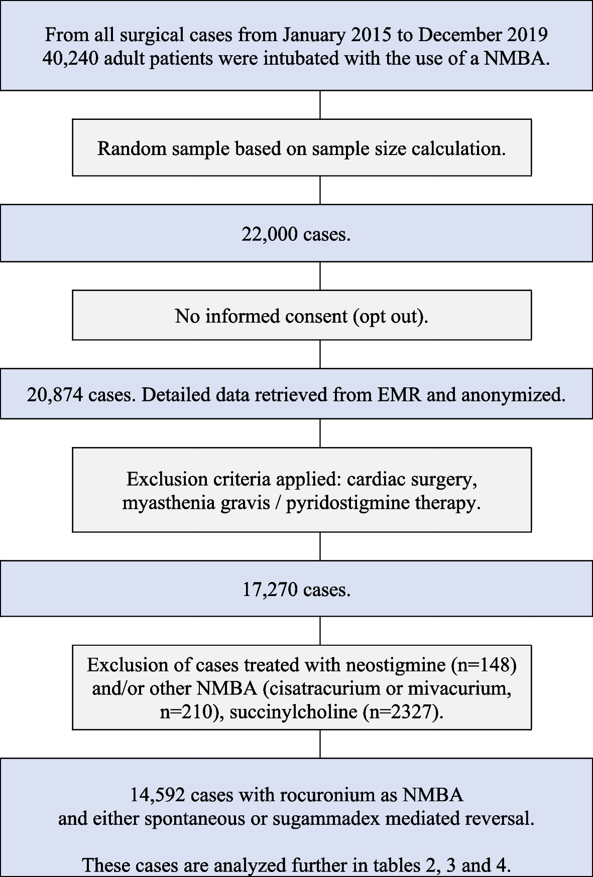
To ensure the safety of airway management, the induction of general anesthesia was performed in preparation for a difficult mask ventilation and tracheal intubation. Our primary backup plan, in case of ventilation and intubation failure, was to awaken the patient. This strategy included preoxygenating the patient for over 5 min and preparing 16 mg/kg of sugammadex for the rapid reversal of rocuronium (Gambee et al. 1987, Sørensen et al. 2012). Our approach also included the administration of a single dose of propofol and a continuous infusion of remifentanil, each characterized by short half-life and short context-sensitive half-time, respectively. Therefore, we anticipated that spontaneous ventilation would resume well before the onset of hypoxemia. Additionally, in the event of patient emergence and oxygenation failure, an ENT surgeon was on standby to perform an emergency tracheostomy.
Second, intubation was performed with videolaryngoscopy instead of flexible bronchoscopy. Precise manipulation of the flexible tip of the bronchoscope is difficult, especially in the presence of supraglottic masses (Choi et al. 2010, Liew et al. 2015). In this case, manipulating a thin flexible bronchoscope to pass between the masses, push them aside, and advance into the glottic opening could require multiple attempts, increasing the risks of bleeding, damage, or displacement of the masses. In addition, during intubation using a flexible bronchoscope, the tip of the ETT is not visible. Therefore, it would be unclear whether the tumors were being displaced beyond the vocal cords due to the tube insertion. In this case, due to the narrow space between the bilateral masses and the glottal opening, advancing the ETT over the flexible bronchoscope could push the masses into the glottis. This poses a risk for serious complications, such as bronchial obstruction and collapse. Therefore, we chose to use a videolaryngoscope instead of a flexible bronchoscope for tracheal intubation. The videolaryngoscope offers better control because the curved blade tip can be manipulated more easily in the desired direction and placed more accurately within the intended anatomical structure (Choi et al. 2010). We carefully placed the tip of videolaryngoscope blade in the vallecula, which was free of pathology. This method allowed for full visualization of the ETT tip on the monitor. Furthermore, the styletted ETT afforded better directional control compared to the flexible bronchoscope. As a result, the ETT tip was precisely advanced under video control through the narrow space between the bilateral masses and into the glottic opening, leading to safe and successful intubation in just one attempt.
In conclusion, tracheal intubation using a videolaryngoscope after induction of general anesthesia, including neuromuscular blockade, may be feasible in patients with supraglottic masses who do not exhibit stridor or dyspnea. However, the decision must be based on a comprehensive preoperative evaluation and adequate preparation. It is also essential to have prearranged strategies in place to address potential challenges related to inadequate oxygenation and unsuccessful tracheal intubation. Integral to these preparations is the collaboration of a multidisciplinary team, which encompasses every stage from evaluation and planning to the execution of the procedure, thereby ensuring safe and effective airway management.


留言 (0)