Table S1. StAx and SAH-p53–8 peptides and their respective negative controls with alkene containing amino acids highlighted in red and blue. Mutated amino acids in negative control peptides are highlighted in bold.
Table S2. List of MSA peptides used in this study with their chemical formula, molecular weight, calculated and found mass (m/z) for charged ions.
Figure S1. HPLC traces of purified MSA peptides measured at 280 nm.
Figure S2. Competition fluorescence polarization assays using MSA peptides with (A) different orientations and (B) varied linker lengths showing that neither peptide orientation nor linker length appeared to affect the measured binding properties. Error bars represent S.D. of triplicate experiments.
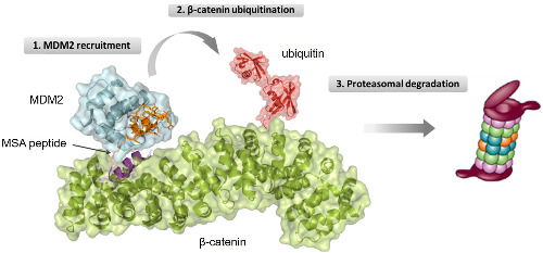
Figure S3. SAH-p53–8 and StAx-35 competed with MSA-2 in the MDM2 pull-down assay, indicating that the recruitment of MDM2 to β-catenin is a reversible process and both SAH-p53–8 and StAx-35 portion in the MSA peptides contribute to the binding.
Figure S4. Multifunctional stapled peptides with varied orientations and linker lengths exhibit different pull-down activity.
Figure S5. In vitro ubiquitination assays using (A) K-Ras and (B) c-myc protein as substrates indicated that the multifunctional peptides specifically promote poly-ubiquitination on β-catenin.
Figure S6. Confocal microscopy images showing subcellular localization of FITC-labeled peptides in HeLa cells after 3 hours treatment at 5 μM.
Figure S7. Endogenous β-catenin levels in SW480 cells upon treatment of (A) StAx-35 and (B) SAH-p53–8 indicated that neither of the unconjugated stapled peptides promotes degradation of β-catenin in SW480 cells.
Figure S8. (A) The endogenous β-catenin levels in SW480 cells were unchanged upon co-treatment of MSA-3 and the proteasome inhibitor MG-132. (B) Quantitative assessment of β-catenin levels in (A) normalized by corresponding β-actin in each sample. (C) Equal loading of each sample was verified by Coomassie staining. Error bars represent the S.D. of triplicate samples, ns = not significant, n = 3.
Figure S9. Luciferase reporter assays with SAH-p53–8 and StAx-35 indicated that neither of the non-conjugated stapled peptide causes significant suppression of β-catenin-dependent luciferase expression. Error bars represent S.D. of triplicate samples.

留言 (0)