
記住我

Rice is among the most widely spread food crops on earth, as the current world production is estimated at around 500 million tons annually.1 Importantly, along with the produced food, approximately 800 million tons of production residues annually arise, which are mainly rice straw and rice husk (RH).2 The common practice to “manage” the post-harvest waste is the open burning after the plant material has been left and dried on the fields, as it is cheap and straightforward, and the resulting ashes serve as fertilizer. However, studies demonstrated that the use or rice husk ashes for plant nutrition is not especially beneficial since other fertilizing technics, for example incorporation of rice straw in soil, have similar or better effects.3 Additionally, this practice causes massive air pollution in all rice-growing regions including China, India, Philippines and Vietnam.4 This pollution poses not only a health risk for humans,5 but also has a negative environmental impact due to the massive emissions of CO2 and particulate matter. Yet, to worsen things, studies also indicated a correlation between high air pollution levels and low rice yields.6 Hence, it is necessary to find alternative methodologies of waste treatment in order to decrease the significant health and environmental burden caused by rice production. Beside solving major challenges in logistics and political implementation, a valid and useful alternative must be presented in order to obtain acceptance from the most affected persons, the rice farmers, and convince them to abandon traditional methods.
For almost one century, there have been efforts to develop and demonstrate applicable methods of utilization of waste from the rice production chain.7 A vast majority of these approaches are dealing with the use of rice husk ash (RHA), which is the product of the often incomplete burning of RH. The calorific value of RH is approx. 18.7 MJ kg−1,8 which is less than half of biodiesel.9 The burning is, therefore, unsatisfactory in terms of energy efficiency and produces significant amounts of CO2, fine particles and silicon-containing waste.10 Nevertheless, it has to be noted that RHA can be used as an additive in concrete due to its high silica content. Even though it increases the water demand of the resulting raw material, this approach has already been demonstrated to be applicable.11 In recent years, many rather complicated methods have been developed to separate the two main compounds of RH, carbon and silica, in order to use them as bio-derived feedstock for silicon-based technologies. The obtainable products are solar-grade silicon,12 alkoxysilanes or orthosilicates (important building blocks in the semiconductors and silicone industry),13 electrode materials14 or light-emitting amorphous SiO2 nanophosphors.15 Carbon from rice husk can further be used as catalyst for production of biofuel additives,16 but the main potential lies in the use as absorbent for waste water treatment,17 due to its high surface area and good adsorption properties. An advantage of this approach is that the carbon must not necessarily be isolated from RHA, as it can be modified or used directly. The removable compounds span from organic pollutants to inorganic molecules like phosphate,18 nitrate19 or various relevant heavy metal ions like Cu2+, Ni2+, Cd2+, Mn2+, Co2+, Hg2+, Pb2+ or Cr6+.20
Next to the removal of organic and inorganic compounds from wastewater, the removal of microbial pathogens to weaken the antimicrobial resistance crisis might be a promising usage of rice husks in future. Even though highly active antibiotics have been a tool to efficiently control bacterial infection since the commercial introduction of Penicillin in the 1940s, we are currently facing a global antimicrobial resistance crisis, caused by the intense and wrong usage of antibiotics in combination with the lack of novel compounds.21 Every year, approximately 700,000 people die of infections caused by resistant pathogens in contrast to 8,200,000 by cancer and 1,200,000 by traffic accidents. It has been estimated that there will be around 10,000,000 deaths caused by resistant pathogens in 2050.21a, 22 Whether novel classes of antibiotics should be in investigation or rather alternative methods, is debatable.23
An alternative approach to commonly used antibiotics is the usage of silver nanoparticles (AgNP), since they are known to have antimicrobial properties.24 Every infection which can be avoided makes the use of “old-school” antibiotics obsolete and therefore lowers the risk of pathogens evolving resistances.25 In aqueous solutions, AgNP release silver ions (Ag+) which have numerous antimicrobial activities.26 Even though AgNPs themselves have antimicrobial properties, it is preferable to use silver ions instead as they are active in trace amounts, which is beneficial since silver can indeed be toxic in higher concentrations to cells of higher animals as well.27 They interact with the membrane and cell wall proteins of the microorganisms, resulting in damage to those structures along with a disturbed proton gradient, leading to cell death.28 If the silver ions reach the cytoplasm, they can inhibit the electron transport chain, damage DNA and RNA, inhibit DNA replication, denaturate the 30S ribosomal subunit resulting in inhibited protein synthesis and induce the formation of reactive oxygen species (ROS).28, 29 The numerous mechanisms of action result in a wide spectrum of target organisms, including (drug-resistant) bacteria, fungi and viruses. In addition, silver nanoparticle resistance of microorganisms is rarely reported.26b Moreover, it has been shown that the combination of silver nanoparticles and antibiotics significantly improves the antimicrobial effect of the antibiotics.29, 30 The wide spectrum of target organisms and the low risk of resistance development have made silver nanoparticles promising targets in current research.24a, 24b, 26, 31 The combination of an antimicrobial material like AgNPs and a material with excellent ability to adsorb pollutants, like RHA, leads to a composite which offers multifunctionality as a filter material. The need of cheap and easily manufacturable materials for both water and air sanitation is undeniably a key aspect to secure the health of millions of humans, since on the one hand, approx. 780 million people do not have access to clean water on a daily basis (status 2014),32 while, on the other hand, air pollution and airborne pathogens are extremely problematic, like recently demonstrated by the devastating COVID-19 pandemic,33 in both private households and indoor working places,34 as well as in professional healthcare environments.35 Fortunately, AgNPs can be and are applied against pathogens in water or on surfaces as well as against airborne pathogens.36 Moreover, since ancient times, the use of silver has not been associated with health risks (when not used in disproportionate amounts).37
State of the art in AgNP preparation is the reduction of an oxidized silver source by chemical, physical, biological or photochemical techniques,38 requiring an external reduction agent which reacts with AgI species. This approach leads to the production of stochiometric amounts of waste which need to be removed after the synthesis. The AgNPs prepared in such a way can be used as colloids, in solution or can be fixed on a solid supporting material which enables easier separation from the treated subject and recycling.
The preparation of supported AgNPs (fixed on a solid supporting material) commonly involves generation of the particles followed by deposition on a previously prepared suitable support, resulting in a multistep process that produces significant amounts of waste.
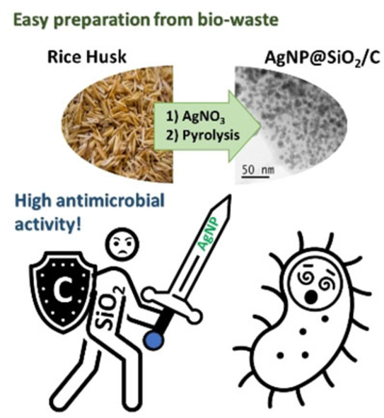
In this work, we present a straightforward method which combines preparation of support and AgNPs in one step, resulting in an excellent atom economy. The direct application of Ag+ ions on the lignocellulosic framework offered by RH is followed by carbothermal reduction yielding carbon/silica composites doped with AgNPs (Ag@RHA). This is, to the best of our knowledge, the simplest yet reported method for the preparation of antimicrobial systems with AgNPs as active agent and rice husk as support precursor. This presents a more sustainable alternative to previous reports on the preparation of “bio-derived” antimicrobial agents. Usually, RH is only used as the feedstock for the preparation of supporting material like active carbon,39 pseudowollastonite,40 silica-carbon nanoparticles,41 pure silica or MCM-41 nanoparticles. In all the previously reported methodologies, silver is impregnated on the prepared support in a subsequent step, therefore producing significant amounts of waste during the preparation.
Results and DiscussionAll tested Ag@RHA were prepared by wet impregnation of previously shredded RH with different amounts of AgNO3 followed by pyrolysis at 600 °C. The resulting pyrolyzed material is made mainly of carbon, silicon, oxygen and contains low amounts of nitrogen and phosphorus as well as sulfur and iron (Table S1 and Figure S1, Supporting Information). X-ray diffraction (XRD) analysis (Figure S1) revealed a broad peak from 15 to 30° suggesting the existence of an amorphous silica phase and/or carbon phase. Additionally, reflexes at 26.6°and 50.1° fitting to a crystallin hexagonal α-SiO2 phase are also present. The peak shapes for cubic silver reflexes at 38.1°, 44.3°, 64.5° and 77.5° match those of similar materials previously reported by our group,42 which indicates the existence of a bimodal Ag crystallite phase. Beside these three phases, two weak peaks at 35.6° and 62.7° indicate the existence of cubic Fe2O4 which can commonly be observed in RHA due to the high iron content in the plant.43 The average silver crystallite size was calculated using whole powder pattern fitting-Scherrer methodology for the raw material, containing 20 m% of silver and was found to be 14 nm. The surface area was investigated using Brunauer-Emmett-Teller (BET) isotherm analysis; a BET surface area of 260.7 m2 g−1 was found (Figure 1A) with a small hysteresis for the desorption. The cumulative volume of pores between 2 nm and 100 nm width is with 0.05 cm3 g−1, only half of the t-Plot micropore volume of 0.104 cm3 g−1 from 0.25 to 2 nm pore width. The clearly visible pore volume and area maximum around 0.6 nm underlines the microporous character of the prepared material.

A) BET-isotherm quantity ad and desorption plot and B) plot of pore volume and area vs. pore width.
IR measurements (Figure S2) revealed vibrations at 454, 571, 798 and 1067 cm−1 which can be correlated with Si−O bonds, confirming the presence of high amounts of silica in the material.44 A broad signal around 1543 cm−1 corresponding to carbon-carbon double bond vibrations was observed, which is a common feature in RHA.45
Moreover, the morphology of the AgNPs was investigated with scanning transmission electron microscopy (STEM), since previous studies suggested a correlation of the particle shape and antimicrobial activity, even though the reported particle size was up to 20-fold higher.46 The STEM images of Ag@RHA with the highest silver concentration revealed a spherical shape (Figure 2A, Figures S3A and 3B) with a diameter of about 10 nm with a few outliers reaching 50 nm and a full crystalline structure of the the AgNPs (Figure 2B and Figure S3C).

Microscopy images of Ag@RHA with highest silver concentration: A) Annular bright field STEM image of spherical shaped AgNPs and B) high-angle annular dark field STEM image displaying the crystalline character of AgNPs.
A series of composites (Ag@RHA) containing different amounts of silver was prepared to evaluate the minimal amount of metal needed to get a significant antimicrobial activity (Table 1). Nine samples have been prepared, starting from 28 wt % of AgNO3 in the RH (which correspond to 4.880(4) wt % of Ag in the pyrolyzed material), to 0.001 wt % of AgNO3 which corresponds to an Ag amount lower than the limit of detection (and quantification) of the applied inductively coupled plasma-optical emission spectrometry (ICP-OES) method.
Table 1. AgNO3 content in the reaction mixture, Ag content in the pyrolyzed material (Ag@RHA), Ag content in the cotton disc and in the agar used in antimicrobial activity tests.ii
AgNO3 added to RH [wt %][a]
Ag after pyrolysis [wt %][a]
Ag at cotton disc [wt %][a]
Ag in agar [wt %][a]
1
28
4.880(4)
0.069(3)
<0.001[b]
2
20
3.472(8)
0.047(5)
<0.001[b]
3
17
2.945(0)
0.033(4)
<0.001[b]
4
13
2.241(3)
0.031(5)
<0.001[b]
5
2.9
0.478(5)
<0.001[b]
<0.001[b]
6
1.4
0.10(2)
<0.001[b]
<0.001[b]
7
0.75
0.03(8)
<0.001[b]
<0.001[b]
8
0.015
<0.001[b]
<0.001[b]
<0.001[b]
9
0.001
<0.001[b]
<0.001[b]
<0.001[b]
10
0
<0.001[b]
<0.001[b]
<0.001[b]
[a] Measured with ICP-OES [b] limit of quanitification at 0.001 m%.For the antimicrobial activity assessment, the pyrolyzed material (Ag@RHA) was suspended in methanol (20 mg ml−1) and 10 μl of the suspension were transferred on a cotton disc (6 mm diameter) which was placed on the agar containing the microbes (Figure S4). Interestingly, no detectable amounts of Ag were found in agar after the antimicrobial tests, suggesting that leaching and diffusion of silver did take place only in minute quantities (Table 1).
The antimicrobial test results revealed a high sensitivity of all Gram-positive bacteria (Enterococcus faecium, Staphylococcus aureus), Gram-negative bacteria (Klebsiella pneumoniae, Acinetobacter baumannii, Pseudomonas aeruginosa, Escherichia coli), and the yeast (Candida albicans) whose growth were inhibited (Figure 3). The activity is clearly related to the silver content on the material and not the pure support, since no inhibition was detected when biochar without silver was used (prepared from RH under the same conditions). Most microorganisms were sensitive against the AgNPs made with an initial AgNO3 content of 0.015 wt % (Table 1, entry 8). E. faecium was sensitive to a lesser extent, but clearly inhibited in growth at an initial Ag content of 1.4 wt % (Figure 3A). Two different strains of E. coli (DSM 498 and 787) have been tested, but only the 498-strain showed clear sensitivity against the AgNPs (Figure 3G). The 787-strain showed a slight decrease in cell density but no clear zone of inhibition (not shown). The analyzed Ag contents in the cotton discs and agar were in every case <0.001 wt % as measured by ICP-OES (Table 1), showcasing the low amounts of silver necessary to achieve antimicrobial activity of the prepared material.

Antimicrobial effect of Ag@RHA to bacterial and yeast test strains. The bar charts show the zones of inhibition (ZOI) induced by different silver concentrations against (A) Enterococcus faecium, (B) Staphylococcus aureus, (C) Klebsiella pneumoniae, (D) Acinetobacter baumannii, (E) Pseudomonas aeruginosa, (F) Escherichia coli, and (G) Candida albicans. MeOH was used as negative control. Gentamicin was used as positive control for bacterial cells and Amphotericin B for the yeast. The cotton discs had a diameter of 6 mm. The error bars show the standard deviation n=3.
The antimicrobial activity of AgNPs against the ESKAPE pathogens (E. faecium, S. aureus, K. pneumoniuae, A. baumannii, P. aeruginosa, and E. coli) as well as C. albicans has been shown by several studies.40, 47 Salouti and colleagues showed a decreased number of colony forming units of a wound infected with S. aureus after treatment with AgNPs on a mouse model.48 The combination of rifampicin with AgNPs was shown to increase the antimicrobial activity against methicillin-resistant S. aureus and K. pneumoniae as compared to rifampicin alone.49 Furthermore, the antibacterial effect of AgNPs was shown against methicillin-resistant S. aureus.50 Transmission electron microscopy imaging revealed a thinning and permeabilization of the cell wall causing cell lysis.
Importantly, our Ag@RHA materials display antimicrobial activities at much lower silver concentrations compared to other studies. For instance, Azam and colleagues produced silver-doped CaSiO3 (synthesized from rice husk ash as a limestone precursor) and showed good antimicrobial activity against E. coli and S. aureus with Ag contents between 1–5 wt %.40 In contrast, we showed clear zones of inhibition with Ag contents below 0.001 wt % in the agar indicating a much higher bioactivity of the RHA-supported AgNPs.
The antimicrobial test results revealed a similar sensitivity of Gram-positive and -negative bacteria against Ag@RHA. Due to a thinner cell wall, it can be argued that Gram-negative bacteria might be more sensitive against AgNPs.51 Other studies argue that Gram-negative bacteria are less sensitive against AgNPs because of the negatively charged lipopolysaccharides of their outer membrane which very likely interact with the positively charged silver ions and prevent the ions from entering the cells.28b
Next to bacterial pathogens, the Ag@RHA showed clear antifungal effects against C. albicans (Figure 3F). This result is supported by other studies showing similar results. Kim and colleagues showed, by transmission electron microscopy imaging, that AgNPs result in a damage of the cell membrane of C. albicans.47b Using flow cytometry, they demonstrated that the cells got stuck in the G2/M phase of the cell cycle. Next to yeasts, the antifungal effect of Ag@RHA was also shown against several fungi, for example Aspergillus niger, Aspergillus fumigatus and Fumigatus soleni, indicating a wide antifungal spectrum.47a
Additionally, the conditions during the antimicrobial test have been reproduced with 50 mg of Ag@RHA to assess the stability of the prepared material and to investigate its applicability. The recovered Ag@RHA sample was analyzed by XRD. The average crystallite size was calculated at 12 nm using Scherrer's method (Table S2), showing no change compared to the freshly prepared material. Notable is the appearance of reflexes at 27.8°, 32.2°, 46.2°, 54.7° and 62.9°, relating to a cubic Na2O phase (Figure S1), which is most likely the result of the contact with the aqueous agar at 37 °C for 24 h rather than the result of using methanol in the preparation of the suspension.
ConclusionIn summary, we have successfully developed a simple method for obtaining highly antimicrobial silver nanoparticles supported on carbon/silica composites derived from agricultural (rice) bio-waste. The high antimicrobial activity was demonstrated by the good results obtained even when extremely low amounts of silver were loaded on the support. The materials were characterized before and after the antimicrobial tests, where no significant change was detected by X-ray diffraction analyses. This suggests a possible long-term stability and recycling potential. In conclusion, the herein described silver-containing rice husk-based antimicrobial materials are suitable for tackling some of the challenges discussed above, as they can be used as multifunctional filters or coating materials in water sanitation and medicinal applications, thanks to its stability and good antimicrobial activity, even with minimal amounts of silver deposited on the surface.
Experimental Section General Preparation ProcedureThe RH samples were received from Vietnamese farmers at the Red River Delta (Nam Ðinh Povince, 20°23′27.7“ N 106°15′34.7” E) and Mekong Delta (An Giang Provice, 10°24′55.0“ N 105°11′52.3” E).The RH was received pre-dried, and was shredded with a cutting mill SM 200 (rotation and speed 1000 rpm, 2 mm sieve sizes) and milled to a fine powder in a ball mill PM 200 (in steel cups with steel balls at 400 rpm for 3 h). The samples were mixed with different amounts of AgNO3, 50 ml ethanol and stirred for 24 h at 21 °C. The solvent was removed with a rotary evaporator and the sample was placed in an aluminium oxide crucible which was placed inside a quartz tube furnace. The samples were heated to 600 °C with 10 °C min−1, respectively, and the temperature was maintained for 1 h. The furnace was continuously flushed with nitrogen (50 ml min−1); after cooling to 25 °C, the material was stored under ambient conditions.
General Agar Diffusion Test ProcedureThe material was not soluble in any common solvent. Therefore, suspensions with MeOH were prepared (20 mg ml−1) and homogenized using FastPrep™ (Lysing Matrix D, 20 s, 6 m s−1; MP Biomedicals, Santa Ana, California, USA). The antimicrobial activity of the suspensions was tested using the antimicrobial disc diffusion test. We used the ESKAPE pathogens Enterococcus faecium (DSM 20477), Staphylococcus aureus (DSM 799), Klebsiella pneumonia (DSM 30104), Acinetobacter baumannii (DSM 30008), Pseudomonas aeruginosa (DSM 1117), Escherichia coli (DSM 498 and 787) and the yeast Candida albicans (DSM 10697) as test organisms. All strains were purchased at the German Collection of Microorganisms and Cell Cultures (DSMZ, Braunschweig, Germany).
The strains were pre-cultured before use overnight at 37 °C on Mueller Hinton Agar II (2.0 g L−1 beef heart infusion, 17.5 g L−1 acid casein hydrolysate, 1.5 g L−1 starch, 17.0 g L−1 agar; Becton Dickinson, Franklin Lakes, New Jersey, USA). For each organism, a solution with an absorbance of OD600 0.125 was prepared. The whole surface of a Mueller Hinton agar II plate was covered with the cell solution using a soaked cotton swab. 10 μl of the test suspension was transferred to the cotton discs (6 mm diameter) and the discs were transferred to the agar plate after the MeOH was evaporated. The negative controls were cotton discs with MeOH only, the positive controls were Gentamicin (10 μg, Sensi-DiscTM GM-10, Becton Dickinson, Franklin Lakes, New Jersey, USA) for bacterial strains and Amphotericin B (ROTI®Antibiotika Disks Amphotericin B (AP) 100 Units, Carl Roth, Karlsruhe, Germany) for the yeast. Organisms were incubated for 24 h as described above and the zones of inhibition were measured.
AcknowledgementsWe would like to thank all the Vietnamese farmers and local authorities at the rice husk sample collection sites (Nam Dinh and An Giang provinces) for their help and hospitality. Also, thanks go to Dr. Hendrik Lund and Reinhard Eckelt for performing the XRD and BET surface measurements, respectively, and for their helpful commends and discussions. Furthermore, we would like to acknowledge the funding for the SUVALIG project from the German Federal Ministry of Education and Research (BMBF, 031B0707B) and the RoHan SDG Graduate School funded by the German Academic Exchange Service (DAAD, 57315854) and the German Federal Ministry for Economic Cooperation and Development (BMZ) inside the framework “SDG Bilateral Graduate school programme”. Financial support from the Leibniz Society and the Leibniz Science Campus ComBioCat is also gratefully acknowledged.
Conflict of interestThe authors declare no conflict of interest.
The data that support the findings of this study are available in the supplementary material of this article.
As a service to our authors and readers, this journal provides supporting information supplied by the authors. Such materials are peer reviewed and may be re-organized for online delivery, but are not copy-edited or typeset. Technical support issues arising from supporting information (other than missing files) should be addressed to the authors.
Filename Description open202100239-sup-0001-misc_information.pdf650.9 KB Supporting InformationPlease note: The publisher is not responsible for the content or functionality of any supporting information supplied by the authors. Any queries (other than missing content) should be directed to the corresponding author for the article.
1 1aB. Singh, Waste and Supplementary Cementitious Materials in Concrete, Elsevier, Amsterdam (Netherlands) 2018, pp. 417– 460; 1bM. Simone, K. Menzie, S. MacDonald, S. Haley, S. Shagam, S. Shagam, S. Meyer for United States Department of Agriculture, Report: “WASDE – 616” 2021, ISSN: 1554–9089. 2L. Domínguez Escribá, M. Porcar, Biofuels Bioprod. Biorefin. 2010, 4, 154– 159. 3 3aP. Ly, Q. D. Vu, L. S. Jensen, A. Pandey, A. de Neergaard, Paddy Water Environ. 2015, 13, 465– 475; 3bE. M. Hanafi, H. El Khadrawy, W. Ahmed, M. Zaabal, World Appl. Sci. J. 2012, 16, 354– 361; 3cM. Gummert, N. V. Hung, P. Chivenge, B. Douthwaite, Sustainable Rice Straw Management, Springer Nature, Basingstoke (UK) 2020, p. 102; 3dG. Singh, S. Jalota, B. Sidhu, Soil Use Manage. 2005, 21, 17– 21; 3eH. Pathak, R. Singh, A. Bhatia, N. Jain, Paddy Water Environ. 2006, 4, 111– 117. 4K. Lasko, K. Vadrevu, Environ. Pollut. 2018, 236, 795– 806. 5 5aM. F. Khan, M. T. Latif, W. H. Saw, N. Amil, M. Nadzir, M. Sahani, N. Tahir, J. Chung, Atmos. Chem. Phys. 2016, 16, 597– 617; 5bS. You, Y. W. Tong, K. G. Neoh, Y. Dai, C.-H. Wang, Environ. Pollut. 2016, 218, 1170– 1179. 6 6aA. Wahid, R. Maggs, S. Shamsi, J. Bell, M. Ashmore, Environ. Pollut. 1995, 90, 323– 329; 6bS. Ishii, F. M. Marshall, J. Bell, A. M. Abdullah, Water Air Soil Pollut. 2004, 154, 187– 201. 7E. C. Beagle, Rice-husk Conversion to Energy, FAO, Roma (Italia), 1978, p. 184. 8U. B. Deshannavar, P. G. Hegde, Z. Dhalayat, V. Patil, S. Gavas, Mater. Sci. Technol. 2018, 1, 175– 181. 9P. Dey, S. Ray, A. Newar, Fuel 2021, 283, 118978. 10T. Chungsangunsit, S. H. Gheewala, S. Patumsawad, Int. J. Eng. 2005, 6. 11 11aS. N. Raman, T. Ngo, P. Mendis, H. Mahmud, Constr. Build. Mater. 2011, 25, 3123– 3130; 11bV. Malhotra, Concr. Int. 1993, 15, 23– 28; 11cX. Liu, X. Chen, L. Yang, H. Chen, Y. Tian, Z. Wang, Res. Chem. Intermed. 2016, 42, 893– 913; 11dY. Zou, T. Yang, Rice Bran and Rice Bran Oil, Elsevier, Amsterdam (Netherlands) 2019, p. 207– 246. 12J. C. Marchal, D. J. Krug III, P. McDonnell, K. Sun, R. M. Laine, Green Chem. 2015, 17, 3931– 3940. 13 13aM. Asuncion, I. Hasegawa, J. Kampf, R. Laine, J. Mater. Chem. A 2005, 15, 2114– 2121; 13bR. M. Laine, J. C. Furgal, P. Doan, D. Pan, V. Popova, X. Zhang, Angew. Chem. Int. Ed. 2016, 55, 1065– 1069; Angew. Chem. 2016, 128, 1077– 1081. 14 14aS.-S. Huang, M. T. Tung, C. D. Huynh, B.-J. Hwang, P. M. Bieker, C.-C. Fang, N.-L. Wu, ACS Sustainable Chem. Eng. 2019, 7, 7851– 7861; 14bL. Wang, J. Xue, B. Gao, P. Gao, C. Mou, J. Li, RSC Adv. 2014, 4, 64744– 64746. 15 15aL. L. Devi, C. Basavapoornima, V. Venkatramu, P. Babu, C. Jayasankar, Ceram. Int. 2017, 43, 16622– 16627; 15bM. K. Chhina, K. Singh, Ceram. Int. 2020, 46, 9370– 9379. 16T. S. Galhardo, N. l Simone, M. Gonçalves, F. v C Figueiredo, D. Mandelli, W. A. Carvalho, ACS Sustainable Chem. Eng. 2013, 1, 1381– 1389. 17Z. Shamsollahi, A. Partovinia, J. Environ. Manage. 2019, 246, 314– 323. 18S. Mor, K. Chhoden, K. Ravindra
留言 (0)