This study was conducted to evaluate the long-term outcome in patients undergoing pancreaticoduodenectomy (PD) followed by adjuvant chemoradiotherapy for distal cholangiocarcinoma (DCC) in a high-volume center and to identify the prognostic impact of clinicopathologic factors.
Materials and MethodsA total of 132 consecutive patients who met the inclusion criteria were retrieved from the institutional database from January 1995 to September 2009. All patients received adjuvant treatments at a median of 45 days after the surgery. Median follow-up duration was 57 months (range, 6 to 225 months) for all patients and 105 months for survivors (range, 13 to 225 months).
ResultsThe 5-year locoregional recurrence-free survival (LRRFS), distant metastasis-free survival (DMFS), disease-free survival (DFS), and overall survival (OS) rates were 70.7%, 55.7%, 49.4%, and 48.1%, respectively. Univariate analysis revealed poorly differentiated (P/D) tumors and lymph node (LN) metastasis were significantly associated with DMFS and OS. Additionally, preoperative carbohydrate antigen 19-9 level was significantly correlated with DFS, LRRFS, and DMFS. Upon multivariate analysis for OS, P/D tumors (p=0.015) and LN metastasis (p=0.003) were significant prognosticators that predicted inferior OS. Grade 3 or higher late gastrointestinal toxicity occurred in only one patient (0.8%).
ConclusionAdjuvant chemoradiotherapy after PD for DCC is an effective and tolerable strategy without significant side effects. During long-term follow-up, we found that prognosis of DCC was mainly influenced by histologic differentiation and LN metastasis. For patients with these risk factors, further research should focus on improving adjuvant strategies as well as other treatment approaches.
Key words: Distal cholangiocarcinoma, Pancreaticoduodenectomy, Adjuvant chemoradiotherapy
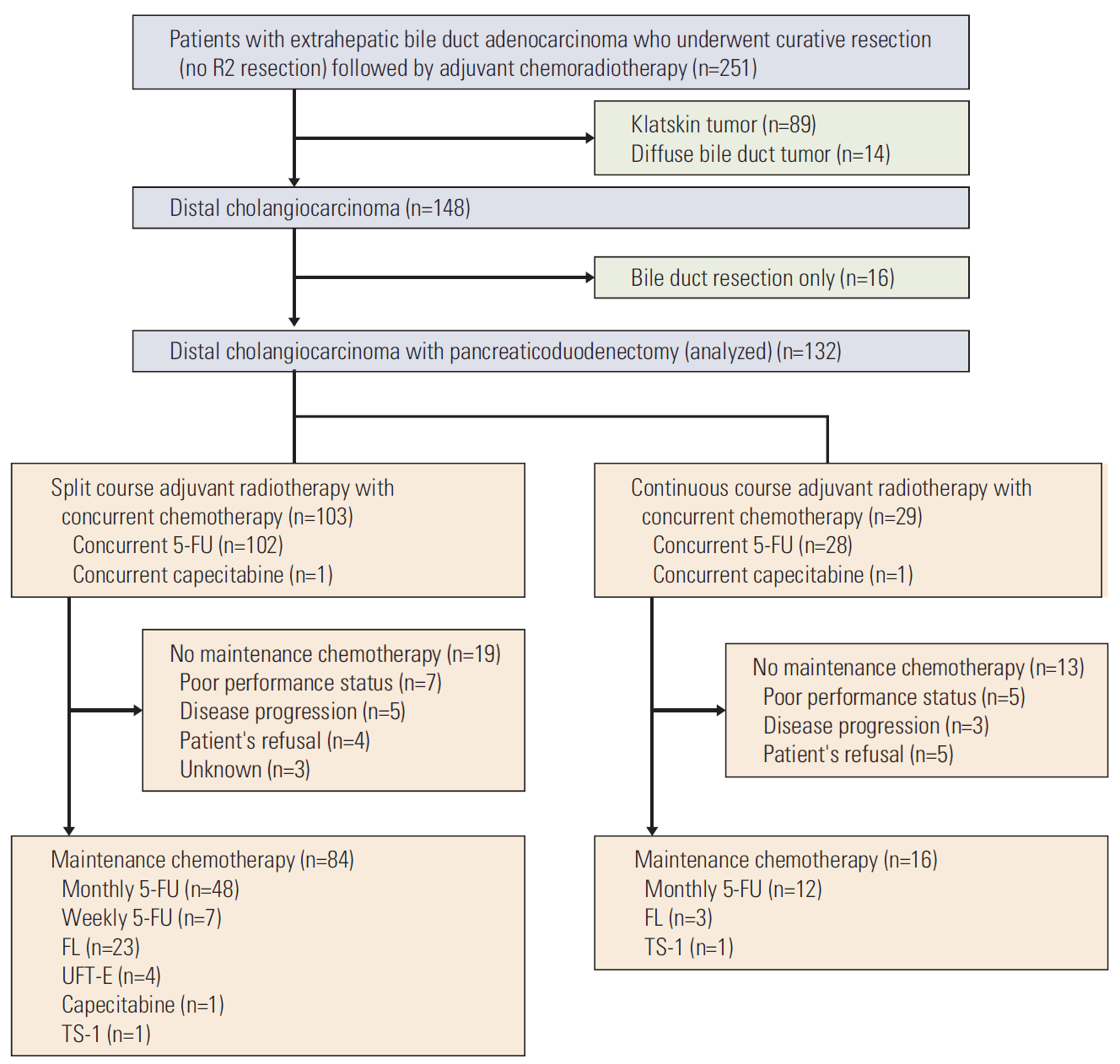
Introduction Extrahepatic bile duct (EHBD) cancer is an uncommon disease in Western countries; however, about 5,000 cases are newly diagnosed every year in Korea (crude incidence rate, 10.6 per 100,000 in 2013) [1]. Although there have been considerable advancements in surgical management and multidisciplinary approaches, its aggressive nature has not been changed. Despite curative resection, which is typically pancreaticoduodenectomy (PD) for distal EHBD, more than half of patients can experience recurrence pancreaticoduodenectomy, leading to poor overall 5-year survival rates ranging from 20% to 50% [2-7]. Because there have been no randomized trials, adjuvant strategies for EHBD cancer following resection have been controversial, particularly with respect to the application of postoperative radiotherapy. However, as we previously reported, our institution has consistently provided adjuvant chemoradiotherapy (CRT) for locoregionally advanced diseases (T2-4 or node positive) and microscopic residual disease (R1 resection) after surgery [8]. Tumor location is of clinical importance and may affect survival outcomes. Distal cholangiocarcinoma (DCC) is defined as bile duct tumors between the cystic duct and the ampulla of Vater (except Klatskin tumors and ampulla of Vater cancer), which include mid common bile duct tumors (between the junction with the cystic duct and the junction with the pancreas) and distal (intrapancreatic) bile duct tumors. DCC usually shows improved outcomes compared with perihilar tumors, which may be because of earlier clinical symptoms. Nevertheless, its prognosis remains poor. Moreover, the prognostic factors for these rare tumors have not been widely established because they only comprise about 30% of EHBD cancer occurrences [9,10]. Many previous studies showed weakness in heterogeneous treatments and insufficient follow-up duration [2-7]. Therefore, we evaluated the long-term outcome of patients undergoing PD followed by adjuvant CRT for DCC in a high-volume center to identify the prognostic impact of clinicopathological factors. Materials and Methods 1. Study population In our institution, most patients with resected EHBD cancer received adjuvant CRT (except those who had pT1N0 disease with R0 resection, poor performance status, or refused further treatment). Therefore, we searched for consecutive patients who underwent curative resection (except R2 resection) followed by adjuvant CRT for EHBD cancer from January 1995 to September 2009 in the institutional database of Seoul National University Hospital. Initially, 251 patients were found; however, 89 with Klatskin tumors, 14 with diffuse bile duct tumors, and 16 who only underwent bile duct resection were serially excluded (Fig. 1). As a result, a total of 132 DCC patients treated with PD followed by adjuvant CRT were eligible for this analysis. Institutional review board approval (H-1403-047-565) was obtained before data collection. 2. Surgery and adjuvant treatmentsResections included PD or pylorus-preserving PD, each of which were combined with regional lymph node (LN) dissection. TNM stage was re-evaluated during data collection according to the American Joint Committee on Cancer staging, seventh edition.
All patients received adjuvant treatments at a median of 45 days after surgery (range, 24 to 89 days). Preoperative chemotherapy or radiotherapy was not performed. In 103 patients, a total of 40 Gy in daily fractions of 2 Gy was delivered to the tumor bed and regional LNs (pericholedochal, retrocaval, and aortocaval). Patients had 2 weeks of planned rest after 20 Gy. Concomitant 5-fluorouracil (5-FU, 500 mg/m2/day intravenous bolus) was administered for the first 3 days of each 2 weeks of radiotherapy. A continuous course of radiotherapy was administered to 29 patients with a median dose of 50.4 Gy (range, 50 to 55.8 Gy) at 1.8-2.0 Gy per fraction. Concomitant fluoropyrimidine-based chemotherapy (intravenous 5-FU, 500 mg/m2/day or oral capecitabine) was administered during the first 3 days of the first and fifth week of radiotherapy. 5-FU–based maintenance chemotherapy was also administered to 100 patients after the completion of radiotherapy. Of the 100 patients, 60 patients received 5-FU (500 mg/m2/day) for 5 days every 4 weeks, 26 received 5-FU with leucovorin for 5 days every 4 weeks, seven received 5-FU (500 mg/m2) once weekly, four received enteric-coated tegafur/uracil, two were administered had TS-1, and one received oral capecitabine. Maintenance chemotherapy was not offered to 32 patients because of poor performance status after CRT (n=12), patient refusal (n=9), disease progression (n=8), or for unknown reasons (n=3).
3. Assessment of recurrence and toxicityAll patients were regularly followed up for surveillance for recurrence. Patient follow-up was completed by May 2014. Routine exams including appropriate imaging methods (abdomino-pelvic computed tomography [CT], ultrasonography, or magnetic resonance imaging) and serum tumor marker (carbohydrate antigen 19-9 [CA 19-9]) assessment were performed at prespecified intervals (typically every 3 months up to 2 years, followed by every 6 months) or when there were any suspicious findings suggestive of recurrence. Most recurrences were clinically diagnosed by imaging studies such as CT on a region of interest and/or positron emission tomography without pathologic confirmation. Recurrence patterns were classified as locoregional recurrence (LRR), distant metastasis (DM), or both. LRR was defined as recurrence in the tumor bed, anastomosis sites, or regional LN area. DM was defined as recurrence in the nonregional LN area or in other organs. Peritoneal seeding was determined by expert radiologists based on the results of CT analysis of ascites, thickening of the peritoneum (either smooth or nodular), or scalloping of the liver or splenic surface. Treatment toxicities caused by primary surgery or adjuvant CRT were also evaluated retrospectively through medical records. Gastrointestinal radiation toxicity was evaluated using the Radiation Therapy Oncology Group (RTOG) criteria.
4. Statistical analysesLocoregional recurrence-free survival (LRRFS), distant metastasis-free survival (DMFS), disease-free survival (DFS), and overall survival (OS) were measured from the day of surgery to the time of LRR, DM, any failure or death from any causes, respectively, during the follow-up period. Patients who were alive or free of recurrence were censored at the time of the last follow-up. The actual survival rates were determined by the Kaplan-Meier method and compared using the log-rank test. The Cox proportional hazards model was used for multivariate analysis to estimate the hazard ratio and to adjust for potential confounding factors. Factors found to be significant upon univariate analysis or thought to be clinically relevant were subjected to both forward and backward stepwise selection multivariate analysis. A p-value of < 0.05 was considered statistically significant. The differences in characteristics between groups were compared using a chi-square test. Data were analyzed using the SPSS ver. 18.0.1 (SPSS Inc., Chicago, IL).
Results 1. Patient and tumor characteristics Overall patient and tumor characteristics are shown in Table 1. The median age of all patients was 62 years (range, 34 to 77 years). Over half of all patients (61.4%) had T3 disease; however, no patients had T4 disease. The median tumor size was 2 cm (range, 0.8 to 8.5 cm). Resection margins were microscopically involved by invasive carcinoma in 14 patients (10.6%), despite the absence of macroscopic residual lesions during PD. Pathologically, tumors were identified as well-differentiated (W/D) adenocarcinoma in 15 patients, moderately differentiated (M/D) adenocarcinoma in 94 patients, and poorly differentiated (P/D) adenocarcinoma in 19 patients. The median number of LNs dissected during surgery was 15 (range, 1 to 45). LN metastases were observed in 43 patients (32.6%). In these patients, the median number of metastatic LNs was one (range, 1 to 9), and only nine patients had four or more LN metastases. 2. Recurrence patterns and a case of delayed recurrenceMedian follow-up duration was 57 months (range, 6 to 225 months) for all patients and 105 months for survivors (range, 13 to 225 months). Overall, 66 patients (50.0%) experienced recurrences. LRR occurred in 34 patients (25.8%) and DM occurred in 58 patients (43.9%). Time to recurrence was similar between patients with LRR (median, 13 months) and DM (median, 12 months). Only 13 patients (9.8%) had an initial isolated local recurrence. In patients with DM, the most common site was the liver (30 patients, 51.7%), followed by peritoneal seeding (18 patients, 31.0%), nonregional LNs (three patients, 5.2%), and others.
Of the 57 patients who did not develop recurrence during the first 5 years after surgery, only one developed late recurrence at 121 months after surgery. After a pylorus-preserving PD for mid common bile duct tumor (pT3N1M0-R1), she was disease-free for 121 months before LRR (hepaticojejunostomy site) and DM (liver and nonregional LN) were detected simultaneously. She refused further treatments and died 5 months later.
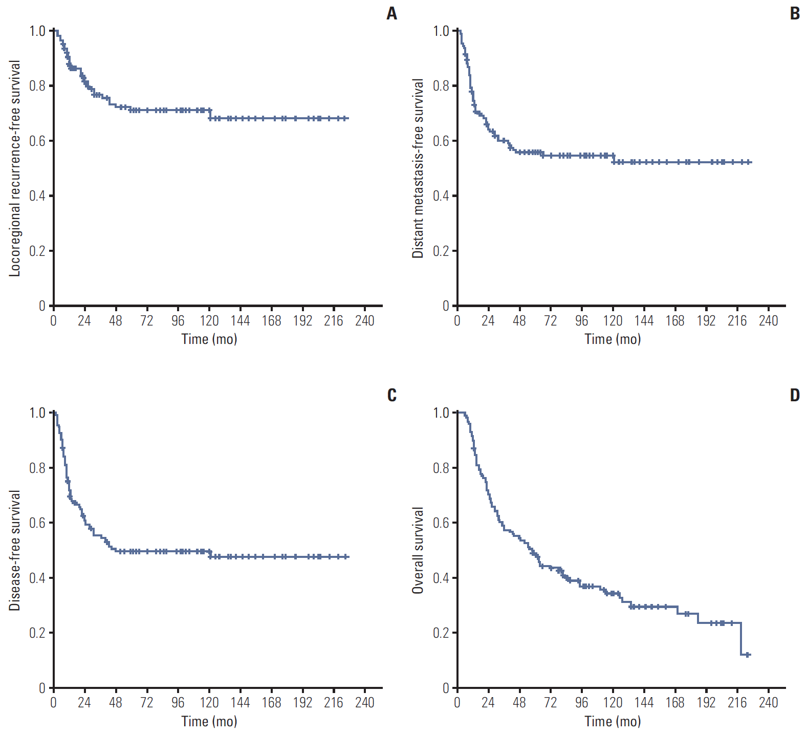
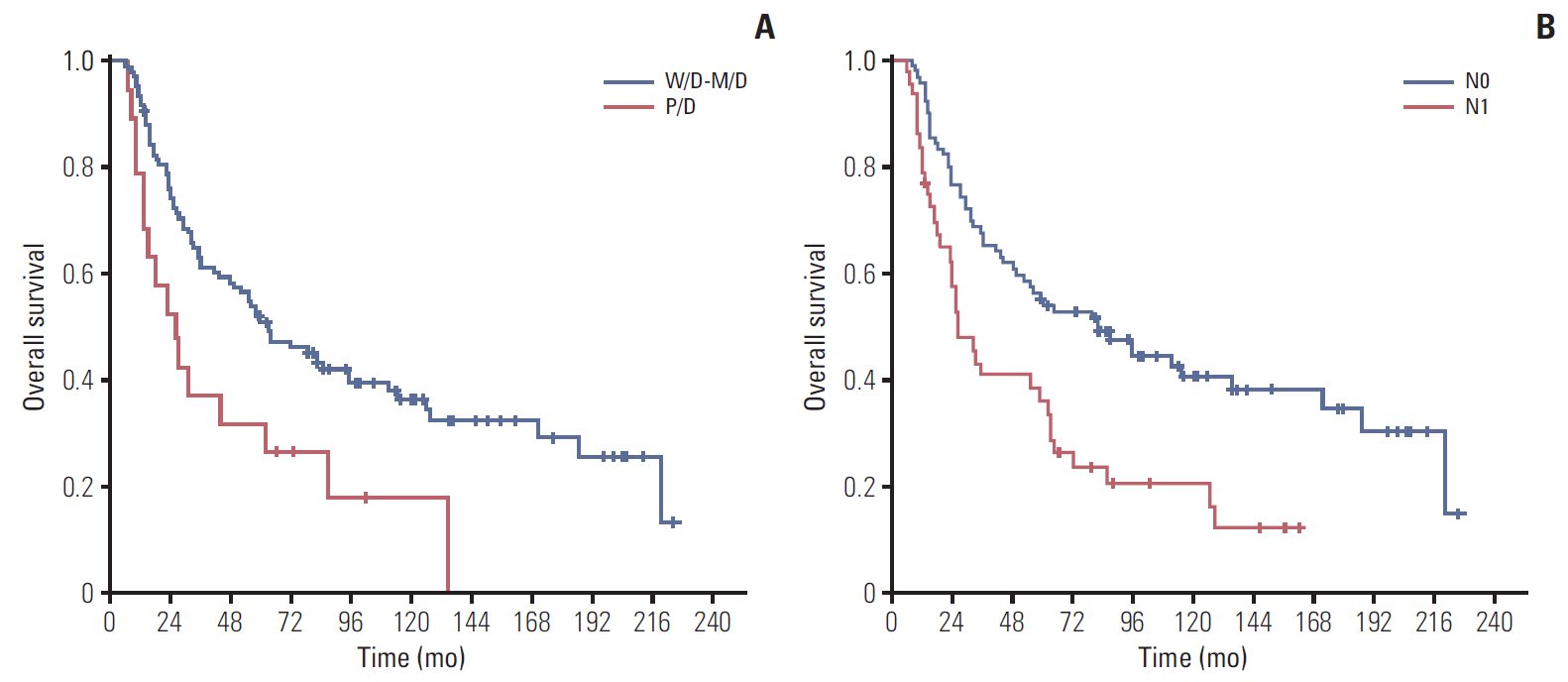
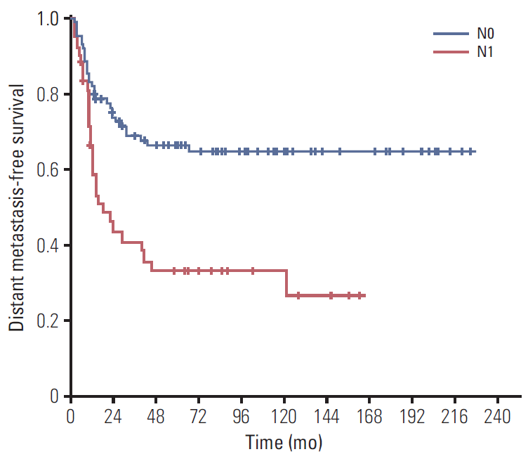
3. Analysis of LRRFS, DMFS, DFS, and OS During the follow-up period, 89 patients (67.4%) died. The 5-year LRRFS, DMFS, DFS, and OS rates were 70.7%, 55.7%, 49.4%, and 48.1%, respectively (Fig. 2). Among all patients, the 10-year OS was 34.1% with 19 actual 10-year survivors (14.4%). Among the group of recurred patients, median OS after recurrence was 7 months (range, 1 to 78 months). The results of the univariate analyses of LRRFS, DMFS, DFS, and OS are shown in Table 2. Upon univariate analysis, P/D tumors and LN metastasis were significantly associated with DMFS and OS, but not with LRRFS. Preoperative CA 19-9 level was also significantly correlated with DFS (p=0.014), LRRFS (p=0.006), and DMFS (p=0.032). R1 resection had marginal significance for LRRFS (p=0.077) and OS (p=0.067), but not for DMFS (p=0.312). Unusually, patients with poor performance status showed better survival outcomes without statistical significance (Table 2); however, this seemed due to several characteristic imbalances between the two groups (Supplementary Table 1). Multivariate analysis incorporating residual disease, LN metastasis, and histologic differentiation demonstrated that P/D tumors (hazard ratio [HR], 2.015; p=0.015; backward selection) and LN metastasis (HR, 1.933; p=0.003; backward selection) were significant prognosticators that predicted inferior OS (Table 3). The 5-year OS rates of patients with W/D-M/D tumors and P/D tumors were 51.0% and 31.6%, respectively (p=0.007) (Fig. 3A). The 5-year OS rates of patients with N0 and N1 disease were 53.9% and 36.0%, respectively (p=0.001) (Fig. 3B). Multivariate analysis of LN metastasis, preoperative CA 19-9 level, and histologic differentiation for DMFS revealed that LN metastasis was the only significant prognosticator that predicted inferior DMFS (HR, 2.345; p=0.005; backward selection) (Fig. 4), while preoperative CA 19-9 level and histologic differentiation had borderline significance (Table 4). In addition, multivariate analysis of LRRFS including preoperative CA 19-9 level and residual disease revealed that preoperative CA 19-9 (HR, 3.898; p=0.011; backward selection) was an independent adverse predictor, whereas R1 resection was not (p=0.305). 4. Treatment toxicitiesTreatment toxicities were also comprehensively evaluated using retrospective medical records. Following PD, the most common complication was delayed wound healing (seven patients, 5.3%), although stomy site leakage (four patients), fluid collection (three patients), postoperative cholangitis (two patients), intra-abdominal abscess (two patients), postoperative bleeding (one patient), and postoperative pancreatitis (one patient) were also observed.
During CRT, acute gastrointestinal toxicity occurred in 88 patients (66.7%), with RTOG grade 1 being observed in 37 (28.0%) and grade 2 in 51 (38.6%) patients. The most common acute symptoms were nausea and abdominal pain, which were tolerable and alleviated after supportive treatments. Grade 3 or higher toxicity did not occur during CRT. During follow-up, only one patient (0.8%) experienced grade 3 late gastrointestinal toxicity. This patient underwent small bowel resection because of an adhesive ileus at 43 months after the end of a radiotherapy session, without evidence of LRR.
DiscussionThis retrospective study was conducted to evaluate the long-term outcome of DCC in patients who underwent PD followed by adjuvant CRT. Although our study only analyzed a selected group of patients, the results suggest that adjuvant CRT for DCC following PD is an effective and tolerable treatment strategy without significant side effects.
As expected, long-term survival was still unsatisfactory; nevertheless, our finding of a 5-year OS rate of 48.1% was comparable or slightly better than the results of previously reported studies [2-5,7,11]. Tumor differentiation and LN metastases were identified as important predictors of survival in our study, as demonstrated in other studies [2-5,7,12]. In particular, a meta-analysis conducted by Zhou et al. [13] revealed that LN metastasis was associated with shorter OS (risk ratio, 2.35; 95% confidence interval, 1.89 to 2.93; p 14]. Therefore, the number of metastatic LNs in our patients may not be an underestimated value. The poor outcome after surgery and adjuvant CRT suggests that approaches other than upfront surgery may need to be considered, especially in cases of LN-positive disease at the time of preoperative work-up. Even after curative resection, there may be a need for more effective adjuvant strategies for patients with LN metastasis. A positive resection margin has been considered as an adverse prognostic factor in some studies [6,7,11,13]; however, its prognostic impact has not been fully determined because of inconsistent results among studies and diverse definitions. Incidences of a positive resection margin in EHBD cancer after curative resection can vary from 10% to as high as 70%, which can be explained by differences between surgeons regarding the principle of the operation or the definition of a positive resection margin, such as whether to include carcinoma in situ [15-19]. Despite the controversy, the effects of R1 resection on survival outcomes was not significant in our DCC patients who underwent adjuvant CRT. Previous studies of adjuvant CRT also demonstrated that a comparison between patients with R0 and R1 resection showed no significant differences in OS and LRRFS [20,21]. These findings corresponded with the results of our study. Therefore, adjuvant CRT may improve outcome with R1 resection in EHBD cancer and also in DCC. Our study had a LRR rate of 25.8%, lower than the reported range from 35% to 74% even after complete resection in the previous studies [3,7,22,23], demonstrating the important role of radiotherapy in terms of locoregional control. However, the major pattern of failure was shifted to DM, as we previously reported for EHBD cancer patients who underwent adjuvant CRT [8]. Even after maintenance chemotherapy, the DM rate was too high, which worsened patient survival and quality of life. Conversely, in the era of targeted therapy, newly developed targeted agents and immunotherapeutic agents have shown some promising results [24,25]. Therefore, to treat DM, which is not well controlled by conventional chemotherapy, and to further improve treatment outcome of this lethal disease, our efforts should be focused on incorporating new chemotherapeutic agents in a combined perioperative approach with radiotherapy. To the best of our knowledge, no randomized controlled trials have evaluated the role of adjuvant CRT in EHBD cancer and in DCC. Given the lack of evidence supporting the application of adjuvant CRT, our favorable long-term results could help researchers establish the application of adjuvant CRT. A recent multicenter study in Korea also demonstrated that adjuvant CRT was associated with significantly improved relapse-free survival and OS in patients with R0-resected DCC [26]. Moreover, our findings showed that acute gastrointestinal toxicity caused by adjuvant radiotherapy was mild, and that late toxicity (grade 3 or more) only occurred in one patient (0.8%), even after a long-term follow-up. Other recent studies also reported that few cases of severe gastrointestinal complications were associated with radiotherapy [21,27]. Unlike perihilar tumors, radiosensitive adjacent organs such as the liver may be less affected by adjuvant radiotherapy in the case of DCC. Recent advances in radiotherapy techniques may further lower its unintended toxicity. However, further prospective trials are needed to evaluate the efficacy and safety of adjuvant CRT for DCC. For a median follow-up of 57 months, about three-fifths of our patients had a recurrence but recurrence 5 years after surgery was rarely observed. These results were somewhat different from those of a previous study by Jang et al. [28], who reported that late recurrence after 5 years was not uncommon in EHBD cancer. However, about one-third of the patients in that study received hepatobiliary resection or bile duct resection for proximal EHBD tumor, while only one-third of the patients received adjuvant treatments. Unfortunately, further comparison of this aspect with other retrospective series was difficult because a limited number of studies have mentioned late recurrences after 5 years, possibly due to insufficient follow-up. Furthermore, some studies have suggested that late recurrence likely resulted from residual carcinoma in situ after resection, which had less malignancy and slower growth than invasive carcinoma [19,29]. However, the effects of residual carcinoma in situ on postoperative recurrence or survival have not been described in most studies and thus remain unclear. Accordingly, further studies of the characteristics of late recurrence after PD are required for the adequate management of DCC patients during a long-term follow-up.It should be noted that there are several limitations to this study. Specifically, its retrospective nature might be a significant weakness; however, we thought that it would be difficult to conduct a prospective study because of the rarity of DCC. Moreover, although our institution offered adjuvant CRT to almost all patients who underwent PD, the patients evaluated in this study were selected ones who both underwent surgery and received adjuvant therapy. In addition, treatment-related toxicity might be underestimated because it was not considered prospectively. Finally, heterogeneous details of adjuvant CRT during a relatively long study period (for example, radiotherapy dose or type of maintenance chemotherapy) might also influence clinical outcomes.
ConclusionIn conclusion, we reported the actual long-term outcome of DCC in patients who underwent PD followed by adjuvant CRT. During long-term follow-up, we found that prognosis of DCC after PD was mainly influenced by histologic differentiation and LN metastasis. For patients with these risk factors, further research should focus on improving adjuvant strategies as well as other treatment approaches.
Electronic Supplementary Material Supplementary materials are available at Cancer Research and Treatment website (http://www.e-crt.org). Conflicts of InterestConflict of interest relevant to this article was not reported.
AcknowledgmentsByoung Hyuck Kim was supported by an institutional R&D project funded by the Armed Forces Medical Research Institute (2015-AFMRI-02). Kyubo Kim was supported by the Ewha Womans University Research Grant of 2016.
Fig. 1.CONSORT diagram of study patients. 5-FU, 5-fluorouracil; FL, 5-fluorouracil+leucovorin; UFT-E, enteric-coated tegafur/uracil.
 Fig. 2.
Locoregional recurrence-free survival (A), distant metastasis-free survival (B), disease-free survival (C), and overall survival (D) in patients treated with pancreaticoduodenectomy followed by adjuvant chemoradiotherapy for distal cholangiocarcinoma.
 Fig. 3.
Overall survival curves according to histologic differentiation (A) and N stage (B). W/D, well-differentiated; M/D, moderately differentiated; P/D, poorly differentiated.
 Fig. 4.
Distant metastasis-free survival curves according to N stage.
 Table 1.
Summary of patient characteristics
Characteristic No. (%) (n=132) Age (yr) ≥ 60 82 (62.1) < 60 50 (37.9) Sex Male 92 (69.7) Female 40 (30.3) Performance (ECOG) 0-1 111 (84.1) 2 21 (15.9) Tumor location Mid common bile duct 65 (49.2) Distal (intrapancreatic) bile duct 67 (50.8) Type of surgery Pancreaticoduodenectomy 46 (34.8) Pylorus-preserving pancreaticoduodenectomy 86 (65.2) Residual disease R0 118 (89.4) R1 14 (10.6) Histologic differentiation W/D, M/D 109 (82.6) P/D 19 (14.4) Unknown 4 (3.0) Tumor size (cm) ≥ 2 90 (68.2) < 2 41 (31.1) Unknown 1 (0.7) Pathologic T stage T1-2 51 (38.6) T3 81 (61.4) Lymph node metastasis Yes 43 (32.6) No 89 (67.4) Overall stage T1N0 2 (1.5) T1N1 2 (1.5) T2N0 34 (25.8) T2N1 13 (9.8) T3N0 53 (40.1) T3N1 28 (21.2) Perineural invasion Yes 103 (78.0) No 28 (21.2) Unknown 1 (0.7) Preoperative CA19-9 (U/mL) ≥ 37 71 (53.8) < 37 35 (26.5) Unknown 26 (19.7) Table 2.Univariate analysis for LRRFS, DMFS, DFS, and OS
Variable No. of patients 5-Yr LRRFS (%) p-value 5-Yr DMFS (%) p-value 5-Yr DFS (%) p-value 5-Yr OS (%) p-value Age (yr) < 60 50 67.4 0.552 49.9 0.275 44.1 0.387 44.0 0.775 ≥ 60 82 72.8 59.1 52.6 50.7 Performance (ECOG) 0-1 111 67.0 0.190 52.6 0.219 45.4 0.107 44.6 0.108 2 21 80.5 71.4 71.4 66.7 Tumor location Mid 65 72.3 0.905 52.7 0.657 45.5 0.455 43.1 0.094 Distal 67 69.5 58.5 53.1 53.2 Residual disease R0 118 72.6 0.077 57.0 0.312 51.7 0.104 51.3 0.067 R1 14 50.0 41.3 26.8 21.4 T stage T1-2 51 67.1 0.606 52.7 0.675 43.6 0.286 45.1 0.563 T3 81 72.8 57.5 53.1 50.1 Tumor size (cm) ≥ 2 90 71.6 0.926 51.4 0.306 47.7 0.657 47.8 0.526
留言 (0)