
記住我


The antiphospholipid syndrome (APS) was first described in the mid-1980s among patients with systemic lupus erythematosus (SLE) [1], but was later also recognized in the absence of autoimmune diseases. APS is defined by clinical and laboratory criteria. Clinical classification criteria comprise objectively verified arterial, venous, or microvascular thrombosis and/or defined manifestations of obstetric morbidity. Laboratory criteria consist of confirmed positive tests for antiphospholipid antibodies (aPL), including anticardiolipin (anti-CL) and/or anti-β2glycoprotein-I (anti-β2GPI) antibodies and/or a positive functional lupus anticoagulant (LA) test [2]. Both IgG and IgM isotypes are included in the criteria, but IgG aPL are more strongly associated with events and thus regarded as more pathogenic [3]. IgA antibodies are not included in the present criteria [2], but have been associated with thrombotic events, especially in patients with SLE [4]. Many researchers believe the plasma protein β2GPI with its five domains to be the main aPL antigen. Conformational changes occur when β2GPI binds to cardiolipin and other negatively charged phospholipids, thus exposing antigenic sites [5].
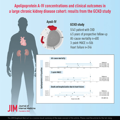
Long-term anticoagulation usually effectively prevents new and recurrent thrombotic events in APS, but because many patients remain undiagnosed, they do not receive adequate treatment [6-8]. The most frequent thrombotic manifestation in APS is venous thromboembolism, while among arterial events, stroke is more commonly reported than ischemic heart disease [9]. Yet, a recent review estimated the frequency of aPL in patients with myocardial infarction (MI) to be as high as 11% [10], though this figure is based mainly on early studies using poorly standardized aPL determinations. However, we recently confirmed this prevalence using modern standardized methods in a large multicenter study. We showed that 11% of 805 patients with a first-time MI were positive for either anti-CL or anti-β2GPI antibodies of the IgG isotype 6–10 weeks after their acute event, as compared to only 1% in individually age-, gender-, and region-matched controls [11]. Another recent study showed positive associations between aPL, subclinical carotid atherosclerosis and cardiovascular events in a large unselected Italian population [12]. Despite these findings, aPL are seldom tested and are not recognized as a risk factor for myocardial infarction in the general population.
The widespread use of rapid coronary angiography in patients with MI has revealed that about 6% of all patients presenting with and fulfilling current criteria of MI have no evident obstructive coronary artery disease, that is, >50% stenosis. This is termed MI with nonobstructive coronary arteries (MINOCA) and is a syndrome requiring further workup aimed at finding a definite diagnosis [13]. In a similar cohort with MINOCA as in the present study, we recently showed that 35% of these had Takotsubo syndrome, 22% actual MI, 17% myocarditis, while 23% showed a normal myocardium [14]. Two case reports of the total seven cases have suggested that SLE is a possible cause of MINOCA [15, 16], but in a recent registry-based study we could not confirm that MINOCA was overrepresented in SLE patients with MI [17]. Lacking further evidence, a position paper issued by the European Society of Cardiology in 2017 recommended that APS should be considered in MINOCA [13]. Since then, Pasupathy et al. have demonstrated that the levels of anti-CL antibodies were similar and that LA was negative in 25 patients with MINOCA and 25 patients with MICAD [18].
The aim of the present study was to explore the prevalence of aPL in patients with MINOCA and MICAD and to compare these with healthy controls. We used standardized methods to assess prothrombotic aPL. Specifically, we measured anti-CL and anti-β2GPI of IgA/G/M isotypes in well-characterized patients from the Stockholm Myocardial Infarction with Normal Coronaries (SMINC) study [19]. To assess possible effects of aPL on the coagulation system, we also measured activated protein C–protein C inhibitor (APC–PCI) complex as a marker of hypercoagulability. As aPL are frequent in patients with SLE, we also investigated a set of the most common anti-nuclear antibodies (ANA).
Materials and methodsThe study was performed in accordance with the Declaration of Helsinki and Good Clinical Practice and was approved by the Stockholm Ethics Committee. We used a cross-sectional case–control design to address the study objectives. Patients fulfilling the universal definition of acute MI, with no or <30% stenosis on coronary angiography were recruited from all five coronary care units serving the Stockholm urban area. From June 2007 to May 2011, 176 patients with MINOCA were screened. Inclusion criteria were 35–70 years of age and sinus rhythm at admission. Exclusion criteria included history of structural or coronary heart disease, pacemaker, severe chronic obstructive pulmonary disease, and severe renal failure (serum creatinine >150 μmol/L). Of these, 152 patients underwent cardiac magnetic resonance imaging to exclude myocarditis and cardiomyopathy. Details on patient selection and definitions can be found in a previous publication [19]. After all inclusion and no exclusion criteria were met, 100 patients with MINOCA, and in parallel 100 age- and gender-matched patients with MI with significant stenosis and 100 healthy controls, were included. Stored sample volume sufficient for the present biochemistry analyses was missing in two patients with MINOCA and one with MICAD.
Inflammatory conditions were defined as asthma, chronic obstructive pulmonary disease, rheumatic diseases, hepatitis, primary biliary cirrhosis, pancreatitis, diverticulitis, collagenous colitis, chronic dental infections, and thromboembolic disorders such as previous thromboembolic diagnoses and known coagulopathy. To exclude pulmonary embolism, the first 100 patients underwent computed tomography (CT) of the chest, but because all these investigations turned out negative, the protocol was changed to measurement of D-dimer, and CT only in case of high suspicion of pulmonary embolism [19].
Vascular measurementsThe degree of arteriopathy was assessed by intima-media thickness (IMT) from images of the left and right common carotid artery (CCA) using an ultrasound scanner (Vivid 7; General Electric [GE], New York, NY) equipped with a 12-MHz transducer. The CCA far wall IMT was measured from each side using semiautomatic analysis software, as previously described [19]. Endothelial function was determined using EndoPAT (Itamar-Medical Ltd) and the postocclusion to preocclusion ratio, called reactive hyperemia index (RHI), as previously described [19]. RHI measures endothelial function in peripheral arteries, where a higher number reflects a better function. In patients, IMT and RHI were determined 3 months after the acute event.
Laboratory analysesBlood samples were taken 3 months after the acute event in MINOCA and MICAD patients and at a similar time in healthy controls. Autoantibodies (IgA, IgG, IgM) targeting cardiolipin (anti-CL) and β2glycoprotein-I (anti-β2GPI) and specific anti-nuclear antigens (ANA) (dsDNA, nucleosomes, ribosomal P, Smith [Sm], Smribonucleoprotein [SmRNP], RNP 68, Sjögren's syndrome antigen A/B [SSA]/Ro52, SSA/Ro60, SSB) were analyzed by multiplexed bead technology using the BioPlex 2200 system (Bio-Rad, Hercules, CA, USA) according to the specifications of the manufacturer. The coefficient of variation (CV) was <10.0% for all isotypes; for ANA-specificity 8%–10%, and <8% for aPL IgG also in the very low range. The cutoff for anti-CL and anti-β2GPI antibodies was set at the 99th percentile of population-based controls, according to APS criteria [2]. Double aPL positivity was defined as positivity for at least one of IgG or IgM of anti-β2GPI and at least one of IgG or IgM anti-CL. IgA antibodies, which are not included in the APS criteria, were not considered when determining double aPL positivity. Cutoffs for ANA subspecificities were set according to the manufacturer.
The concentration of the APC–PCI complex was measured with a previously described immunofluorometric sandwich assay [20]. The method utilizes two mouse monoclonal antibodies that bind a unique neoepitope in PCI that is formed as a result of complex formation between APC and PCI.
StatisticsContinuous variables with a normal distribution are reported as means with standard deviations (SD), whereas aPL titers are reported as medians with interquartile ranges due to substantial skewing. Dichotomous variables are reported as counts with percentages. Student's t-test was used for normal distributions and the Mann–Whitney U test for nonnormal distributions for comparison between two groups. The chi-square test and Fisher's exact two-sided tests were used for comparison of dichotomous variables between groups. ANOVA (overall) with Student's t-test as post hoc was used for comparisons between three groups. For comparing antibody titers between three groups, the nonparametric Kruskal–Wallis test was used and the Wilcoxon test was applied for post hoc analyses between groups. SPSS version 25 (IBM, Armonk, NY, USA) was used for statistical analyses. A p-value of <0.05 was considered significant.
ResultsCompared to MICAD patients, MINOCA patients were less often current smokers, and they had a lower prevalence of diabetes mellitus and hyperlipidemia. MINOCA patients had a higher prevalence of inflammatory conditions than MICAD patients. When compared to healthy controls, MINOCA patients had a higher prevalence of all cardiovascular risk factors (smoking, hypertension, hyperlipidemia, and diabetes mellitus), but comparable concentrations of hs-CRP (Table 1).
Table 1. Clinical characteristics of the three groups MINOCA (n = 98) MICAD (n = 99) Healthy controls (n = 100) p-Value Mean age, years 58.0 ± 8.2 58.6 ± 8.1 58.9 ± 8.2 0.76 Female sex 71% 72% 72% 1.0 BMI, kg/m2 25.6 ± 4.6 27.0 ± 4.7 25.2 ± 3.6 0.02 Current smoker 20% 33% 7% <0.0001 Ex-smoker 30% 34% 40% 0.30 Hypertensiona 37% 47% 17% <0.001 Hyperlipidemiaa 9% 20% 4% 0.001 Diabetes mellitusa 4% 10% 0% 0.003 IGT or DM 38% 51% 20% <0.001 Thromboembolismb 6% 1% 2% 0.09 Any inflammatory diseasec 30%¤ 20% 10% 0.002 Hemoglobin (g/L) 137.4 ± 12.9 140.4 ± 13.8 136.6 ± 13.2 0.16 Platelets (109/L) 249.0 ± 61.0 248.7 ± 50.5 243.5 ± 52.5 0.76 CRP (mg/L) 7.94 ± 25.0 5.00 ± 12.5 n/a Relative troponin 111 ± 177 273 ± 563 n/a Hs-CRP* (mg/L) 2.24 ± 3.93 2.12 ± 2.45 1.68 ± 2.48 0.06 NT-proBNP* (ng/L) 133.2 ± 127.8 277.5 ± 462.2 71.8 ± 83.0 <0.001 Cystatin C* (μmol/L) 0.86 ± 0.17 0.89 ± 0.16 0.84 ± 0.15 0.02 Cholesterol* (mmol/L) 5.10 ± 0.96 5.35 ± 1.08 5.64 ± 1.01 0.003 Triglycerides* (mmol/L) 1.05 ± 0.51 1.43 ± 0.64 0.97 ± 0.57 <0.001 LDL* (mmol/L) 3.03 ± 0.85 3.44 ± 0.93 3.64 ± 0.92 <0.001 HDL* (mmol/L) 1.57 ± 0.55 1.28 ± 0.38 1.56 ± 0.51 <0.001 IMT* (mm) 0.71 ± 0.13 0.73 ± 0.15 0.70 ± 0.12 0.34 RHI* (units) 2.23 ± 0.67 2.08 ± 0.61 2.27 ± 0.64 0.09 Note: Values are expressed as proportions or mean ± standard deviation. All values determined at the index event except those indicated by *, which were measured at 3 months follow up. p-Values are for Kruskal–Wallis test or chi-square test as appropriate (overall). Abbreviations: BMI, body mass index; CRP, C-reactive protein; DM, diabetes mellitus; HDL, high-density lipoprotein; hs, high sensitive; Hs, high sensitivity; IGT, impaired glucose tolerance; IMT, intima-media thickness; LDL, low-density lipoprotein; MICAD, myocardial infarction with coronary artery disease; MINOCA, myocardial infarction with nonobstructive coronary arteries; NT-proBNP, N-terminal prohormone of brain natriuretic peptide; RHI, reactive hyperemia index. Relative troponin was calculated as peak troponin I or T divided by the 99th percentile upper reference limit. aDefined as on treatment for the diagnosis. bPrevious thromboembolic diagnoses and known coagulopathy. cAsthma, chronic obstructive pulmonary disease, rheumatic diseases, hepatitis, primary biliary cirrhosis, pancreatitis, diverticulitis, collagenous colitis, and chronic dental infections. Relative troponin: ratio to normal level cutoff. Further details on the cohort have been published previously [19].The prevalence and titers of aPL of the IgG isotype were significantly higher both in patients with MINOCA and MICAD when compared with controls, and aPL IgG positivity (defined as either anti-CL IgG and/or anti-β2GPI IgG) was twice as frequent among patients with MICAD than MINOCA, though this difference was not significant (p = 0.21). We observed no group differences regarding aPL of IgM and IgA isotypes (Figure 1, Table 2). Of ANA subspecificities, only anti-SSB differed between groups and occurred more commonly among controls (p = 0.04) (Table 2).

Antiphospholipid antibodies (aPL) titers in the three groups. p-Values are based on Wilcoxon as post hoc test for comparisons between groups. Dotted lines indicate positivity, set at 99th percentile of local population controls.
Table 2. Titers of aPL, proportion-positive aPL and ANA subspecificities, and levels of APC–PCI MINOCA (n = 98) MICAD (n = 99) Healthy controls (n = 100) p-Value Anti-β2GPI IgG 1.3 (1.3–1.3) 1.3 (1.3–1.3) 1.3 (1.3–1.3) 0.002 Proportion positive 6.1% 11.1% 0.0% 0.0003 Anti-β2GPI IgM 1.4 (0.5–3.3) 1.3 (0.5–3.1) 1.1 (0.6–3.4) 0.99 Proportion positive 0.0% 2.0% 1.0% 0.36 Anti-β2GPI IgA 0.7 (0.5–1.4) 0.7(0.5–1.4) 0.6 (0.5–1.1) 0.42 Proportion positive 3.1% 1.0% 3.0% 0.55 Anti-CL IgG 1.5 (1.5–1.5) 1.5 (1.5–1.5) 1.3 (1.3–1.3) 0.0007 Proportion positive 6.1% 11.1% 0.0% 0.0003 Anti-CL IgM 1.8 (0.6–3.9) 1.3 (0.6–4.2) 1.3 (0.6–1.8) 0.78 Proportion positive 0.0% 1.0% 1.0% 0.61 Anti-CL IgA 0.8 (0.4–1.4) 0.7 (0.4–1.4) 0.6 (0.4–1.2) 0.77 Proportion positive 2.0% 1.0% 2.0% 0.81 Proportion double aPL positive 6.1% 12.1% 1.0% 0.006 Anti-dsDNA 0.9 (0.9–1.0) 1.0 (0.9–1.0) 0.9 (0.9–2.0 0.42 Proportion positive 4.1% 2.0% 1.0% 0.35 Anti-nucleosomes 0.1 (0.1–0.1) 0.1 (0.1–0.1) 0.1 (0.1–0.1) 1.00 Proportion positive 3.0% 3.0% 3.0% 1.00 Anti-ribosomal P 0.1 (0.1–0.1) 0.1 (0.1–0.1) 0.1 (0.1–0.1) 0.07 Proportion positive 1.0% 0.0% 4.0% 0.05 Anti-Sm 0.1 (0.1–0.1) 0.1 (0.1–0.1) 0.1 (0.1–0.1) 0.77 Proportion positive 2.0% 1.0% 1.0% 0.78 Anti-SmRNP 0.1 (0.1–0.1) 0.1 (0.1–0.1) 0.1 (0.1–0.1) 0.36 Proportion positive 2.0% 2.0% 0.0% 0.19 Anti-RNP 68 0.1 (0.1–0.1) 0.1 (0.1–0.1) 0.1 (0.1–0.1) 0.60 Proportion positive 1.0% 1.0% 0.0% 0.60 Anti-SSA/Ro52 0.1 (0.1–0.1) 0.1 (0.1–0.1) 0.1 (0.1–0.1) 0.36 Proportion positive 4.1% 4.0% 1.0% 0.28 Anti-SSA/Ro60 0.1 (0.1–0.1) 0.1 (0.1–0.1) 0.1 (0.1–0.1) 0.31 Proportion positive 6.1% 9.1% 4.0% 0.34 Anti-SSB 0.1 (0.1–0.1) 0.1 (0.1–0.1) 0.1 (0.1–0.1) 0.04 Proportion positive 1.0% 1.0% 6.0% 0.04 APC–PCI μg/L 0.24± 0.15 0.24 ± 0.11 0.27 ± 0.20 0.265 Abbreviations: Anti-β2GPI = anti-β2glycoprotein-I, Anti-CL = anticardiolipin, double aPL positivity is defined as positivity for at least one of IgG or IgM of anti-β2GPI and at least one of IgG or IgM anti-CL, dsDNA = double stranded DNA, Sm = Smith, SmRNP = Smith Ribonucleoprotein, RNP = Ribonucleoprotein, Sjögren's syndrome Antigen A/B (SSA)/Ro52, SSA/Ro60, SSB, APC–PCI = activated protein C–protein C inhibitor complex. Titers are given as median (interquartile range) and proportion positive as% with the exception of APC–PCI which is given as mean ± standard deviation. P-values are calculated by Kruskal-Wallis test (overall), except for APC–PCI where p was calculated with ANOVA.aPL IgG-positive MICAD patients had higher levels of the APC–PCI complex as compared to aPL IgG-negative patients (Figure 2 and Table S1).

Activated protein C–protein C inhibitor complex (APC–PCI) in myocardial infarction (MI) subgroups stratified by antiphospholipid antibodies (aPL) status, and in controls. p-Values were calculated with ANOVA (overall), with Student's t-test as post hoc for comparisons between groups.
DiscussionIn the present study, well-characterized patients with both MINOCA and MICAD were more often positive for and had higher titers of aPL IgG than healthy controls. The proportion of aPL IgG positivity was twice as high among patients with MICAD compared to MINOCA, though this difference did not reach significance. In general, we confirmed a more frequent occurrence of aPL IgG, but not IgM or IgA, in patients with MI as compared to controls [11]. We observed no differences between MINOCA and MICAD patients regarding hemostasis as determined by the APC–PCI levels on a group level. Interestingly, among aPL IgG-positive MICAD patients, we detected signs of hypercoagulability, as measured by increased levels of the APC–PCI complex, indicating that aPL IgG was not only associated with MI but also with functional disturbances of the coagulation system.
A major objective for this study was to determine whether patients with MINOCA had a more frequent occurrence of aPL, as the pathogenesis of these MIs is varied and includes a potential role for thrombosis [15]. Patients with MICAD have a higher plaque burden and thus more substrate for thrombosis, which may potentially be more harmful in patients with aPL. This contrasts with patients with MINOCA, where a previous study indicates that the plaque burden does not differ from healthy controls [21]. Among patients with SLE, 30%–40% are known to have aPL [22]. In a recent large registry-based study, patients with SLE without AMI before 1996 (n = 4.192) and 10 matched controls without SLE and AMI for each patient were identified. During a 20-year follow-up in the Swedish national patient registry, AMIs occurred at younger ages in patients with SLE and the incidence was approximately twice that of controls, but the proportion of MINOCA was similar in both groups [17]. According to our results, aPL IgG occurred in 6% of MINOCA patients and were thus more common than in controls, but numerically less frequent than in MICAD (11%).
Though no direct comparisons with controls have been made previously, a Polish study by Stepien et al. investigated acquired and inherited thrombophilia in 84 MINOCA patients and 84 age-matched patients with cryptogenic stroke. Higher frequencies of aPL than in our cohort were detected both in patients with MINOCA (15.5%) and cryptogenic stroke (10.7%), though no comparisons with MICAD or controls were made. Their samples were taken 8 months after the acute event, as compared to 3 months in our cohort, and the patients were on average 12 years younger with more frequent traditional risk factors, such as smoking, hyperlipidemia, hypertension, and diabetes [23]. In contrast, a small study that compared 25 MINOCA patients with 25 MICAD patients did not detect any anti-CL antibodies or positive LA tests [18]. Another small study from New York investigated 40 patients diagnosed with APS and acute coronary syndromes who had been subject to cardiac catheterization. Among them, eight cases (20%) were diagnosed with MINOCA, but they had no control group [24]. Davies and Hunt [15] also reported five clinical cases of young adults who suffered a first MI with normal coronary angiograms, all with positive LA tests. Together these data indicate that aPL may distinguish a small but separate subset of MINOCA.
The association between aPL and accelerated atherosclerosis is debated. We report that aPL of the IgG isotype are positive in 11% of patients with MICAD, confirming our previous report from a large multicenter study of patients with a first-time MI [11]. These results indicate that aPL may be more common in MICAD than in MINOCA, suggesting an association with coronary atherosclerosis. Interestingly, we did not observe any association with carotid IMT or endothelial function, as previously reported [25]. Furthermore, both IMT and RHI were similar in MICAD patients with and without aPL. These findings are consistent with some previous studies, where IMT in patients with APS and controls was similar [26, 27], though others report small increases in IMT in APS patients, compared to controls [28, 29]. In contrast, there are reports of positive associations between aPL and atherosclerotic plaques. Kravvariti et al. investigated carotid and femoral plaques in patients with primary APS, APS secondary to SLE, and diabetes mellitus. They found that the occurrence of plaques was 2.5 times more frequent as compared to matched controls in both the APS and the diabetes groups [30]. One meta-analysis reported that aPL are more common in patients with carotid plaques, increased IMT, and endothelial dysfunction [25]. The results of the present study are inconclusive regarding the association between aPL and atherosclerosis/arterial injury, as aPL were numerically more frequent in patients with coronary atherosclerosis, that is, MICAD, than in MINOCA, whereas aPL did not impact IMT or measurements of endothelial function. One possible cause for this inconsistency is that IMT and the endothelial function test do not mirror coronary atherosclerosis, as discussed by Spence et al. [31].
We observed higher levels of APC–PCI complex in the aPL IgG-positive group than in the aPL IgG-negative MICAD subgroup. Protein C is a vitamin K-dependent zymogen of a serine protease that regulates coagulation. Protein C is activated (APC) in the capillary bed by thrombin, a process which requires two endothelial cell receptors, thrombomodulin (TM) and endothelial cell protein C receptor (EPCR). The prime inhibitor of APC in plasma is the protein C inhibitor (PCI), which is present at 10,000 times higher concentrations than APC. Thus, the concentration of the APC–PCI complex reflects the APC concentration. The concentration is elevated in hypercoagulable states such as deep venous thrombosis [32], where it has been demonstrated to be a more specific but less sensitive biomarker than the commonly used D-dimer [33]. APC–PCI has also been demonstrated to be sensitive, though less specific, for separating ischemic (thrombotic or embolic) from hemorrhagic stroke [34]. To our knowledge, APC–PCI complexes have not been evaluated in the context of APS. However, the protein C pathway was recently demonstrated to be pivotal in an APS mouse model. Muller-Calleja et al. demonstrated that some aPL can, through binding to endothelial cell surface complexes of lysobisphosphatidic acid (LBPA) and the EPCR receptor, cause both systemic inflammation and activation of toll-like receptor 7 (TLR7), which contributes to interferon-α signaling and continuous enhanced production of aPL. Blocking the interaction between aPL and the LBPA–EPCR complexes protected the investigated mice from aPL-induced thrombosis [35].
Blood samples in this study were collected 3 months after the acute event and they were not confirmed by a second measurement. From the case–control design, it is neither possible to know if the antibodies were present before the MI nor if they are persistent or transient. It is possible that the observed antibodies are transient and part of an immunological reaction to myocardial damage. However, we did not see any association with the size of the MI, as measured by troponin levels. Prior studies with repeated measures favor the concept that the antibodies are persistent in patients with MI [36, 37]. Moreover, two prospective nested case–control studies have demonstrated that anti-CL IgG antibodies precede and predict MI. Vaarala et al. studied hyperlipidemic men in the Helsinki Heart Study [38], and Wu et al. investigated 50-year-old men followed prospectively during 20 years [39]. It is known that transient aPL can be induced by infections [40], but these aPL are generally not believed to be associated with thrombotic events, despite lack of evidence [41]. The assumption that transient aPL are harmless should therefore be questioned, as suggested by de Groot and Urbanus [42].
Based on an extensive literature review, the European Alliance of Associations for Rheumatology (EULAR) recently published recommendations for the management of APS defining a “high-risk aPL profile” as either double or triple aPL positivity—defined as positivity for two or three of the following tests: anti-β2GPI (either the IgG or IgM isotype), anti-CL (either the IgG or IgM isotype), and the LA test—or persistently high aPL titers. The aPL IgG-positive patients in this study were all double positive as anti-CL IgG and anti-β2GPI IgG correlated perfectly. However, as participants were only sampled once, we do not know if they also fulfilled the persistency requirement stating that positivity should be confirmed on at least two occasions with
留言 (0)