
記住我

Hepatocellular adenomas (HCAs) are rare, benign hepatic tumors in children, with limited imaging data available for pediatric cases.
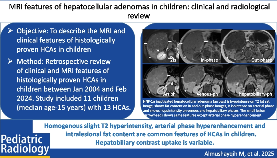
ObjectiveTo describe the magnetic resonance imaging (MRI) and clinical features of histologically proven HCAs in children.
Materials and methodsSingle-center retrospective review of pathology-proven HCA from January 2004 to February 2024. Patients with available pre-intervention imaging in our PACS were included. Two independent readers reviewed the imaging studies. The features were summarized using descriptive statistics and inter-reader agreement was assessed using Cohen’s kappa.
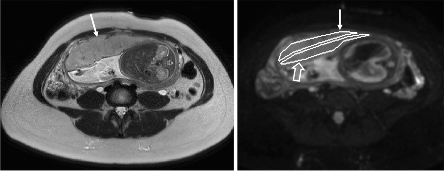
ResultsThis study included 11 children (6 boys and 5 girls; median age 15 years) with 13 pathologically proven HCAs. Three patients had type 1a glycogen storage disease. Five patients had a single lesion, while six had multiple lesions. The lesions were well-defined with a median average diameter of 3.6 cm. Most were homogenously T1 iso-intense (61.5%) and mildly hyperintense (76.9%) on T2-w fat saturated images. The atoll sign was present in two lesions. Intralesional fat was observed in 69.2% of cases: microscopic in eight lesions and macroscopic in one. Hemorrhage occurred in three (23.07%) lesions and necrosis in one (7.7%). Nine out of 10 (90%) lesions showed arterial phase hyperenhancement, and only 3/10 (30%) lesions retained contrast on hepatobiliary phase. In total, 6/13 (46.1%) lesions showed washout, and all received hepatobiliary agent. One lesion ruptured with the hemoperitoneum. Of the 11, 63.6% of patients underwent percutaneous biopsy and 36.4% underwent surgical resection.
ConclusionMR imaging features are nonspecific, but homogenous slight T2 hyperintensity, arterial phase hyperenhancement, and intralesional fat content are common features. Hepatobiliary contrast uptake is variable.
Graphical Abstract
留言 (0)