The present study demonstrated the capability of compressed sensing-based Cine-MRI to dynamically visualize swallowing events closely associated with the anatomical structures of the head and neck, and the duration of these swallowing events is concurrently influenced by the swallowing function and the location of the lesion in patients with dysphagia. Additionally, the area under the ROC curve of combined Cine-MRI time parameters for predicting mild and severe dysphagia after treatment was 0.806, which highlight that compressed sensing-based Cine-MRI may evaluate the severity of dysphagia.
Traditionally, MRI has been utilized primarily for static qualitative research in imaging. However, in the context of this study, dynamic MRI was employed to assess the swallowing function of patients, marking a departure from conventional usage. This dynamic MRI technology, previously applied to functions such as the temporomandibular joint [15], extraocular muscle structure and eye movement [16], joint range of motion [17], heartbeat, and aortic blood flow [18, 19], compensates for the deficiencies in simple structural imaging. Swallowing is an intricate and continuous process, necessitating the close coordination of various structures and organs. In normal circumstances, the swallowing action is consistent, and the process is completed swiftly. The Videofluoroscopic Swallow Study (VFSS) is a widely utilized technique in the clinical diagnosis of dysphagia [4]; however, its diagnostic accuracy is limited due to inadequate visualization of soft tissue structures. The advantage of dynamic CT lies in its high spatial resolution, which however necessitates the oral administration of contrast agents containing iodine ions or barium sulfate. Cine-MRI possesses the advantage of soft tissue imaging, and there is no ionizing radiation. It permits multiple follow-ups during the rehabilitation treatment. To achieve continuous dynamic observation under magnetic resonance equipment, a scanning sequence with a high time resolution is essential for evaluating swallowing movement in stages. In this study with normal volunteers, the entire swallowing process took place within 1.5 s. Previous research has indicated that a time resolution too low (e.g., 800 msec [20]) cannot adequately represent this process or identify the specific causes of abnormal swallowing [21]. The present study advances the field by utilizing bFFE sequence, based on compressed sensing, has a real-time resolution of 74 msec and 13.5 frames per second, and the total imaging time is brief, with the scanning time for 200 images being only 15 s. This approach not only ensures image quality but also achieves superior Cine acquisition speed, eliminating motion artifacts and capturing the anatomical structure movement and food mass progression throughout the swallowing stages.
The utilization of Cine-MRI during swallowing enables the acquisition of various functional parameters pertaining to sphincter movements involved in the process, including oro-lingual (OTT), soft palate and posterior pharyngeal wall (PR1), tongue base and posterior pharyngeal wall (PR2), epiglottis (epiglottis inverted) as well as cricopharyngeal muscles (EOT). Additionally, it allows for the observation and quantitative analysis of water bolus passage through the pharynx (PTT), ascending movement of the larynx (LAT) and descending movement of the larynx (LDT). Although our method deviates from sequences used in previous research, the Cine-MRI-related parameters derived from normal patients are congruent with measurements obtained by Zhang et al. [14]. Previous studies necessitated the employment of blueberry juice, a natural paramagnetic contrast agent [22]. The image contrast of the bFFE sequence is contingent upon the ratio of the T2 value to the T1 value of the tissue, is sensitive to pure water, and manifests as a high signal. This allows high signal and contrast images, offering a patient-friendly and clinically practical alternative. Considering the constraints faced by head and neck cancer patients, such as intolerance to irritant food or the impracticality of preparing blueberry juice, this approach using pure water promotes the incorporation of this sequence into routine clinical examinations.
Based on the comparison classified by the location of the primary tumor, we discovered that OTT, PR1, LAT, and LDT were closely associated with the location of the primary tumor, and the corresponding parameters of Cine-MRI corresponding to the location of the primary tumor presented a significant increase. This might be because the radiotherapy and chemotherapy for head and neck cancers have a more severe influence on the tissues in the region where the tumor is located (such as fibrosis, edema, or nerve injury), thereby subsequently causing a significant decline in the swallowing function of this area. According to our defined Cine-MRI parameters, OTT represents swallowing events occurring in the oral cavity, PR1 represents swallowing events occurring in the oropharynx, while LAT and LDT represent swallowing events taking place in the larynx. These parameters obtained through Cine-MRI, which represent various stages of swallowing, can effectively identify the primary areas responsible for swallowing delay, determine specific swallowing issues experienced by patients, and aid in selecting targeted rehabilitation programs to restore patients’ swallowing function. For instance, individuals with dysphagia exhibit a marked delay in oral transit time (OTT), indicating that the primary impairment in swallowing occurs during the oral phase. Targeted training of tongue function not only enhances swallowing capabilities within this phase but also facilitates the restoration of swallowing movements in subsequent phases [23, 24]. In addition, the frequency of practicing the Masako maneuver should be increased when there is a significant delay in PR2, while the Mendelsohn’s maneuver should be practiced more frequently in cases of significant delays in LAT and LDT [25]. Meanwhile, we anticipate that Cine-MRI can be employed for future follow-up assessments, enabling the evaluation of rehabilitation efficacy through the observation of alterations in OTT, PR1, LAT, and LDT.
The 36 patients in this study exhibited significant time delays compared to normal volunteers throughout the entire swallowing process. Although only OOT and EOT exhibited statistically significant differences between patients with severe dysphagia and those with mild dysphagia, the majority of parameters demonstrated delayed results in patients with severe dysphagia. This observation suggests that patients with dysphagia after radiotherapy and chemotherapy for head and neck cancer have swallowing dysfunction at all stages of the swallowing process, and the severity of dysphagia may be proportional to the extent of temporal delay observed in different phases of swallowing. The complexity of swallowing as a sensorimotor task necessitates the precise coordination of various anatomical structures [12]. After the completion of the oral phase, the sequential process of soft palate elevation and closure of nasal passages takes place first, followed by laryngeal elevation and opening of the esophagus, all occurring rapidly within a concise time frame [26], and the entire process of swallowing may be impacted if there is a delay in either stage. Simultaneously, the treatment-induced conditions such as edema, fibrosis, nerve damage, and pain can result in diminished muscle motor coordination and impaired nerve perception throughout the entire process of swallowing. Thus, the comprehensive diagnosis and evaluation of dysphagia necessitates the thorough examination of all structures involved in swallowing, as well as the assessment of each phase within the swallowing process.
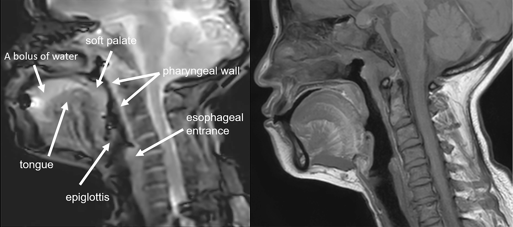
Despite significant advancements in temporal resolution, anatomical visualization, and contrast agent optimization, the primary limitations of this study reside in its low spatial resolution and restricted acquisition to a single slice. The Cine-MRI technique based on compressed sensing enables visualization of the tongue, soft palate, pharyngeal wall, epiglottis, esophageal entrance. However, these structures may appear blurred due to the limited spatial resolution. Therefore, it is necessary to combine conventional MR images for further clarification. Additionally, the limitations in spatial resolution have an impact on the accurate depiction of intricate anatomical structures, such as the glottis. And it may be more advantageous to obtain images simultaneously from multiple directions and planes for dysphagia assessment. It is our anticipation that the aforementioned limitations can be resolved through future optimization of the reconstruction algorithm and signal sampling technique utilized in Cine-MRI.
In conclusion, the combination of the bFFE sequence and compressed sensing for Cine-MRI offers a radiation-free and high temporal resolution approach, holding promising potential for visualizing and quantitatively analyzing specific delays in the swallowing process. The quantitative data obtained through this method can be valuable in differentiating the severity of dysphagia.


留言 (0)