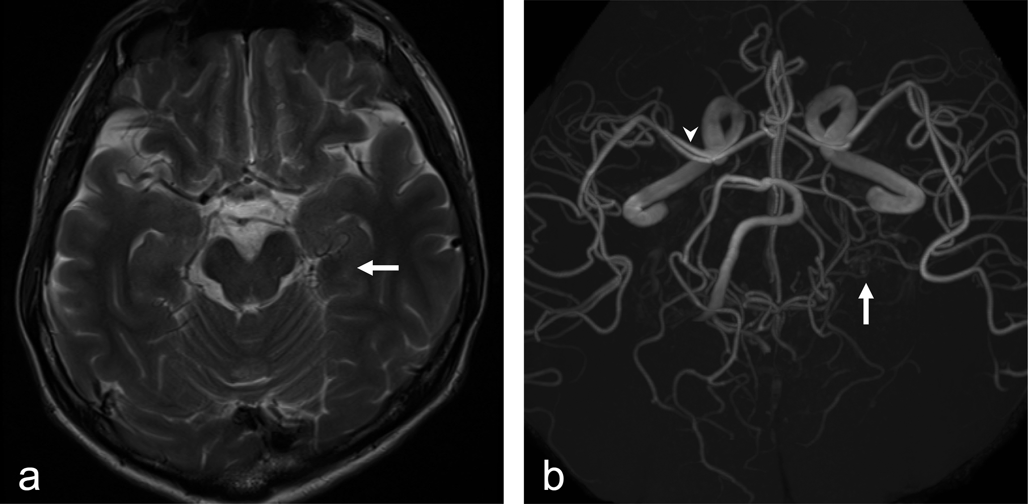
The Primitive ophthalmic artery (POA) is formed through a series of complex embryological processes [4, 8].
According to Padget's and Lasjaunias' hypotheses, the POA in this case is the dorsal ophthalmic artery (DOA) [4, 8]. In either hypothesis, the anastomosis, called the arterial ring, between the VOA and DOA may have been inadequate, suggesting that the POA persisted from birth in this case. It is thought that after birth, the ophthalmic artery that remained behind disappeared as the tissues were nourished by normal OA alone. Although there have been case reports of duplicate ophthalmic arteries, none that we have been able to locate indicate that the primitive ophthalmic arteries disappeared after birth [1,2,3, 5,6,7, 9, 10]. In this case, the primitive ophthalmic artery regressed naturally during follow-up, without any specific treatment or intervention.
In the first place, contrast-enhanced computed tomography (CT) or magnetic resonance imaging (MRI) are rarely performed in children unless shunting diseases such as dural arteriovenous fistula or neoplastic lesion is suspected. Additionally, it is suggested that in children, there may be instances of developmental immaturity, which raises the possibility that the natural regression of primitive ophthalmic arteries (POAs) may exist, even if it has not been confirmed through imaging or literature.
In any case, it might be helpful to be aware of the presence of a normal variant, as in the present case, to aid in proper diagnosis.
In this case, she was 2 years old at the time the primitive ophthalmic artery was noted on MRA. Therefore, it is a limitation of this case report that no imaging studies such as computed tomography angiography (CTA) or digital subtraction angiography (DSA) were performed due to the effects of radiation exposure.


留言 (0)