
記住我


After institutional review board approval from the ethics committee of Peking University Third Hospital (no. M2019193), consecutive patients who were diagnosed with FAIS and labral tear and underwent hip arthroscopic treatment in our institute between July 2020 and July 2021 were retrospectively reviewed. The inclusion criteria were as follows: patients who were diagnosed with (1) FAIS by clinical findings (persistent hip pain and positive physical examinations), plain radiographs (alpha angle > 55° and/or lateral center–edge angle (LCEA) > 40°), (2) labral tear by magnetic resonance imaging (MRI) [9, 10], (3) underwent hip arthroscopic treatment, and (4) had preoperative MRI and postoperative MRI at least 6 months following arthroscopy. The exclusion criteria were as follows: (1) patients with preoperative FHCI and (2) history of prior hip surgery. Informed consent was obtained from all participants.
Surgical techniqueAll surgeries were performed by one senior surgeon (Y.X.) with over 10-years of experience in hip arthroscopy using a standard supine approach as described by Gao et al. [11]. In brief, a detailed inspection of the central compartment was performed to assess the acetabular rim, acetabular labrum, articular cartilage, and ligamentum teres after interportal capsulotomy. Labral repair, labral debridement, femoral osteoplasty, or acetabuloplasty was performed according to the intraoperative findings. Capsular closure was routinely done at the end of surgery.
Data measurementsSupine anteroposterior hip radiographs, cross-table lateral radiographs, and CT were obtained for all patients preoperatively. Preoperative alpha angle and LCEA were measured as described by previous studies [12, 13].
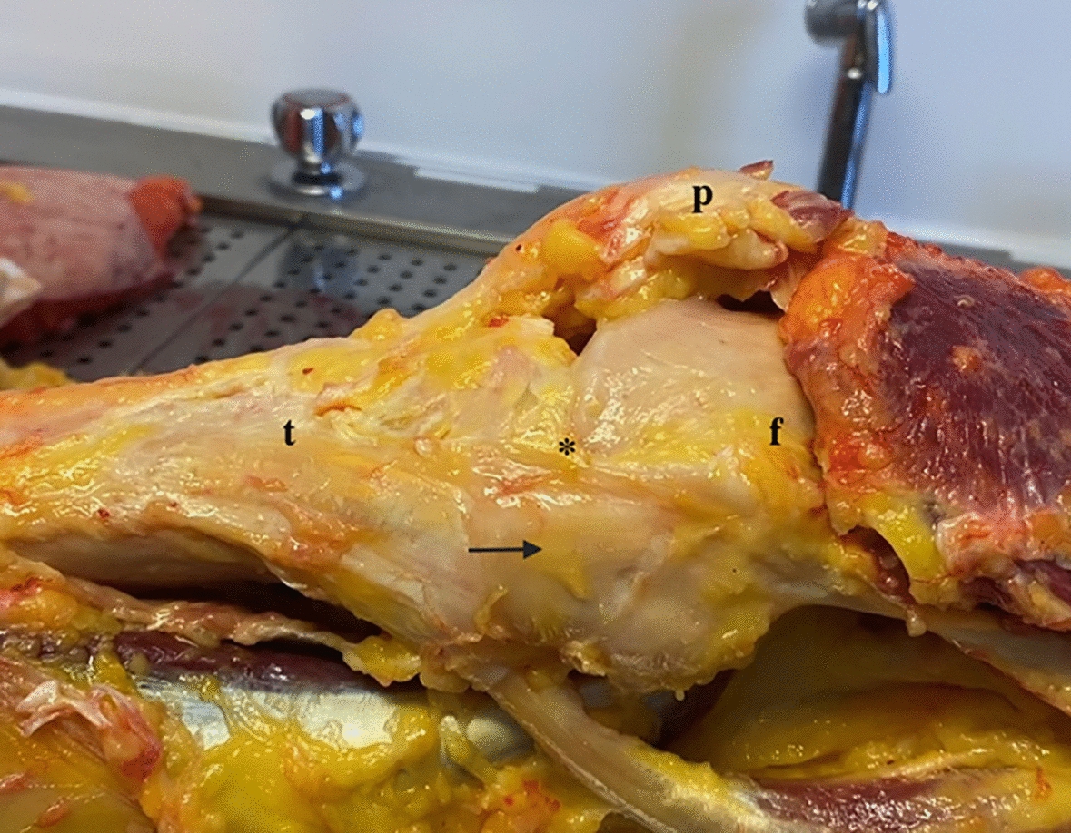
Patients underwent MRI preoperatively (the day before surgery) and at least 6 after arthroscopy (at clinic follow-up). As described by Gao et al. [14], the hip MRI was performed with a 3.0 T MR scanner (Magnetom Trio with TIM system, Siemens Healthcare) and a dedicated flexible surface coil around the affected hip joint. Fat-saturated proton density (FSPD) sequence and T2-weighted sequences were performed in the axial, coronal, and oblique sagittal planes respectively. Postoperative FHCI was identified as a flattening of the head sphericity or concavity with a small area of hyperintensity of the cartilage or underlying bone observed in the oblique sagittal or coronal plane on MRI (Fig. 1). Observer A with more than 5 years of experience with hip MRI and arthroscopy and observer B with more than 10 years of experience with hip MRI and arthroscopy analyzed all MRI scans. Any disagreements in findings were deferred to the corresponding author for final determination. The evaluations were performed twice by both surgeons to determine the intraobserver and interobserver reliability. The length of the acetabular hip labrum was measured at lateral and anterior anatomic sites along the acetabular rim as described by previous studies [15,16,17].
Fig. 1
A Postoperative FHCI identified as concavity with a small area of hyperintensity of underlying bone observed on MRI. B Postoperative FHCI identified as a flattening of the head sphericity observed on MRI. C Postoperative FHCI observed in hip arthroscopy. White arrow, the area of postoperative femoral head cartilage injury. FHCI femoral head cartilage injury, L labrum, F femoral head
Clinical outcomesPreoperative and postoperative patient-reported outcomes (PROs) were obtained via questionnaires. PROs included visual analog scale (VAS) for pain and modified Harris Hip Score (mHHS). The PROs at final follow-up were evaluated at the same time as the MRI follow-up. For the 0–10 VAS, 0 means no pain and 10 means the worst pain. For the 0–100 mHHS, 0 means the poorest function and 100 means the most satisfied function. Complications or revision hip arthroscopy were recorded. For the mHHS, the minimal clinically important difference (MCID) was defined as 8 by Kemp et al. [18], and the patient acceptable symptom state (PASS) was defined as 74 by Chahal et al. [19].
Statistical methodsContinuous variables with a normal distribution in the baseline data between groups were examined using the independent samples t-test. Mann–Whitney U test was used for non-normally distributed data. The two-tailed paired t-test was used to evaluate significance between preoperative and postoperative variables. Percentages were compared between patients with and without FHCI using the chi-squared test. The intraobserver and interobserver reliability was evaluated by calculating the kappa coefficient (k). P values < 0.05 were considered statistically significant. Confidence intervals (CIs) were set at 95%. All statistical analyses were performed with SPSS Statistics, version 22 (IBM).
留言 (0)