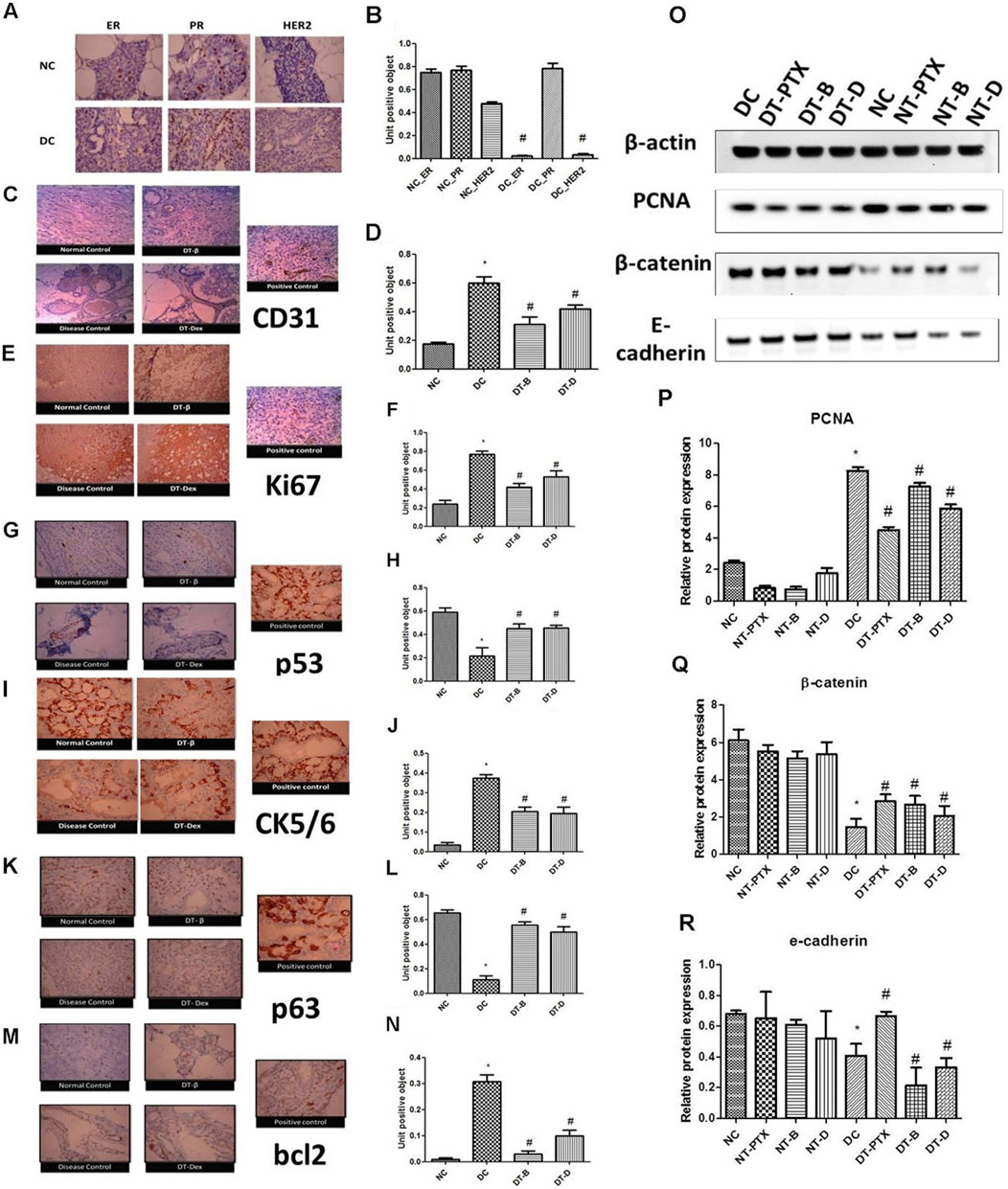
In the published article, there was an error in (Figures 2, 4) as published. These errors occurred in preparation of composite figures from individual images, which were inadvertently placed. The corrected (Figures 2, 4) appear below.

Figure 2. (A) Representative PCR bands in various treated cells, (B) α-interferon gene expression study in MDA-MB-231 cells, (C) α-interferon gene expression study in HEK293 cells, (D) β-interferon gene expression study in MDA-MB-231 cells, (E) β-interferon gene expression study in HEK293 cells, (F) γ-interferon gene expression study in MDA-MB-231 cells and (G) γ-interferon gene expression study in HEK293 cells. Determination of β-interferon release in various cell lines; (H) β-interferon release in MDA-MB-231 cell line and (I) β-interferon release in HEK293 cell line. # Significantly different from control (P < 0.05), Values expressed as Mean ± SEM. Control and con – untreated HEK293 and MDA-MB-231 cells, P1 and P – 1 μM paclitaxel, P2 – 5 μM paclitaxel, D1 and D – 1 μM DEAE-Dextran, D2 – 5 μM DEAE-Dextran, B and β – β-interferon treated cells.

Figure 4. (A, B) Immunohistochemistry studies for ER, PR, and HER2 in DMBA induced mammary gland model. NC – control rats, DC – positive control, ER – estrogen antibody staining, PR – progesterone antibody staining, HER2 – HER2 antibody staining, magnification X100. (C, D) Immunohistochemistry studies of CD31 in DMBA induced mammary gland model. Positive control of liver section was used, magnification X100. (E, F) Immunohistochemistry studies of ki67 in DMBA induced mammary gland model. Positive control of breast carcinoma was used, magnification X100. (G, H) Immunohistochemistry studies of p53 in DMBA induced mammary cancer model. Positive control breast cancer sections were used, magnification X100. (I, J) Immunohistochemistry studies of CK5/6 in DMBA induced mammary cancer model. Positive control of lung squamous cell carcinoma slide was used, magnification X100. (K, L) Immunohistochemistry studies of p63 in DMBA induced mammary cancer model. Positive control as breast cancer section was used, magnification X100. (M, N) Immunohistochemistry studies of bcl2 in DMBA induced mammary gland model. Positive control of tonsil section was used. Control animals, positive control, DT-B – rats treated with β-interferon, DT-Dex – rats treated with DEAE-Dextran, magnification X100. Determination of protein expression by Western blot analysis; (O) representative Western blot bands, (P) determination of PCNA protein expression, (Q) determination of β-catenin protein expression, and (R) determination of E-cadherin protein expression. ∗Significantly different from control animals (P < 0.05), # Significantly different from positive control (P < 0.05), each group consists of six animals, Values expressed as Mean ± SEM. NC – Control animals, DC – positive control, DT-D – 100 mg/kg DEAE-Dextran treated, DT-PTX – 30 mg/kg paclitaxel treated, DT-B – β-interferon treated, NT-D100 – normal treated with 100 mg/kg DEAE-Dextran, NT-PTX – normal treated with 30 mg/kg paclitaxel and NT-B – normal treated with β-interferon.
The authors apologize for this error and state that this does not change the scientific conclusions of the article in any way. The original article has been updated.
All claims expressed in this article are solely those of the authors and do not necessarily represent those of their affiliated organizations, or those of the publisher, the editors and the reviewers. Any product that may be evaluated in this article, or claim that may be made by its manufacturer, is not guaranteed or endorsed by the publisher.
Keywords: DEAE-Dextran, β-interferon, TNBC, anti-proliferative, apoptosis, angiogenesis, VEGF, NOTCH1
Citation: Bakrania AK, Variya BC and Patel SS (2024) Corrigendum: Role of β-interferon inducer (DEAE-Dextran) in tumorigenesis by VEGF and NOTCH1 inhibition along with apoptosis induction. Front. Pharmacol. 15:1433625. doi: 10.3389/fphar.2024.1433625
Received: 16 May 2024; Accepted: 27 November 2024;
Published: 13 December 2024.
Edited and reviewed by:
Olivier Feron, Université catholique de Louvain, BelgiumCopyright © 2024 Bakrania, Variya and Patel. This is an open-access article distributed under the terms of the Creative Commons Attribution License (CC BY). The use, distribution or reproduction in other forums is permitted, provided the original author(s) and the copyright owner(s) are credited and that the original publication in this journal is cited, in accordance with accepted academic practice. No use, distribution or reproduction is permitted which does not comply with these terms.
*Correspondence: Snehal S. Patel, c25laGFscGhhcm1hNTNAZ21haWwuY29t; c25laGFsLnBhdGVsQG5pcm1hdW5pLmFjLmlu; Anita K. Bakrania, YW5pdGFiYWtyYW5pYUBnbWFpbC5jb20=
留言 (0)