
記住我
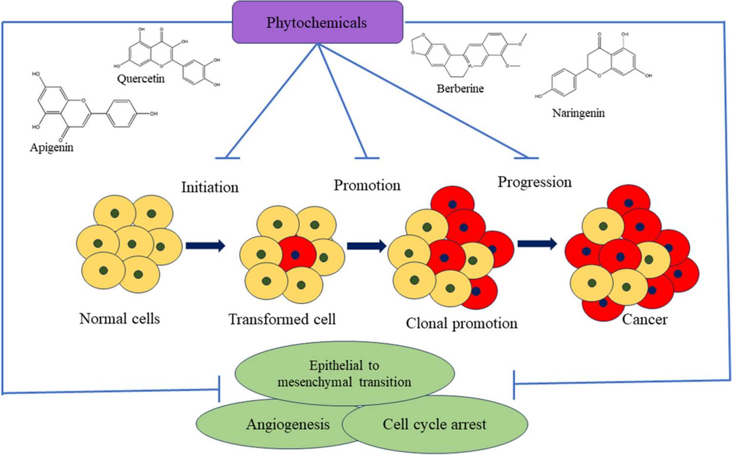


Figure 1 presents the effects of dictamnine on body weight, FBG, and serum AGE levels in rats with GDM. Comparing with the control, the GDM rats demonstrated a reduction in body weight and an increase in both FBG and serum AGE levels. Nevertheless, the treatment of dictamnine at concentrations of 15 and 30 mg/kg led to a considerable increase in the body weight of GDM rats. The treatment of dictamnine also resulted in reduced FBG and AGE levels in the GDM rats.
Fig. 1
Effect of dictamnine (1) on the bodyweight, serum advanced glycation end products, and fasting blood glucose levels in the experimental rats. The mean ± SD of triplicates were represented by each bar, and statistical analysis is performed using SPSS software. Values are evaluated using one-way ANOVA and Tukey’s post hoc analysis. *p < 0.01 compared to the control; **p < 0.05 compared to the streptozotocin-induced gestational diabetes mellitus group
Effect on Fetus and Placental WeightsThe effects of dictamnine on the fetus weight, placental weight, and placental index of the GDM rats are given in Fig. 2. When compared with the control, the GDM rats demonstrated an elevated placental weight and index while decreasing their fetus weight. Interestingly, the 15 and 30 mg/kg of dictamnine treatment revealed a remarkable reduction in the placental weight and index while remarkably elevating the fetus weight of the GDM rats.
Fig. 2
Effect of dictamnine (1) on the fetus weight, placental weight, and placental index of the experimental rats. The mean ± SD of triplicates were represented by each bar, and statistical analysis is performed using SPSS software. Values are evaluated using one-way ANOVA and Tukey’s post hoc analysis. *p < 0.01) compared to the control; **p < 0.05 compared to the streptozotocin-induced gestational diabetes mellitus group
Effect on Lipid Biomarker LevelsFigure 3 displays the changes in lipid markers in the control and treated rats. The rats with GDM demonstrated elevated total cholesterol, triacylglycerides, VLDL, and LDL while reducing the HDL level when compared to control. Conversely, the dictamnine at 15 and 30 mg/kg concentrations effectively reduced the LDL, VLDL, triacylglycerides, and total cholesterol levels in rats with GDM. The dictamnine also increased the HDL levels in the rats with GDM, providing evidence of its therapeutic efficacy.
Fig. 3
Effect of dictamnine (1) on the lipid profiles of the experimental rats. The mean ± SD of triplicates were represented by each bar, and statistical analysis is performed using SPSS software. Values are evaluated using one-way ANOVA and Tukey’s post hoc analysis. *p < 0.01 compared to the control. **p < 0.05 compared to the streptozotocin-induced gestational diabetes mellitus group
Effect on Antioxidant LevelsThe antioxidant levels, including SOD, GPx, CAT, and GSH, were measured in the experimental rats, and the results are depicted in Fig. 4. The rats with GDM revealed a drastic decrease in their GSH, GPx, CAT, and SOD levels. Nevertheless, the dictamnine treatment at 15 and 30 mg/kg dosages remarkably elevated the GSH, GPx, CAT, and SOD levels in rats with GDM. These results demonstrate the antioxidant potential of dictamnine.
Fig. 4
Effect of dictamnine (1) on the antioxidant levels of the experimental rats. The mean ± SD of triplicates were represented by each bar, and statistical analysis is performed using SPSS software. Values are evaluated using one-way ANOVA and Tukey’s post hoc analysis. *p < 0.01 compared to the control; **p < 0.05 compared to the streptozotocin-induced gestational diabetes mellitus group
Biochemical Marker LevelsFigure 5 displays the levels of biochemical markers, including FINS, hepatic glycogen, serum C-peptide, HbA1c, and FFA, in the experimental rats. Elevated concentrations of FINS, hepatic glycogen, C-peptide, and HbA1c, while reduced FFA levels were noted in the rat with GDM. Remarkably, the dictamnine considerably decreased the concentrations of FINS, hepatic glycogen, C-peptide, and HbA1c levels and also increased the FFA levels in the GDM rats.
Fig. 5
Effect of dictamnine (1) on the biochemical marker levels of experimental rats. The mean ± SD of triplicates were represented by each bar, and statistical analysis is performed using SPSS software. Values are evaluated using one-way ANOVA and Tukey’s post hoc analysis. *p < 0.01 compared to the control; **p < 0.05 compared to the streptozotocin-induced gestational diabetes mellitus group
Serum Inflammatory CytokinesThe levels of cytokines were analyzed in the serum, and the results are presented in Fig. 6. The rats with GDM displayed a significant elevation in IL-6, TNF-α, and IL-1β levels, accompanied by a reduction in IL-10 levels. Remarkably, the dictamnine treatment effectively reduced these pro-inflammatory cytokines and elevated the IL-10 level in the GDM rats.
Fig. 6
Effect of dictamnine (1) on the inflammatory cytokines in the serum of experimental rats. The mean ± SD of triplicates were represented by each bar, and statistical analysis is performed using SPSS software. Values are evaluated using one-way ANOVA and Tukey’s post hoc analysis. *p < 0.01 compared to the control; **p < 0.05 compared to the streptozotocin-induced gestational diabetes mellitus group
Apoptotic ProteinsThe apoptotic proteins such as Bax, Bcl-2, and caspase-3 levels in the pancreatic levels of the experimental rats are presented in Fig. 7. The rats with GDM revealed a significant decrease in the Bcl-2 level, whereas an elevation in the Bax and caspase-3 levels were observed. Captivatingly, the dictamnine treatment at dosages of 15 and 30 mg/kg remarkably enhanced the Bcl-2 level while reducing the Bax and caspase-3 in the pancreatic tissues.
Fig. 7
Effect of dictamnine (1) on the apoptotic protein levels in the experimental rats. The mean ± SD of triplicates were represented by each bar, and statistical analysis is performed using SPSS software. Values are evaluated using one-way ANOVA and Tukey’s post hoc analysis. *p < 0.01 compared to the control; **p < 0.05 compared to the streptozotocin-induced gestational diabetes mellitus group
Molecular MarkersFigure 8 displays the levels of NF-κB p65, VCAM-1, EGFR, MCP-1, NOX-2, and RAGE in the placental tissues. The rats with GDM showed increased levels of NF-κB p65, VCAM-1, EGFR, MCP-1, NOX-2, and RAGE in their placental tissues. Meanwhile, the dictamnine treatment at concentrations of 15 and 30 mg/kg remarkably decreased the levels of these markers in the placental tissues of GDM rats, indicating its salutary effects.
Fig. 8
Effect of dictamnine (1) on the molecular marker levels in the experimental rats. The mean ± SD of triplicates were represented by each bar, and statistical analysis is performed using SPSS software. Values are evaluated using one-way ANOVA and Tukey’s post hoc analysis, *p < 0.01 compared to the control; **p < 0.05 compared to the streptozotocin-induced gestational diabetes mellitus group
HistologyFigure 9 depicts the results of a histological assessment of the pancreatic tissues. The tissues obtained from control rats exhibited no inflammatory signs and exhibited typical cellular structure. Conversely, the pancreas of rats with GDM revealed inflammatory cell infiltrations, shrinkage of islet cells, and enlargement of adipose tissue. The histological changes in the pancreas were effectively reduced by the 15 and 30 mg/kg dictamnine treatments.
Fig. 9
Effect of dictamnine (1) on the pancreas and liver histopathology. Group I: normal pregnant rats; group II: streptozotocin-induced diabetic pregnant rats; group III: streptozotocin-induced diabetic + 15 mg/kg of dictamnine-treated pregnant rats; group IV: streptozotocin-induced diabetic + 30 mg/kg of dictamnine-treated pregnant rats
Gestational diabetes mellitus is a metabolic disorder that occurs during pregnancy and is characterized by an inability to tolerate glucose during pregnancy. The onset of GDM is caused by an increased and abnormal resistance to insulin, which is defined by a decrease and/or reduced sensitivity of insulin receptors in the cells of the body. Consequently, the cells become less receptive to the activity of insulin, thereby resulting in increased levels of glucose in the bloodstream (Sheiner et al. 2019). The pathophysiology of GDM is difficult to predict as it typically presents with minimal or no preceding symptoms. The rising prevalence of GDM is driven by the increasing rates of obesity and the advancing mother age (Preda et al. 2022). The etiology of GDM is known to be influenced by both environmental and genetic variables, though the precise molecular mechanism underlying this process remains unclear. In patients with GDM, insulin secretion is reduced due to an increased rate of IR (Skajaa et al. 2020).
The negative consequences of GDM have significant implications for public health and emphasize the importance of comprehending the fundamental mechanisms involved. The malfunction of β-cells in individuals with GDM causes inadequate insulin secretion, leading to IR and disrupted metabolism (Johns et al. 2018). Presently, several approaches, such as insulin therapy, dietary modifications, and lifestyle interventions, are extensively employed for the management of GDM. Nevertheless, prolonged utilization of these techniques may have an impact on the developing fetus (Zito et al. 2020). Hence, innovative and efficient approaches are advantageous for treating GDM. The present work highlighted that dictamnine effectively reduced the GDM condition in STZ-induced pregnant diabetic rats.
The placenta is indispensable for controlling the transfer of nutrients between the fetus and mother. The placenta undergoes functional and structural modifications to accommodate the increasing needs of the developing fetus and also adjusts in response to variations in the availability of nutrients in the mother (Sferruzzi-Perri et al. 2019). GDM has been observed to impact the development and function of the placenta. Studies have demonstrated that there is an enhanced movement of glucose from the placenta to the fetus in cases of GDM. Additionally, the levels of glucose transporters and genes related to glycolysis are found to be elevated in individuals with GDM (Jayabalan et al. 2019). In this work, the findings revealed increased placental weight and index, while reduced fetus weight was noted in the rats with GDM. Interestingly, the dictamnine treatment effectively increased the fetus weight and reduced both placental weights in the GDM rats.
During the third trimester, patients with GDM experience IR, as well as reduced rates of insulin production and glucose intake. The reduced secretion of insulin and the increased state of IR disrupt the balance of insulin in the mother, leading to the progression of maternal glucose intolerance and promoting GDM. While the role of IR is important in the onset of GDM, the precise mechanism by which insulin resistance is initiated remains unclear. The downregulation of insulin signaling is controlled by the inflammatory markers produced by adipose tissue (Lempesis et al. 2023). The present results showed that dictamnine successfully decreased both FINS and blood glucose levels in the GDM rats, which proves dictamnine improved IR and glucose intake in the GDM condition.
Hyperglycemia disrupts the normal inflammation and oxidative stress mechanisms in the development of GDM, leading to enhanced inflammatory cytokine levels. The abnormality in metabolites induces a rapid and intense pro-inflammatory response, leading to systemic IR. Research has demonstrated that placental inflammation plays a critical role in the development of GDM by influencing the fetal environment during pregnancy (Hoffmann et al. 2022). TNF-α, IL-6, and IL-1β are essential cytokines that play a critical role in inflammation. TNF-α is an early component of inflammation and can trigger the IL-6 release as a starting factor. IL-1β is a prominent cytokine that has a role in controlling insulin signaling (Gehrke et al. 2022). Patients diagnosed with GDM have markedly elevated inflammatory markers, including IL-6 and TNF-α, compared to individuals with a normal pregnancy. Consequently, they experience a chronic inflammation. Zhang et al. found that GDM patients had elevated levels of inflammatory factors, which resulted in a chronic state of inflammation. The present study revealed that GDM rats had increased TNF-α, IL-6, and IL-1β levels while reduced IL-10 levels were observed. Interestingly, the dictamnine treatment successfully decreased the pro-inflammatory cytokines while enhancing the IL-10 cytokine levels in the rats with GDM.
Pregnant women in the late gestational stage experience a 30% increase in the production of glucose in order to meet their energy needs while fasting. Nevertheless, in women diagnosed with GDM, there is a considerable impairment in the functioning of β-cells, resulting in a rise in IR and ultimately causing hyperglycemia during pregnancy (Bochkur Dratver et al. 2022). As a result of the heightened IR, there is an increase in the levels of lipids in the bloodstream. During an earlier human investigation, researchers identified changes in lipid metabolism that promoted the accumulation of fat in the initial phases of pregnancy. These changes were marked by reductions in triacylglycerides and total cholesterol, or LDL, and an elevation in HDL levels in the bloodstream. On the other hand, during the later stages of pregnancy, there are alterations in lipid metabolism that encourage the release of fat, resulting in higher levels of triacylglycerides and LDL. Nevertheless, GDM is distinguished by dyslipidemia, which manifests as increased triglyceride levels and decreased HDL concentrations in the bloodstream (Mulder et al. 2024; Wang et al. 2019). The current findings reveal that the dictamnine treatment successfully increased HDL levels while reducing triacylglycerides, cholesterol, LDL, and VLDL levels in the rats with GDM. These outcomes suggest the beneficial activities of dictamnine on the regulation of STZ-induced dyslipidemia in GDM rats.
The placenta is a more vascularized organ that has a large number of mitochondria and a high generation of ROS and metabolism. Thus, pregnancy is considered a pre-diabetic condition characterized by increased oxidative stress. It has been indicated that pregnancy exacerbated by obesity and GDM intensifies oxidative stress within the womb, resulting in elevated production of ROS in both the body and placenta (Ramirez-Emiliano et al. 2017). Maternal oxidative stress occurs at the same time as aberrant blood flow in the uterine artery and/or improper functioning of the placenta, which affects the transfer of nutrients from mother to fetus, hence impacting fetal growth (Bugatto et al. 2018). Oxidative stress, a significant contributor to the onset of diabetes, plays a pivotal role in the progression of IR (Calvo et al. 2024). Patients with GDM had high oxidative stress levels and diminished antioxidant levels throughout the 26–32 weeks of pregnancy (Lyu et al. 2022). The present findings highlighted that the dictamnine successfully mitigated the oxidative stress response in the GDM rats by enhancing antioxidant levels. These findings proved the antioxidant properties of dictamnine.
There is a association between heightened oxidative stress and the occurrence of apoptosis and mitochondrial malfunction. Apoptosis is triggered by caspase activation, an elevation in Bax expression, and a diminution in Bcl-2 expression (Sharma et al. 2022). Caspases are a group of enzymes that have a significant function in apoptosis. Cell apoptosis is initiated through two mechanisms, namely the extrinsic and intrinsic processes, which can occur simultaneously. The extrinsic route, referred to as the death receptor cascade, is activated by the binding of a stressor to its specific receptor. TNF-α ligands are both examples of substances that can promote cell death. Their interaction triggers the activation of caspases 8 and 9, which then activate the executioner caspases, resulting in the beginning of apoptosis (Huang et al. 2019). The intrinsic pathway, commonly referred to as the mitochondrial pathway, is characterized by mitochondrial malfunction. The intrinsic process involves the formation of a pore on the mitochondrial membrane by Bcl-2 proteins. As a result, cytochrome c is able to exit the mitochondria and enter the cytosolic compartments (D’Arcy 2019). The present study clearly proved that the dictamnine treatment remarkably increased the Bcl-2 protein level while reducing the Bax and caspase-3 protein levels in the pancreatic tissues of the rats with GDM, which highlights its anti-apoptotic effects.
In the presence of hyperglycemia, there is a rise in the development of AGEs, which triggers the AGE-RAGE signaling activation. This cascade enhances NF-κB expression, which leads to inflammation by increasing the secretion of ICAM-1 and VCAM-1 and releasing inflammatory markers (Bansal et al. 2023). ROS is produced along this pathway, causing oxidative stress and attracting inflammatory cytokines (Antonetti et al. 2021). The NOX2 enzyme facilitates ROS accumulation, which in turn promotes oxidative damage in insulin-sensitive cells (Lee et al. 2020). A prior investigation demonstrated that the levels of NOX2 were enhanced while the levels of endogenous antioxidants were reduced in mice with HFD/STZ-induced diabetes, indicating an increased state of oxidative stress (Yan et al. 2018). The present work indicated drastic elevations in the levels of various molecular markers such as NF-κB p65, VCAM-1, EGFR, MCP-1, NOX-2, and RAGE in the placental tissues of the rats with GDM. However, the treatment with dictamnine successfully reduced these marker levels.
留言 (0)