
記住我


The CPC granules used in this study are Chitai® (Shanghai Rebone Biomaterial, Shanghai, China). Briefly, they were synthesized by combining dicalcium phosphate anhydrous (DCPA) with equimolar tetra calcium phosphate (TECP). To obtain the high-purity TECP to achieve a solid-to-solid reaction between calcium phosphate and calcium carbonate, the temperature was set at 1500 degrees Celsius for 8 h. Ammonium hydrogen phosphate [(NH4)2HPO4] and calcium nitrate [Ca (NO3)2] were set at an acidic pH to prepare Dicalcium phosphate dihydrate (DCPD, CaH- PO4.2H2O). DCPA was obtained by evaporating the crystallization water in DCPD at 120 degrees Celsius for 5 h. The scaffolds were prepared at a specific ratio of TECP: DCPA (73.21: 26.79 in weight, aiming to have a 1.67 Calcium/ Phosphor molar ratio to achieve the same ratio of bone tissue), in which NaCl granules ~ 500 μm in size were incorporated and combined with 100 μL saturated NaCl solution in water to form the cement mire. The mire was then cast into a mold under the pressure of 2 MPa for one minute, to avoid H2O loss at the high pressure or granule deformation at low pressure. Then the granules were prepared into 0.25 to 1 mm CPC-granule samples weighing 0.25 g each, which will be implanted in the dorsal sites of the rats.
Preparation of BMP-2@CPC granulesThe abovementioned CPC granules were sterilized by ethylene oxide vapor. Recombinant human BMP-2(rhBMP-2, Shanghai Rebone Biomaterial, Shanghai, China) in acetic acid solution was doped onto CPC granules and stayed 4 h until fully absorbed. Afterward, the granules were lyophilized and stored at − 20 degrees Celsius for later use. The final amount of rhBMP-2 loaded in CPC is about 1 mg of rhBMP-2 in 1 g CPC.
Topography characterization of CPC and BMP-2@CPCThe BMP-2@CPC granules were sputter-coated with gold particles before topography, then the topography of the CPC and BMP-2@CPCB granules was observed by scanning electron microscopy (SEM, JSM-6360LV, JEOL, Japan).
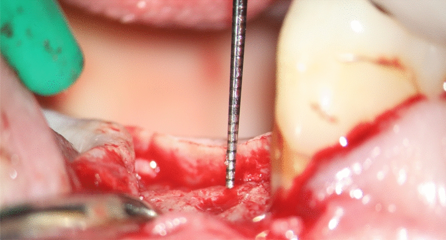
In-vivo investigation and histological evaluationA total of six seven-week-old male C57 (C57BL/6) rats (17 ~ 19 g weight) were used for subcutaneous bone formation assessments. Rats were housed in steel hanging mesh cages, provided with a standard chow, and had no limited access to water. Experiments on rats and animal care were ethically approved by the Laboratory Animal Management and Welfare Ethical Review Committee of Zhejiang Chinese Medical University (ZSLL-2018-038) and ACTA Ethical Committee (202012). In this animal study, each rat was implanted with CPC or BMP-2@CPC at the left or right dorsal subcutaneous site randomly, which was consistence with a “split-mouth” design. Thus, for each kind of material, there were six sites for the following assessments (n = 6 per group, two dorsal subcutaneous sites per animal). General anesthesia (inhalation of isoflurane) was performed before surgery, and 0.25 g CPC or BMP-2@CPC granules were randomly implanted subcutaneously in the dorsal sites (simple cut and suture). For this purpose, rats were placed in a dorsal position and immobilized. The backs of the rats were shaved and disinfected. Dermal incisions were made in a longitudinal direction parallel to the spine. After implantation of CPC or BMP-2@CPC, interrupted sutures were used to close the skin.
Five weeks after the operation, the rats were sacrificed, and the implanted CPC and BMP-2@CPC granules samples were retrieved together with a minimum quantity of the surrounding tissue for chemical fixation and embedding, as previously reported [19]. With a systematic random-sampling strategy [20], the samples were sectioned into 7–9 slices of 600 μm thickness vertical to the short axis and with 1 mm intervals from each other. Of each sample, six slices were mounted on Plexiglas holders and then polished to 400 μm. The slices were then stained with McNeal’s Tetrachrome, basic Fuchsin, and Toluidine Blue O.
Biocompatibility of implanted CPC and BMP-2@CPC was evaluated based on the volume of fibrous capsule tissue and multinucleated giant cells (MNGC) volume density. In addition, osteoinductivity was assessed according to the density of bone and the osteoblast cells under the skin of rats. The total volume of the subcapsular space embraced by the fibrous connective tissue capsule was taken as a reference volume [21]. Within the fibrous capsule of each slice, the cross-sectional area of bone, fibrous connective tissue, and residual CPC were estimated using the Cavalieri/point-counting estimator technique [22]. The mean volume of bone, fibrous connective tissue, and residual CPC of each retrieved tissue of CPC or BMP-2@CPC groups were calculated by multiplying the sum of the cross-sectional area of bone, fibrous connective tissue, and residual CPC by the fixed distance between slices, defined as volume = (scale length / actual length × sieve length) ^ 2) × counted points × width of slice. The volume density of each tissue mentioned above was obtained by dividing the tissue volume by the total volume of the subcapsular space (reference volume).
Two small areas per slice were randomly selected to represent each slice's osteoblasts and MNGCs density (Fig. 1). Briefly, a grid was put on the slices to cover most of the tissue, high magnification photos at 200 × of two randomly selected areas were printed, and cell volume density was determined by a point-counting technique and calculated to represent the osteoblasts and MNGCs density [21]. A grid with 18 subareas is placed over the histology picture (scale bar, 2 mm), covering most of the tissue. Two random numbers will decide the randomly selected two subareas per slice. The two selected subareas will be analyzed in 200 × magnification, and the results from two randomly selected areas will represent this slice.
Fig. 1
Randomization method for Cavalieri-counting for osteoblasts and MNGCs in each slice
Retrospective evaluation of BMP-2@CPC in maxillofacial surgerySince there are no clinical evaluations of the short- and long-term adverse events of BMP-2@CPC in maxillofacial surgery, a retrospective descriptive study was carried out to analyze the potential adverse events of BMP-2@CPC in this clinical condition. Inclusion criteria were: (1) Oral facial bone disorders treated with sinus floor elevation, bone regeneration after cyst enucleation surgery, guided bone regeneration (GBR), alveolar ridge preservation and (2) complete clinical record in medical histories available. Exclusion criteria were: (1) Patients for whom the status at certain time points could not be verified by either the treating physician or any relative; (2) incomplete medical records; (3) isolated nasal bone fractures and dental fractures. All the associated data were collected from medical records from the Department of Maxillofacial Surgery in the Affiliated Dental Hospital of Jiamusi University. The used BMP-2@CPC in these maxillofacial operations is approved by the Chinese Food and Drug Administration (CFDA No. 2013: 34-60199), and the retrospective study protocol was approved by the ethical committee of the Academic Center for Dentistry Amsterdam (2020281). As the institute delivered the data that did not include the privacy information of the patients, the requirement for informed consent was waived.
The general workflow of this clinical study can be found in Fig. 2. For the inclusion criteria, subjects had to have BMP-2@CPC implantation in maxillofacial areas. The cut-off date of data collection was August 1, 2018, and all data were collected in a de-identified fashion and entered into a standardized case report form that included demographic information (gender, race, and age), diagnosis, surgical procedure, amount of implanted BMP-2@CPC, and any adverse events. Patients were followed up for up to 2 years.
Fig. 2
Workflow of this retrospective clinical study
For a short-term evaluation, the inflammation after surgery was detected using complete blood cell counting, mainly focused on the number of white blood cells (WBC) and neutrophil–lymphocyte-ratio (NLR). In addition, the patient’s body temperature, pain grade, and any self-reported adverse events were recorded.
留言 (0)