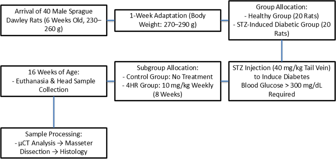
In this study, the size of the contralateral submandibular gland was significantly increased post-operatively after the ipsilateral submandibular gland extirpation (Fig. 3A). This increase was also noted in the radiation therapy group, indicating that the presence or absence of radiation therapy did not affect post-operative changes in submandibular gland volume (Fig. 3D). These changes were not significantly different in gender and aging (Fig. 3B, C).
After the unilateral removal of a salivary gland, the contralateral gland may undergo compensatory hypertrophy, characterized by increased proliferation and enlargement of acinar cells [11, 12]. This observation is consistent with our findings (Fig. 3A). If compensatory hypertrophy and the subsequent increase in saliva flow do not occur, xerostomia may develop. Various approaches are available for managing post-surgical xerostomia. Although artificial saliva can be used, its effects are often short-lasting [13]. Another option is the submandibular gland transfer, which involves relocating the contralateral submandibular gland to the submental area. This method has been reported to be more effective than pilocarpine [14]. However, this procedure may not be feasible for oral cancer patients, as it can lead to a loss of facial symmetry [15]. An alternative approach involves relocating the gland near the parotid area, which has shown success in avoiding radiation exposure [14]. Recently, intensity-modulated radiotherapy (IMRT) has been employed to spare the parotid gland, though there is limited literature on its use for submandibular gland sparing [4]. The effect of IMRT could be a favorable outcome factor [16]. Despite the advancements in dose-reducing IMRT, new technologies are still needed to better preserve saliva production and improve the overall quality of life for patients [9].
As individuals age, salivary glands naturally undergo atrophy, leading to a reduction in both size and function. This atrophy can potentially limit the capacity for compensatory hypertrophy in the remaining gland after one is removed. In contrast, younger patients may have a greater capacity for glandular compensation due to more robust cellular regeneration and overall physiological resilience. In this study, there was no significant difference in post-operative volume change between the groups aged over 70 and those under 70 (Fig. 3B). Aging significantly influences salivary flow, with the maximum increase in salivary flow rate observed in individuals aged 20–29 years [17]. Additionally, from birth to early adulthood, all salivary glands grow consistently in size [18]. However, as people age, studies have shown that increased cell death and reduced salivary function are major contributors to xerostomia in elderly individuals [19]. The median age in this study was 70 years, and much younger individuals were not included, which might explain the insignificant differences observed between the age groups.
In this study, the volume change ratio in males was larger than that in females; however, the difference between the groups was not statistically significant (Fig. 3C). Contrarily, a study conducted in Nepal observed that the volume of salivary glands in females is generally greater than in males, though this difference was also not significant [20]. Regarding salivary flow rates, males exhibited higher rates compared to females under both unstimulated and stimulated conditions [17]. Although this does not directly address gland size, another study found that female prisoners had a higher prevalence of salivary gland diseases compared to male prisoners [21]. Age and gender differences have a significant effect on salivary gland functions which is more apparent in women than in men [17, 22, 23]. These findings suggest that while there are observed differences in salivary gland size and function between genders, the evidence remains inconsistent and inconclusive. Further research is required to better understand these potential anatomical or physiological differences.
Radiation therapy is known to have a significant impact on salivary gland size and function [24]. The exposure of salivary glands to radiation, particularly during treatments for head and neck cancers, can induce atrophy, leading to a reduction in glandular size and a marked decrease in saliva production [25]. This radiation-induced atrophy occurs due to damage to the acinar cells, which are responsible for saliva production, as well as fibrosis and vascular changes within the gland [26]. The extent of atrophy and the consequent reduction in salivary flow can vary depending on factors such as the dose of radiation, the specific glands targeted, and the duration of the treatment [27]. Over time, this atrophy can lead to chronic xerostomia, significantly affecting a patient’s quality of life [25]. In some cases, the damage may be irreversible, with little to no recovery of glandular function. In studies examining post-radiation therapy patients, a significant reduction in salivary gland volume has been observed, confirming the deleterious effects of radiation on these glands [28]. The loss of glandular size and function after radiation therapy highlights the need for protective strategies or interventions to mitigate these effects and preserve salivary gland function. However, there was no significant difference in post-operative salivary gland volume change in the presence of post-operative radiation therapy in this study (Fig. 3D). This might be due to well-organized protective strategies or interventions to mitigate these effects and preserve salivary gland function.
Compensatory increases in saliva flow following salivary gland extirpation have been reported in a few clinical studies [29, 30]. However, this phenomenon is more commonly observed in animal studies [31, 32]. If compensatory salivation occurs, the extent of this change may vary depending on the observation period. In this study, the mean volume change ratio in the group observed for less than 3 months was slightly lower than in the group observed for more than 3 months, but this difference was not statistically significant (Fig. 3E). This suggests that compensatory gland hyperplasia occurs in the early period following unilateral salivary gland removal and is maintained thereafter. Previous reviews have shown that removal of the submandibular gland significantly reduces unstimulated saliva production [3]. Unfortunately, salivary flow was not measured in this study, so the relationship between compensatory volume increase in the submandibular gland and saliva flow remains undetermined. Additionally, a study conducted in Nepal reported slight size variations between the right and left submandibular glands: the mediolateral width of the right gland was slightly greater, while the left gland had a slightly larger volume [20]. In contrast, this study found no significant volume difference between the right and left submandibular glands (Fig. 3F).
The limitations of this study should be acknowledged. First, the sample size of oral cancer patients who underwent ipsilateral neck dissection was small, and the inclusion of younger patients was particularly limited. Although oral cancer is more prevalent in older populations, the potential for compensatory hypertrophy may vary with age, which could influence the study’s conclusions. Including a more diverse age range, particularly younger patients, might yield different outcomes. Additionally, this was a single-center study, which limits the generalizability of the findings. A multicenter study would be necessary to validate these results across different populations and settings. Second, the study was retrospective in nature, which inherently carries certain biases and limitations in data collection and analysis. Future research should focus on prospective studies to provide more robust and controlled data, allowing for a clearer understanding of the factors influencing compensatory hypertrophy and post-surgical outcomes. Third, this study did not include functional assessments such as measuring salivary flow rates or conducting radioisotope studies, which would have provided valuable insights into the functional outcomes of compensatory hypertrophy. These functional analyses are crucial for understanding the clinical significance of the morphological changes observed and should be incorporated into future research. Fourth, different types of radiotherapy may affect post-operative salivary function. In this study, most patients were referred to regional radiotherapy centers where they received IMRT. IMRT delivers precise radiation doses to the tumor or specific areas within the tumor while minimizing exposure to surrounding healthy tissues, such as the salivary glands, thereby reducing the risk of xerostomia [9, 15]. In contrast, other types of radiotherapy, such as 3D conformal radiotherapy or high-dose-rate brachytherapy, are more likely to induce xerostomia [33]. Due to the limited number of patients who received post-operative radiotherapy in this study, we were unable to evaluate the differences in post-operative salivary gland volume changes based on the type of radiotherapy received.


留言 (0)