Patient characteristics
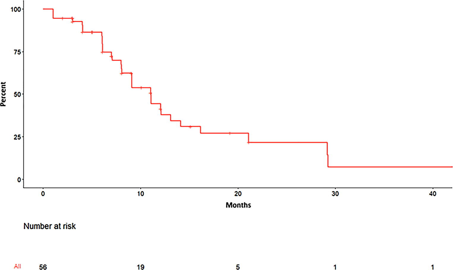
In this study, n = 7 patients with AML (median age: 48.9 years, range 21–67 years, 5 male, 2 female) and n = 4 patients with ALL (median age: 47.8 years, range 22–59 years, 1 male, 3 female) were enrolled, while n = 6 healthy donors (median age: 41.8 years, range 30–56 years, 2 male, 4 female) served as controls. Patients were treated with total body irradiation (TBI) as part of the conditioning regimen prior to an allogeneic hematopoietic cell transplantation. In the patient cohort, pre-transplant minimal residual disease (MRD) or refractory disease was detected in n = 7 patients (64%), n = 1 patient (9%) was of unknown status and n = 3 patients (27%) were MRD negative. The trial was approved by the Institutional Ethics Committee of the University Hospital Frankfurt am Main (protocol code 15/18) and was conducted in accordance with the Declaration of Helsinki. All patients have signed an informed consent.
Blood collection from patients and healthy donors
Peripheral blood was collected from patients with leukaemia via port catheter using 7.5 ml Serum/CAT tubes (Sarstedt, Nümbrecht, Germany) at the Department of Medicine II, Hematology and Oncology, University Hospital, Goethe University Frankfurt am Main. Blood samples were taken before TBI by a photon beam linear accelerator (Synergy, Elekta, Crowley, UK) with doses of 2 × 2 Gy per day for a total dose of 4 Gy at the Department of Radiotherapy and Oncology with an 8‑hour interval between doses and at 24 h after irradiation. All but one patient received chemotherapy between nine and two days prior to TBI. Peripheral blood of healthy donors served as a control.
Isolation of EVs from serum
EVs were isolated from peripheral blood using an ultracentrifugation workflow. In detail, blood from patients with leukaemia or healthy donors was allowed to clot for 30 min at RT and centrifuged at 1000 × g for 15 min to obtain serum. Subsequently, serum was centrifuged at 3000 × g for 15 min at room temperature and supernatant was centrifuged at 30,000 × g for 30 min. Next, supernatant was filtered through a 0.2 µm filter (Carl Roth, Karlsruhe, Germany) and centrifuged at 100,000 × g in an ultracentrifuge (Beckman Coulter, Krefeld, Germany) for two hours. Finally, the pellet was resuspended in 150 µL PBS (Thermo Fisher Scientific, Darmstadt, Germany) and EVs were stored at −80 °C for further analyses.
Quantification of protein concentration of EVs using BCA assay
To determine the protein concentration of isolated EVs, the Micro BCATM Protein-Assay-Kit (Thermo Fischer Scientific, #23235,) was used according to the manufacturer’s protocol. In brief, 150 µl of standard and 149 µl of ddH2O + 1 µl of isolated EVs were pipetted in triplicates in a 96-well plate (Greiner Bio-One, Frickenhausen, Germany), 150 µl Micro BCATM assay working reagent were added, and the plates were incubated for 2 h at 37 °C. Finally, optical densities were measured at 562 nm using a 96-well microplate reader (Infinite M200 Pro, TECAN, Männedorf, Switzerland) and concentrations were determined according to standard protein dilutions.
Characterization of EVs by Western immunoblotting
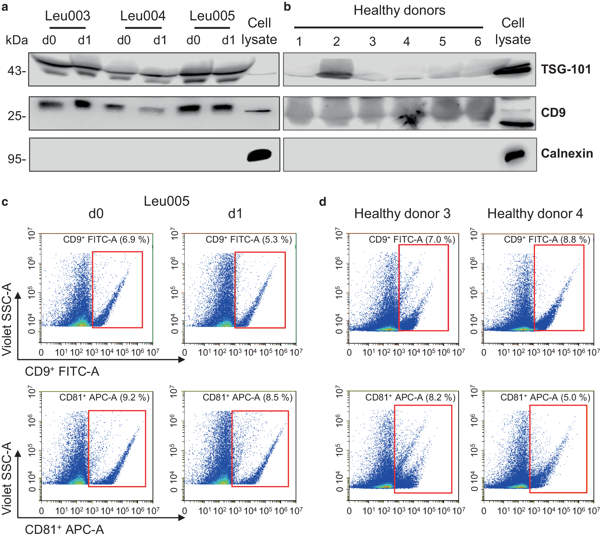
To characterize purified EVs, Western blot analysis was performed. 100 µg of EVs were lysed in modified radioimmunoprecipitation assay buffer (RIPA) as previously described [16], and incubated for 30 min on ice, followed by a heating step for 5 min at 99 °C. The lysed EVs were subjected to SDS-polyacrylamide gel electrophoresis (SDS-PAGE) and transferred to a Nitrocellulose membrane (GE Healthcare, Munich, Germany) using a semidry blotter unit (GE Healthcare). Membranes were incubated over night at 4 °C with the following primary antibodies: anti-Calnexin rabbit (rb) (Abcam, Cambridge, UK, #ab22595, 1:1000, 95 kDa), anti-TSG101 rb (Abcam, #ab125011, 1:1000, 45 kDa), anti-CD9 rb (Abcam, #ab92726, 1:1000, 25 kDa). After incubation with a secondary antibody (goat-anti-rabbit-IgG HRP, Biozol, Eching, Germany, #4050-05, 1:1000) for 1 h at room temperature, detection was accomplished using a Pierce ECL Western blotting substrate (Thermo Fischer Scientific) and the Odyssey Fc imaging system (LI-COR Biotechnology, Bad Homburg, Germany) for documentation.
Validation of EVs by flow cytometric analysis
To further verify isolated EVs by flow cytometry, 40 µg of EVs were stained with CD81 (APC-conjugated, clone 5A6, BioLegend, Amsterdam, The Netherlands, #349509) and CD9 (FITC-conjugated, Thermo Fisher Scientific, clone SN4 C3-3A2, #11-0098-42) for 1 h at 4 °C. Next, EVs were washed with PBS and precipitated at 100,000 × g for 70 min at 4 °C. The pellet was resuspended in 150 µl PBS and measured using flow cytometry (CytoFlex S, Beckman Coulter). The violet side scatter (V-SSC) was used as described in Brittain et al. 2019 [17]. For correct gating of EVs, Gigamix beads (Beckman Coulter) were used to determine the size standard for EVs. In a V-SSC-A/FSC‑A dot plot EVs/beads with a size around 100 to 900 nm were determined. Subsequently, percentages of FITC-positive (CD9) or APC-positive (CD81) EVs/beads were identified in a V-SSC-A/FITC or V‑SSC-A/APC dot plot.
RNA isolation from serum-derived EVs
Total mRNA, including miRNA, was isolated from EVs with an RNeasy Mini Kit (Qiagen, Hilden, Germany) and RNA content was quantified by photospectroscopic analyses (Infinite M200 Pro, TECAN).
Next generation sequencing of EV miRNA cargo
Total RNA was subjected to next generation sequencing (NGS) at Arraystar Inc. (Rockville, MD, USA) using an Illumina NextSeq 500 system. Briefly, samples covering in total six samples of healthy donors and 11 samples from patients prior to and after irradiation were transferred to the company and sequenced. The sequencing quality score revealed sufficient quality for subsequent data analysis in all samples. After quality control, the reads were 3′-adaptor trimmed and filtered ≤ 15 bp reads with cutadapt software. The trimmed reads were aligned to reference genome with bowtie software. Data analysis after trimming was performed with the software miRDeep29 to quantify known miRNA and predict novel miRNAs. Additionally, differentially expressed miRNAs were filtered using R package edgeR10,11. Hierarchical clustering and miRNA target prediction was performed by targetscan. Pathway analysis for the miRNA target mRNAs was performed based on the on the top 10 differentially expressed miRNAs using the latest Kyoto Encyclopedia of Genes and Genomes (KEGG) database [18]. This analysis allows to determine whether the identified miRNA target genes are enriched in specific biological pathways. The p values calculated by Fisher’s exact test are thereby used to estimate the statistical significance of pathway enrichment between the two groups compared. The KEGG database resource was developed to better understand functions of biological systems, including cells and organs, based on information on the molecular level, e.g., large-scale datasets from NGS sequencing approaches or other high-throughput technologies [18]. A schematic workflow of sequencing data analysis (Arraystar Inc. Rockville, USA) is shown in Supplementary Figure S1. Venn diagrams to identify similarities between the differentially expressed miRNAs were created using the InteractiVenn software (www.interactivenn.net, [19]).
Statistical evaluation
EdgeR was used to evaluate differentially expressed miRNAs. The sequencing raw data were fitted to negative binomial model using the quantile-adjusted conditional maximum likelihood (qCML) method as implemented in EdgeR. The differentially expressed genes were then tested by exact test using the model (for details see [20]). The p value is the f‑statistic p value, the q value (false discovery rate, FDR) comprises the FDR-adjusted p value. A p value less or equal to 0.05 and a q value less or equal to 1 were considered statistically significant. For KEGG pathway analysis, p values calculated by Fisher’s exact test were used to estimate the statistical significance of the enrichment of the pathways between the two groups.


留言 (0)