
記住我


Gross post-mortem examination revealed severe vascular lesions in all organs with extensive haemorrhages in heart, skeletal muscles, small and large intestines, and lung. The heart showed acute petechial to ecchymal hemorrhage in the subepicardial, intracardiac and subendocardial region with edema in the area of adipose tissue. The spleen had acute subcapsular petechial hemorrhages. The lymph nodes were hemorrhagic, diffuse and enlarged, especially intestinal lymph node. The liver showed extensive subcapsular edema with hemorrhagic rounded edges. The tongue was cyanotic and tinged with blue Esophagus. The small intestine presented with hemorrhagic walls with edema. On the skin multifocal petechial hemorrhage in the subcutaneous tissue was observed (Additional Fig. 2). Bacteriological examination of the liver and bronchial mucus revealed the presence of gram-positive cocci after enrichment and Streptococcus alactolyticus, respectively.
Histological findings were widely similar to the cases previously assigned to other EEHV genotypes, mainly characterized by haemorrhages, oedema and congestion. Inflammation was nearly absent, and despite intensive investigation, inclusion bodies, which are characteristic for other herpesvirus infections, were not detected.
Molecular analysis on the extracted DNA confirmed the presence of EEHV5 DNA in all tested tissues. Viral genome copies were quantified in infected tissues (Table 1). Attempts to isolate the virus from infected tissues as described before [7] were unsuccessful.
Table 1 Viral DNA copies assayed for the EEHV5 DNA polymerase gene and normalized to cellular DNA in tissues and per ml blood by qPCRWe sequenced the viral genome using DNA extracted from the elephant’s heart where viral DNA was most abundant. Illumina short-read sequencing was employed to determine possible variants and Oxford Nanopore long-read sequencing was used to complement Illumina data and fill in sequence gaps and low-coverage regions. Given that no protocols were available for EEHV5 enrichment, whole-genome sequencing was performed for both technologies, resulting in data containing both viral and host information. To minimize interference from host data, analysis was geared towards a reference-based approach and larger complex structural variants were discarded. As previous molecular analysis placed this isolate within the EEHV5 genotype, obtained sequencing data was first mapped against the only peer-reviewed complete EEHV5 genome: EEHV5A isolate Vijay (henceforth referred to as EEHV5A Vijay). Since the analytical focus was on detection of SNPs and short-spanning nucleotide substitutions, the genomic structure of the generated consensus corresponds directly to the reference’s structure.
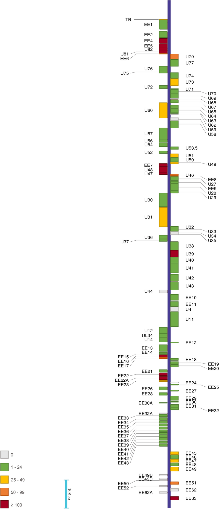
The data in Additional Table 2 shows variants obtained after analysis of the entirety of our sequencing data. Interestingly, the obtained consensus was considerably different from the previously published genome. Using EEHV5 Vijay as a reference we determined 3,881 variants; 444 of those are located outside open reading frames (ORFs) and 45 are located within terminal repeats (TR). Of the 3,881 detected variants, 972 were found in both short-read and long-read data, 393 were only supported by short-read data and 2,516 were only supported by long-read data; within long-read only variants, 620 were supported by manually parsing the mapping of short reads and 1896 were located in regions that short-read data did not cover. A representation of the detected variation across the Vijay reference can be found in Fig. 1. Variants are generally distributed throughout the genome; however, some genes do not harbor any (Additional Table 2).
To gauge the positioning of our data, several phylogenetic trees previously used for placement of EEHV5 isolates in relation to other EEHVs and herpesviruses were recreated (Figs. 2, 3 and 4) [3]. The original publication relied on partial sequences, mostly obtained via Sanger sequencing and to these, the corresponding regions from the EEHV5A Vijay genome as well as from our consensus sequence (factoring in all detected variants) were added. Three DNA-level phylogenetic trees were constructed: two of the trees are based on U38 (DNA polymerase), for placement within the EEHV group only and for comparison with other key herpesviruses, respectively; the last tree is based on U73 (origin-binding protein), also used for comparison of EEHVs with key herpesviruses. Additionally, one protein-level tree was constructed using U81 (uracil DNA glycosylase) and encompassing key herpesviruses. The selected loci and protein segments contain different levels of variation between our data and the references, and contain enough resolution to allow differentiation between EEHV5 isolates.
Fig. 2
DNA level evolutionary relationships between the obtained EEHV5A consensus sequence and EEHV family representatives. The radial phylogenetic tree is based on the U38 (POL) gene and was inferred using the Neighbor-Joining method. All codon positions were included. Ambiguous positions were removed for each sequence pair, leaving 1075 positions in the final dataset. The bar displays the number of nucleotide substitutions per site. Evolutionary analysis was conducted using MEGA11
Fig. 3
DNA level evolutionary relationships between the EEHV5A consensus, EEHV family representatives and other relevant herpesviruses. The linear phylogenetic trees are based on U38 (POL) and U73 (OBP) genes, respectively. Trees were inferred using the Maximum Likelihood method and Jukes-Cantor model. All codon positions were included and ambiguous positions were removed for each sequence pair, leaving 1075 positions in the final dataset for U38 (POL) and 672 positions in the final dataset for U73 (OBP). The percentage of replicate trees in which the associated taxa clustered together in the bootstrap test (500 replicates) are shown next to the branches. Evolutionary analysis was conducted using MEGA11
Fig. 4
Protein level evolutionary relationships between the EEHV5A consensus, EEHV members and Betaherpesvirinae subfamily representatives. The radial phylogenetic tree is based on the U81 (UDG) protein sequence and was inferred using the Maximum Likelihood method and JTT matrix-based model. The final datased included 257 positions. The percentage of trees in which the associated taxa clustered together is shown below the branches. Full evolutionary analysis was conducted using MEGA11
The intra EEHV5 Polymerase (U38) radial tree (Fig. 2) very closely replicates the original. It places the Vijay reference and the consensus with the remaining EEHV5 isolates, as expected. When extending the analysis to include key herpesviruses (Fig. 3), EEHV5A Vijay clusters along with EEHV5A NAP28. Despite our data containing only 18 variants in relation to EEHV5A Vijay, these are sufficient for the consensus to cluster within an EEHV5 group but peripherally from all other isolates. However, when translating the nucleotide consensus sequence into an amino acid sequence there are no differences detected, which argues for high functional conservation of the DNA polymerase as observed across many herpesviruses.
The remaining DNA-level tree, based on the Origin-Binding Protein (U73) (Fig. 3), also places the EEHV5 sequences closely, with EEHV5A Vijay clustering outside the EEHV5A NAP50 and NAP28 branch. Similarly to the U38 tree, EEHV5A consensus is placed peripherally to the remaining EEHV5 isolates. Our data’s U73 sequence contains 25 variants in relation to EEHV5A Vijay, that translate into 4 differences at the amino acid level.
Lastly, the protein-based phylogenetic tree was built using the amino acid sequence of the Uracil DNA Glycosylase (U81) (Fig. 4) and it clusters EEHV5A Vijay along with EEHV5A NAP50 as observed in the original tree. The newly generated consensus is again in a different branch, slightly closer to EEHV5B isolates. In the case of U81, the number of variants relative to the reference is quite high at 173 detected variants.
Overall, the consensus is placed close to the remaining EEHV5 isolates, but in distinct branches from the reference, with the structure of each tree reflecting that of the original trees [3]. Variant and phylogenetic analysis suggest that our data is different from the EEHV5A Vijay genome. At this point, no assertions could be made about how the data relates to EEHV5B genomes.
To assess this, we used a GenBank entry corresponding to a full EEHV5B genome: EEHV5B isolate North American NAP58 Tucker (henceforth referred to as EEHV5B Tucker). However, the manuscript associated with this genome is not yet published, making it a non-peer reviewed entry. Due to the scarcity of genomic information for EEHV5 viruses, we nevertheless decided to use it as a secondary reference. All previous analytical steps were repeated using EEHV5B Tucker as a reference: variant calling was performed and a new consensus sequence was derived (EEHV5B consensus); all phylogenetic analyses were also repeated to include EEHV5B Tucker and the new EEHV5B consensus. As depicted in Additional Fig. 3, genome size differs slightly between EEHV5A Vijay and EEHV5B Tucker—the EEHV5A Vijay genome is 5 kbp longer than EEHV5B Tucker; additionally, there is also substantial inversion between the two, spanning a 103 kbp region. This region, albeit inverted, harbours a consistent and common repertoire between the two genomes, containing most genes from U27 to U82. However, each isolate encodes a few unique proteins scattered across the genome related to gene regulation, as well structural proteins involved in capsid and tegument structuring.
Using EEHV5B Tucker as a reference we determined 3,083 variants (Additional Table 3); 160 of those are located outside ORFs and 24 are located within terminal repeats. Of the 3,083 detected variants, 1258 were detected with both short and long-reads, 14 were only supported by short-read data and 1550 were only supported by long-read data; within these variants, 171 were supported by manually parsing the mapping of short reads and 1379 were located in regions that short-read data did not cover. A representation of the detected variation across the Tucker reference can be found in Additional Fig. 4. For EEHV5B variants are also distributed throughout the genome, with the exception of a few genes that contain no variants.
To gauge the positioning of our data in light of EEHV5B, all previously mentioned trees (Figs. 2, 3 and 4) were re-built with the addition of EEHV5B Tucker and the EEHV5B consensus and can be found in Additional Figs. 5, 6 and 7. The intra EEHV5 Polymerase (U38) radial phylogenetic tree (Additional Fig. 5) continues to very closely replicate the structure of the original tree. As for the U38 tree including other herpesviruses (Additional Fig. 6) EEHV5B Tucker clusters along with EEHV5B NAP58. EEHV5B Tucker and EEHV5B NAP58 are anticipated to cluster together, given that the Tucker genome reference is also labelled as NAP58TW: there is a difference of one amino acid between the two protein sequences, which might be due to the methodology used to obtain the full Tucker genome. Levels of variation for this protein are similar with 18 and 19 SNPs in relation to EEHV5A Vijay and EEHV5B Tucker, respectively.
The Origin-Binding Protein (U73) phylogenetic tree (Additional Fig. 6) places all the EEHV5 sequences closely once more, with EEHV5A Vijay clustering outside the EEHV5A NAP50 and NAP28 branch, followed by EEHV5B NAP58 and finally EEHV-5B Tucker. Although belonging to a similar isolate, the EEHV5B NAP58 sequence is incomplete in comparison with the EEHV5B Tucker genome. Similarly to the U38 based tree, EEHV5A and EEHV5B consensuses are placed peripherally to other EEHV5 isolates and cluster together. Our data’s U73 sequence contains 25 and 24 variants in relation to the EEHV5A Vijay and EEHV5B Tucker respectively, translating into 4 and 2 differences at the amino acid level.
Lastly, the Uracil DNA Glycosylase (U81) tree (Additional Fig. 7) clusters EEHV5B Tucker along with EEHV5B NAP58 and otherwise resembles the original tree and the tree in Fig. 4. Also for this protein, one difference at the amino acid level between EEHV5B NAP58 and EEHV5B Tucker can be seen. The newly generated consensuses are again in different branches, placed slightly closer to EEHV5B isolates. For U81, the number of variants relative to each reference is quite different: for EEHV5A Vijay our data contains 173 variants, whilst for EEHV5B Tucker there are only 3. These translate onto 34 and 1 changing amino acids, indicating that this protein is more similar to the EEHV5B sequence.
In all phylogenetic trees containing EEHV5B Tucker and the EEHV5B consensus, both consensuses are placed close to EEHV5 isolates, but in distinct branches, usually clustering together and indicating that our data is different from the available EEHV5A and B genomes.
Discussion and conclusionsEEHVs continue to be one of the main threats to elephants in captivity. EEHV1A and B are long known to be a major main cause of fatal disease in young (Asian) elephants in captivity, with many cases described in the last three decades [5, 6]. EEHV5, on the other hand, has been associated with non-fatal clinical infection only until the first fatal case was reported in 2011 in the UK. EEHV infections are considered ubiquitous among elephant populations in captivity and in free range [3, 27, 28], however fatal incidences of haemorrhagic disease in young elephants remain peculiar as they seem to contradict the smooth coexistence of virus and host achieved through long standing co-evolution [29].
Here, we report another fatal case associated with EEHV5 in a young Asian elephant (and the first case in continental Europe). Clinical signs appeared for a brief period before leaving little room for diagnostic measures and therapeutic intervention. Haemorrhagic lesions were observed in most of the visceral organs and tissues, which is typical for fatal cases related to EEHV infection [4, 5, 7, 9, 11, 28]. Although typical for herpesvirus infection, we did not observe intranuclear inclusion bodies despite extensive histological search. We detected EEHV5 DNA in various tissues throughout the elephants body, which again resembles findings from previous EEHV associated fatalities [7, 9, 29]. This pan-body tropism of the virus might be attributed to its endotheliotropic nature with subsequent extensive replication, in the endothelial lining of all tissues. Since EEHV5 infection was thus far not associated to severe disease in captive elephants in continental Europe, standard testing protocols for sick Asian elephants do not necessarily include EEHV5 testing. We hope that this report will raise awareness for the potentially fatal nature of EEHV5 infection in Asian elephants and strongly encourage inclusion of EEHV5 in virological diagnostics for this species.
We further continued our efforts and have attempted to isolate EEHV5 on CrFK (feline), ENL-2 (elephant) and elephant fibroblasts (extracted from Raj skin) cell cultures. Unfortunately, our attempts remained unsuccessful: after the 2nd virus passage on each cell line, no viral DNA could be detected from cultured cells. As previously reported [6, 7], virus replication was not supported in cell culture. The reason for the inability to culture EEHVs remains elusive, we speculate that this may be due to the lack of suitable cultured host cells or missing of tissue complexity required for virus replication as provided by the natural host. This inability to support viral replication in cell culture reverberates into the amount of genomic information available for EEHV5 viruses, as there is a lack of robust references and genomic sequences which, in turn, complicates both the analysis of new sequencing data and the establishment of molecular enrichment methods for these viruses. Currently, no wet-lab EEHV enrichment or host depletion methods are available, meaning that EEHV-targeted sequencing is not yet a possibility. With cell culture also being unavailable, only whole-sample sequencing from the tissue of an infected animal is possible. This technique, which was employed in this study, carries a high percentage of host DNA that cannot be separated in wet-lab procedures. In such conditions, no analytical route is without disadvantages, making genomic analysis rather delicate. Although in silico host depletion is an option, it also may hinder the recovery of parts of the viral genome that may closely resemble that of the host; as homology between herpesviruses and their hosts is well documented and host mimicry is a relevant part of the viral evolutionary strategy, these regions may prove relevant to recover [30, 31]. While opting for reference-based approaches and employing stricter mapping parameters, as well as focusing on SNPs and small substitutions, is helpful in minimizing possible “background noise” from host DNA without loss of information on these common genomic areas, it does preclude detection of larger structural variation and structural novelty within the data that can only be found using de novo assemblies. Due to their stringency, mapping-based strategies may then overestimate the similarity between the sequencing data and the references used, undermining potential structural differences. Using a mapping approach on long and short-read next-generation sequencing derived from heart tissue, we could nevertheless determine variation across both the whole 180 kb genome sequence of EEHV5A Vijay, the only published and peer-reviewed reference available to us. Bioinformatic analysis of data obtained in the current study confirms the relatedness of the virus to EEHV5 isolates, but also reveals significant differences between the reference and our consensus sequence: almost 4000 variants were detected all over the genome relative to EEHV5A Vijay. One single gene (glycoprotein H, gH) showed alone 400 SNPs that result in 212 amino acid changes to this protein relative to the reference. This gene is well known to be conserved among all herpesviruses and involved in virus entry and/or cell fusion [32]. The role of gH as well as all other genes of EEHVs is not yet studied. Albeit in lesser quantity, variants at the protein level were also discovered in other typically highly conserved structural genes: for the large tegument protein encoded by U31, 2 amino acid variants were detected; for the DNA packaging protein 2, encoded by U50, 2 amino acid variants were detected; for major capsid protein U57, 5 amino acid variants were detected; for package subunit 1 encoded by U64, 6 amino acid variants were detected; for the portal protein encoded by U76, 2 amino acid variants were detected; for the terminase subunit encoded by U60, amino acid changes involve major structural changes that result in only 388 out of 660 amino acid residues aligning with the reference protein, and within this segment 9 amino acid variants were detected. Our data is thus substantially different from the published EEHV5A genome, at the nucleotide and amino acid levels. We could not, however, discard the possibility that the present data might represent an isolate closer to EEHV5B than EEHV5A. So far, there is only one complete EEHV5B genome sequence available in GenBank, which, at the date of writing this report, is still undergoing submission in a peer-reviewed journal [9]. However, due to the shortage of data regarding EEHV5 whole genomes, with no other EEHV5B sequence available, EEHV5B Tucker was used as a secondary reference in this study, to complement the main body of results derived from using EEHV5A as a reference. Looking at this additional body of results, over 3,300 variants were detected relative to EEHV5B Tucker, highlighting the unique character of this data amongst EEHV5 isolates, regardless of subgroup. When using the obtained 5A and 5B consensus sequences to recreate previously established phylogenetic dendrograms, they are consistently clustered together and placed in the periphery of other EEHV5 isolates, for both DNA and protein-level phylogenies, across proteins with differing counts of variants detected. Along with the results qPCR EEHV testing and variant calling, phylogenetic analysis reveals differences that point to the presence of an EEHV5 isolate in this sample that is seemingly distinct from the EEHV5 genomes described so far, for both 5A and 5B subgroups.
This division of EEHV5 into A and B subgroups was created to highlight the chimeric nature of EEHV5A and 5B genomes, which contain hypervariable sequence patterns among several loci, likely indicating ancient chimeric exchange events between A and B [3]. This chimeric tendency of the viral genome is shared with EEHV1A and EEHV1B, a group subdivided thusly for the same reason, and known to be the most likely cause of fatal haemorrhagic disease in Asian elephants [3]. The genotype EEHV7, also divided into A and B subgroups presumably for the same reason, has recently been implicated in a case of fatal disease in an African elephant, the first involving EEHV7 [33]. Due to the limited number of available EEHV5A and 5B genomes, it is difficult to know the full extent of these possible chimeric exchanges and how ancient or recent they may be, as well as what effect they might have on the virulence of EEHV5 isolates and the severity of disease caused by them. Due to the involvement of EEHV5 isolates in fatal cases of disease, the possible emergence of previously unknown EEHV5 genomes should not be overlooked.
Almost all our knowledge about herpesvirus genes is gained from human herpesviruses and few animal herpesviruses considered to be important for livestock. Lack of funding greatly hinders basic research on animal and exotic herpesviruses and decelerates the development of useful genomic and cell culture tools to address these questions in full.
As such, we leave this available for the scientific community for any possible future work that will provide a basis for better understanding of epidemiology, host–pathogen interaction and evolution of EEHV5 virulence. One possible application of our analysis could be the development of novel SNP-based PCR assays that can allow the detection of EEHV5 strains and help accurately determine the incidence of the virus. The genomic information included here could also be used to design EEHV5 DNA enrichment protocols for re-sequencing and genome walking, eventually allowing for de novo assemblies and thus inference upon larger structural genomic differences that may be present and could not be unveiled using the current methodology.
In conclusion, we have detected and studied the pathology of EEHV5 in fatal case of an Asian elephant. We encourage inclusion of EEHV5 in virological diagnostics of Asian elephants, even if no previous reports of EEHV5 related death or illness are available from a certain region or country. We believe that the reported EEHV5 DNA genomic analysis and consensus sequences will help further to understand the pathogenesis and virulence of EEHVs with respect to developing new diagnostic methods, prophylactic strategies, and implementation of control measures in future.
留言 (0)