Cell culture
H9c2 cells (rat cardiomyocytes) were purchased from the Korean Cell Line Bank (KCLB; Seoul, Korea) and were grown in Dulbecco's modified Engle's mixture (DMEM, Hyclone; Logan, USA) supplemented with 10% fetal bovine serum (FBS, Gibco™; NY, USA) and 1% penicillin/streptomycin (Welgene; Gyeongsangbuk-do, Korea). All cells were cultured in a humidified atmosphere at 37 °C with 5% CO2.
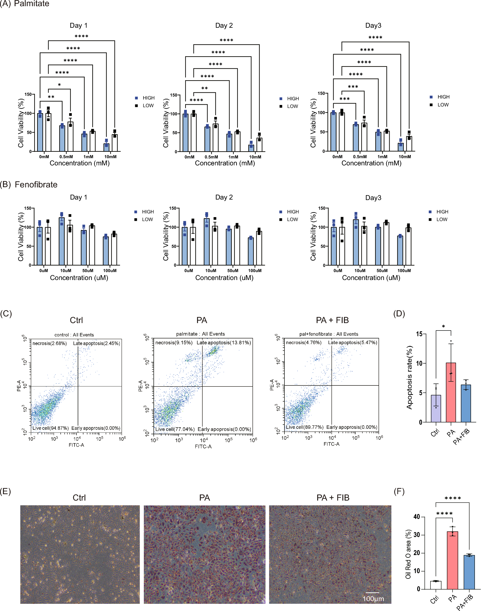
For the experiments, H9C2 cells were seeded in a 6-well plate. Then, cells were cultured in either low glucose DMEM (1 g/L) or high glucose DMEM (4.5 g/L, Himedia; PA, USA) with fatty acid-free Bovine Serum Albumin (BSA) (Goldbio; Missouri, USA). Cells were categorized into three groups: vehicle-treated group (control), palmitate-treated group (palmitate), palmitate plus fenofibrate-treated group (palmitate + fenofibrate). Palmitate acid (Sigma-Aldrich; MA, USA) was dissolved in ethanol at a concentration of 10 mM. Fenofibrate (Sigma-Aldrich) was dissolved in DMSO.
Animals
All protocols requiring the use of animals were approved by all experiments were reviewed and approved by Institutional Animal Care and Use Committee of Gwangju Institute of Science and Technology (GIST) and studies were conducted in adherence to the NIH Guide for the Care and Use of Laboratory Animals. Euthanasia was performed by cervical dislocation while the mice were under a condition of anaesthesia. Male C57BLKS/J-Lepr (db/db) mice were purchased from the Shizuoka Institute for Laboratory Animals, Inc. (Japan SLC; Shizuoka, Japan). Mice were maintained in a 12-h light–dark cycle at ambient temperature (22 ± 1F °C) and fed ad libitum a standard chow diet (SCD) and water. Male mice were randomly divided into two groups: db/db with corn oil (Sigma) (DB_VEH, n = 4), and db/db with fenofibrate (DB_FIB, n = 4). Mice were treated with either corn oil or fenofibrate between 6 and 20 weeks of age. Fenofibrate was dissolved in corn oil and injected orally at regular intervals every day at a dose of 100 mg/kg/day [15]. For type 2 diabetes mice model, male C57BL/6 J mice were purchased from the Shizuoka Institute for Laboratory Animals, Inc. (Japan SLC; Shizuoka, Japan). The 6-week-old male mice were administered an intraperitoneal injection of streptozotocin (STZ, 50 mg/kg body weight) daily for 5 days (n = 8) or vehicle (n = 8). The powder of streptozotocin was dissolved in 0.1 M Na-Citrate Buffer, pH 4.5. Then mice were fed with a high-fat diet ((HFD, 60 kcal% fat, D12492, Research Diets, Inc)) for 16 weeks for inducing type 2 diabetic mellitus (T2DM).
Body weight was recorded once a week, and echocardiography and glucose tolerance test (GTT) were performed one week before harvest, respectively. All experiments were carried out in accordance with the appropriate guidelines. The Institutional Animal Care and Use Committee of Gwangju Institute of Science and Technology approved all animal procedures (GIST-2021–110). Our study did not include any human data or tissue.
MTT assay for cell viability
H9c2 cells were seeded at 2 × 10⁴ cells in each of the 96 wells and incubated overnight. Then, cells were cultured with either different drug concentrations or vehicle for 24 h, 48 h, and 72 h. The Cell proliferation kit I (Roche; IN, USA) was used for MTT (3-[4,5-dimethylthiazol-2-yl]-2,5-diphenyltetrazolium bromide) diphenyltetrazolium bromide) assay as manufacturer’s protocol. Briefly, following drug treatment, the culture medium was replaced with MTT solution. After four hours of incubation, the formed formazan was dissolved in dimethyl sulfoxide (DMSO), and the absorbance at 550 and 600 nm was detected using an automated microplate reader.
Flow cytometric analysis of apoptosis
H9C2 cells were treated for 10 min at room temperature with Annexin-V-FITC (MedSystems Diagnostics; Vienna, Austria) and propidium iodide (Sigma, St. Louis, MO, USA) in binding buffer (Invitrogen™; MA, USA) [16]. The Cytoflex (Beckman counter; CA, USA) flow cytometer was used to evaluate apoptotic cells. For each sample, a total of 2 × 10⁴ events were collected. At least three distinct experiments were used to complete the analyses in triplicate [16]. Apoptosis (Annexin V-FITC positive, PI negative) in necrotic (Annexin V-FITC positive, propidium iodide positive) cells was identified by flow cytometry [16]. Temperature, washing, and resuspension are all factors to consider.
Flow cytometry with oxidized DCFDA for ROS identification
2ʹ,7ʹ-Dichlorofluorescin Diacetate (DCFDA, Sigma) dye was used for determining total intracellular reactive oxidative species (ROS) levels. H9C2 cells were incubated in a plate with a glass bottom for 20 min with 10 μM DCFDA dye. Fluorescence were measured using confocal microscopy (Carl Zeiss, LSM 880 NLO) or semi-quantitatively analyzed using a flow cytometer at 488 nm and 525 nm.
Oil red O staining for H9c2 cells
The amount of intracellular lipids was measured by Oil Red O staining. To make a working solution, Oil Red O solution (Sigma-Aldrich) was dissolved in distilled water (3:2) and diluted. H9c2 cells were fixed in 4 percent paraformaldehyde for 30 min at room temperature, washed three times in PBS, and then incubated with the Oil Red O working solution for 30 min at room temperature.
Western blot analysis
Cell lines and tissue samples was lysed in a RIPA buffer. Total protein concentrations were measured with the DC protein assay kit (Biorad; Hercules, CA). Then proteins were separated by electrophoresis and transferred to PVDF membranes. The membranes were blocked in Tris-buffered saline (TBS) containing 0.05% Tween 20 and 5% non-fat dry milk. Then membranes were incubated with specific primary antibodies (1:1,000) including Sod2 (#66,474, Proteintech; IL, USA), Fatty Acid Synthase (#3180, Cell Signaling Technology; MA, USA), PPARα (#sc-398394, Santa Cruz Biotechnology; TX, USA) and β-Actin (#12,262, Cell Signaling Technology). After washing with TBS containing 0.05% Tween 20, membranes were incubated with HRP-linked secondary antibodies (Cell Signaling Technology) and visualized using the ECL detection kit (Merck Millipore). Signals were captured using a Luminograph II System (ATTO; Tokyo, Japan). Densitometric analysis was performed using Image J software (version 1.53).
Echocardiography
1.5–2.0% isoflurane (inhaled at 3.0 L/min) was used to anesthetize the mice, and their cardiac hypertrophy was assessed using an ACUSON NX3 Elite Ultrasound system (SIEMENS Healthineers; Munich, Germany) with a VF16-5 transducer (16.0 MHz). Echocardiographic measurements were performed as previously described [17]. The dimensions of the left ventricle (LV), the ejection fraction (LVEF), the fractional shortening (FS), the mass of the LV, and the thickness of the LV wall were all measured.
Blood analysis and glucose tolerance test
Blood samples were collected from the mice heart at the time of sacrifice. Aspartate Aminotransferase (AST), Alanine Aminotransferase (ALT), Total bilirubin (T-BIL), and lipid levels were measured at the Korea Testing & Research Institute (KTR; Jeollanam-do, South Korea), an institution authorized to perform non-clinical studies. For the glucose tolerance test, mice were fasted for around 12 h. Mice were intraperitoneally injected with 2 g/kg D-glucose in PBS. A glucometer will be used to determine the blood glucose level in tail vein blood. Blood is obtained by snipping the tail. After the glucose injection, blood glucose levels are monitored at 0, 15, 30, 60, and 120 min.
Histological examination
Cardiac tissues were harvested and fixed in 10% (w/v) neutral buffered formalin and embedded in paraffin. Sections were stained with hematoxylin–eosin (H&E). Masson's trichrome staining was performed for tissue fibrosis analysis. For lipid and fat staining, fresh collected cardiac tissues were embedded in Tissue-Tek Optical Cutting Temperature compound (Sakura; CA, USA). Then, midventricular sections were stained with oil red O and counterstained with hematoxylin.
Quantitative real-time reverse transcriptase-PCR (qRT-PCR) analysis
Total RNA was extracted with the TRIzol® reagent (Invitrogen) according to the manufacturer's protocol. Then, 2ug of total RNA was used to generate complementary DNA with the High-Capacity cDNA Reverse Transcription kit (Thermofisher, MA. USA). To analyze gene expression, real-time PCR was performed using the amfisure qGreen Q-PCR master mix (GenDEPOT; TX, USA). The primer sequences used in this study are provided in Supplementary Table 1.
RNA-sequencing and data analysis
RNA sequencing was performed on an Illumina Hiseq 2000 platform. Raw data was processed using the'edgeR' package in R software (version 4.1) to generate counts per million (cpm). Then processed data were transformed to log2 scale and standardized using quantile normalization. The adjusted log2-cpm is then used in an integrative statistical technique to detect differentially expressed genes (DEGs). The observed T value and log2-median-ratio between two conditions were calculated using the Student's t-test and log2-median-ratio for each gene. To create an overall p-value, the corrected p-values were blended using Stouffer’s method [18]. DEGs were chosen for each comparison based on two criteria: an overall p-value of less than 0.05 and an absolute log2-median-ratio greater than the median of the empirical distribution's 2.5th and 97.5th percentiles.
Gene Set Enrichment Analysis (GSEA) was performed to assess the enrichment of DEGs using the R software's (version 4.1) “clusterProfiler” package [19]. Gene Ontology (GO) analysis, and Kyoto Encyclopaedia of Genes and Genomes (KEGG) analysis were performed using the DAVID program [19]. The commercial QIAGEN Ingenuity® Pathway Research (IPA®, QIAGEN Redwood City, www.qiagen.com/ingenuity) software was used to undertake an upstream regulatory analysis (URA) of DEGs detected in our analysis [20]. The p-value was calculated using Fisher's Exact Test, with 0.05 set as the significant level.
Single cell RNA-sequencing analysis
Heart tissues were dissected from the mice and dissociated into single cell suspensions using Liberase (Roche, 5,401,119,001) according to the manufacturer's protocol. The tissues were harvested in the predetermined volume (1000–1500 mm3), cut into 2–4 mm size pieces, and placed in GentleMACS C tubes with Liberase [1 mL of 1X PBS (Gibco, #10,010,023), Liberase 5 mg/ml]. Tissue dissociation was performed using the 37C_m_TDK_1 program with the GentleMACS Octo Dissociator (Miltenyi Biotec; cat. #30–093-235) and incubated in a water bath at 37 °C. After dissociation, the samples were filtered and centrifuged, the supernatant was discarded, and the sediment was resuspended in PBS.
The cell concentration of the single cell suspension was maintained at 500 cells/ul (total 1 * 10^5 cells) and loaded onto the Chromium Next GEM Chip G Single Cell Kit (10X genomics, #1,000,120). RNA sequencing libraries were prepared using the Chromium Next GEM Single Cell 3' Kit v3.1 (10X genomics, #1,000,268). Individual libraries were diluted to 250–500 pM and sequenced on the Illumina Novaseq6000 sequencing platform using the S2 Reagent Kit (200cyc) in paired-end mode.
Gene expression matrices were generated using cellranger (v.6.1.1) with the mm10 reference genome after removal of environmental RNA using cellbender (v.0.3.0). The gene expression matrix was loaded with Seurat (v.5.0.1) and cells with less than 300 nFeature_RNA, 300 nCount_RNA and more than 80% mitochondrial ratio were excluded. Then, samples were normalized with SCTransform, 3,000 variable.features.n, and then integrated by harmony with 30 PCAs. Nearest neighbors were computed with 30 dimensions of harmony reduction. UMAP was also generated based on the 30 dimensions of harmony reductions.
Human data sources
We used the National Health Insurance Database (NHID) in Korea, established by the National Health Insurance Service in conjunction with the National Health Checkup Program [21]. This database provides longitudinal data on 97% of the South Korean population and includes de-identified sociodemographic information and insurance claims coded according to the International Classification of Diseases, 10th Revision (ICD-10). The National Health Examination Programme includes questionnaires on health status, anthropometric measurements and laboratory data. Our study protocol (2021–11-026) was approved by the Institutional Review Board of Kangbuk Samsung Hospital. As we did not access any personally identifiable information, the requirement for informed consent was waived. The study was performed according to the principles outlined in the Declaration of Helsinki.
Study design and participants
A total of 856,286 patients taking statins in Korea were prescribed fenofibrate between 2010 and 2017. Patients over the age of 40 were selected for this study, while those with a history of heart failure or missing data were excluded. Patients who developed CHF within one year of taking fenofibrate were also excluded, leaving 427,154 patients eligible for analysis. To reduce potential bias and demographic imbalance related to fenofibrate use, a fenofibrate-naive group who were also taking statins was selected from the NHID and matched to the fenofibrate-using group with 1:1 age and sex adjustment (Supplementary Fig. 4 and Table 2). The final analysis included 427,154 patients using fenofibrate and an equal number of patients not using fenofibrate. All patients were followed up until 31 December 2019.
Measurements and definitions
A standardized self-report questionnaire was used to collect information on smoking, drinking and exercise. Heavy drinking was defined as consuming 30 g or more of alcohol per day. Regular physical activity was defined as at least 30 min of moderate-intensity physical activity on five or more days per week, or at least 20 min of vigorous-intensity physical activity on three or more days per week. Those in the lowest income quintile receiving medical assistance were considered to have low household income. Obesity was defined as a body mass index (BMI) of 25 kg/m2 or more. Hypertension was defined as having a blood pressure of 140/90 mmHg or higher or taking antihypertensive medication, as indicated by ICD-10 codes I10-I15. Diabetes was defined as having a fasting plasma glucose (FPG) concentration of 126 mg/dL or higher or being prescribed antidiabetic medication according to ICD-10 codes E11-E14. Chronic kidney disease (CKD) was defined as an estimated glomerular filtration rate (eGFR) of less than 60 mL/min/1.73 m2, calculated using the Modification of Diet in Renal Disease study equation. Blood samples were taken after an overnight fast of at least 8 h to determine glucose, total cholesterol, triglyceride (TG), HDL cholesterol and LDL cholesterol concentrations.
Study outcomes
Incident HF was identified by hospitalization with ICD-10 codes I110, I130, I150, or I971 as the primary diagnosis. Study participants were followed from the start of the study until they were diagnosed with incident HF or until December 31, 2019, whichever came first.
Statistical analyses
Continuous variables were reported as either mean with standard deviation (SD) or median with interquartile range, while categorical variables were reported as numbers (%). Independent samples t-test and χ2 test were used to compare characteristics of participants at baseline. Incidence rates were reported as number of events per 1000 person-years. To examine the associations between fenofibrate and HF incidence, hazard ratios (HRs) and 95% confidence intervals (CIs) were calculated using multiple Cox regression analysis. The analysis was adjusted for potential confounding factors, including age, sex, income, smoking status, history of alcohol consumption, regular exercise, obesity, underlying diseases (such as DM, hypertension, and CKD), HDL cholesterol, TG, LDL cholesterol, and statin intensity. Subgroup analyses were performed to examine the potential effects of fenofibrate use on HF incidence based on underlying medical conditions. All data analyses were performed with SAS version 9.4 (SAS Institute, Cary, NC, USA), and a P value of less than 0.05 was considered statistically significant.


留言 (0)