Treatment options for locally advanced gastric cancer comprise multimodality therapy which consists of surgery and chemotherapy with the use of radiation as indicated. The Japanese Cancer Treatment Guidelines recommend surgery followed by adjuvant chemotherapy in early and locally advanced gastric cancer without bulky lymph nodes, whereas Western studies recommend peri-operative chemotherapy in locally advanced GC [3].
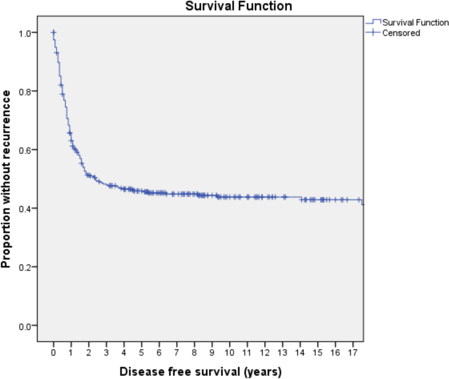
The incidence of positive margins following gastrectomy is variable ranging from 0.9 to 59% in various reported series, and its negative effect on survival outcomes has been demonstrated in many studies [5]. In a systematic review of 19,355 patients, it was shown that T3/T4 tumors, higher N status, diffuse-type gastric cancer, poorly differentiated tumors, lymphovascular invasion, and Bormann type 4 were predictive factors for positive resection margin [6]. In our study, among the patients who had microscopic positive margins, the frequency of T3/T4 disease was 92.3%, poorly differentiated histology was 84.6% and nodal positivity was 92.3% with extra-nodal extension in 50% of them, and lymphovascular invasion in 61.5% patients.
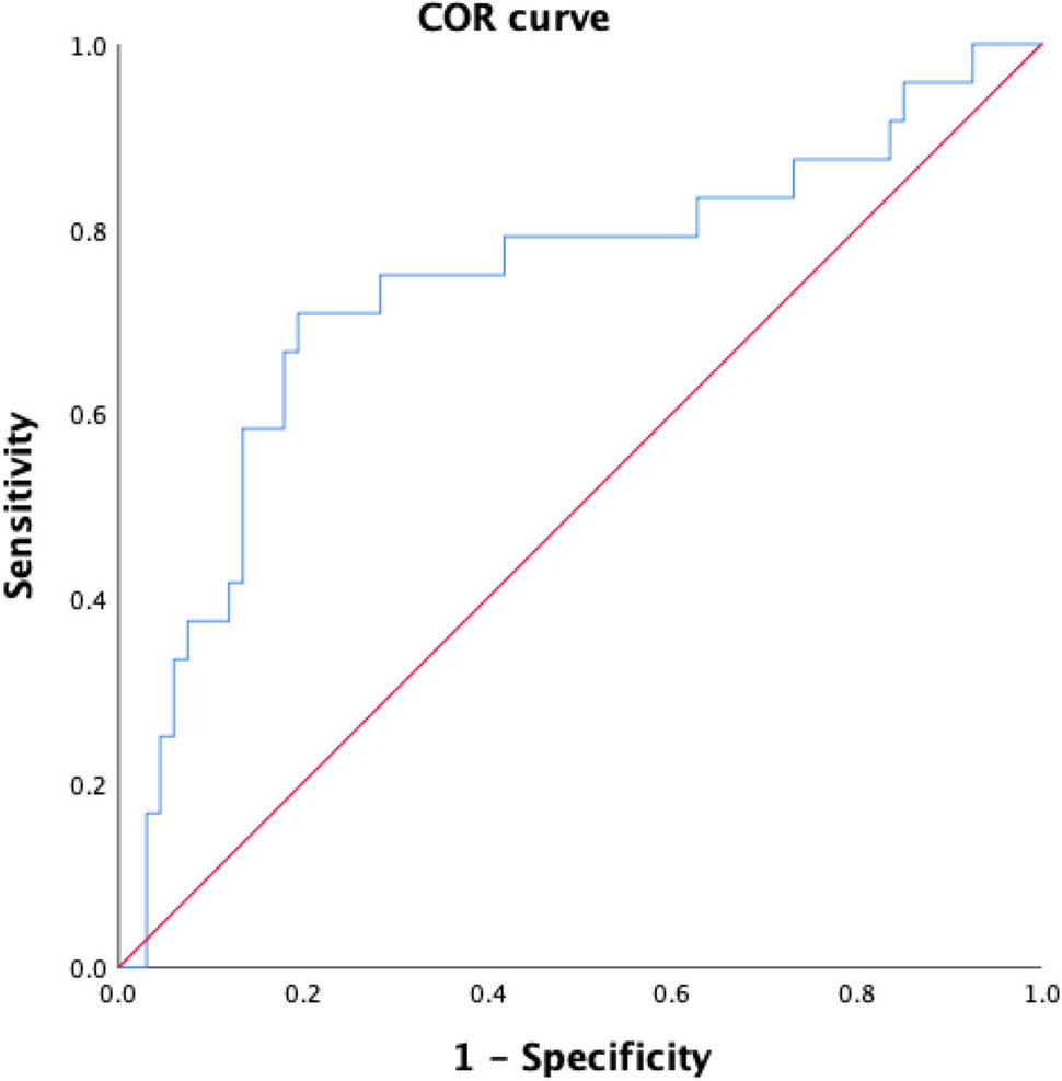
Intra-operative frozen section analysis can be a reliable method to decrease the incidence of positive margins in curative resection of gastric cancer. In a study of 110 patients with gastric cancer, it was reported that routine frozen section analysis of margins was able to detect positive margins in 14% of cases with no false negative result [7]. In another study of 377 patients, intra-operative frozen section analysis prevented positive resection margins. It was also found overall survival following re-resection of frozen section positive patients was similar to frozen section negative patients [8]. Shen et al. in their study of 66 patients with gastric adenocarcinoma of cardia observed that the accuracy, sensitivity, and specificity of an intra-operative frozen section were 97%, 77.8%, and 100%, respectively [9]. In our institute, routine examination of the resected margin with intra-operative frozen section analysis was not done previously.
The impact of a positive surgical margin is still not clear. Figueiredo et al. in their study showed that proximal resection margin was not a prognostic factor as long as R0 resection is achieved [4]. Postlewait et al. in their study of 162 patients with proximal gastric adenocarcinomas found that R1 resection was not associated with decreased survival but only increased local recurrence. However, on multivariate analysis, R1 resection was not independently associated with local recurrence [10]. Contrarily, Wang et al. [11] and Nagata et al. [12] found the microscopic positive resection margin an independent prognostic factor in both univariate and multivariate analysis across all stages. Bickenbach et al. [13] pointed out that R1 resection had no effect on survival in T3–T4 tumors and disease with greater than 3 positive lymph nodes. They observed a local recurrence rate of 16% in patients with R1 resection. Supporting this evidence, Cho et al. (14) and Cascinu et al. [15] found that the R1 resection margin is an independent negative predictor of survival only in N0 cases. In a systematic review of 19,992 patients, the R1 margin was associated with poorer overall survival [5].
There is a difference of opinion among medical practitioners regarding further management of patients with R1 resection. The reason is that there is no randomized controlled trial regarding the best management of patients with microscopic positive margins after gastrectomy. Though surgical re-resection to negative margins is a valid option, it is technically complex and is associated with high risks of mortality and morbidity. Moreover, recommendations in published literature regarding re-operation also vary. In a study which included 47 patients with margin-positive resection, Kim et al. showed that R1 margin status lost its prognostic value in multivariate analysis in all except in patients who had a low nodal burden of disease. They studied the patients who had positive margins on intraoperative frozen section analysis and compared the group of patients who had a re-resection to a negative margin with the group who left a residual resection line microscopically positive disease. Their finding was that the survival outcomes did not differ significantly between the two groups. However, in a subset analysis, when they compared the patients who had fewer than or equal to 5 positive nodes to those with more than five positive nodes, they found a significantly longer overall survival among the former subset. Thus, the presence of more than 5 positive nodes was found to be the primary determinant of survival and not the R1 status [16]. Aurello et al. [17] in their systematic review suggested that surgical re-excision should be considered for patients who have less than three metastatic lymph nodes. Cho et al. [14] also suggested re-excision for R1 cases in the absence of lymph node metastasis. Though there are no definite guidelines for the treatment of patients with R1 resection, it is best to present these cases in front of a multidisciplinary team and plan further treatment on a patient-to-patient basis. Probably patients with early-stage disease stand to benefit maximum from a re-resection. Other treatment options for this subset of patients are observation, chemotherapy, or chemoradiotherapy. None of the patients in our study had re-operation as all our patients were either T3–T4 or had lymph nodal metastasis. Patients receiving chemoradiotherapy had a better OS than patients receiving chemotherapy alone (median OS 35 months vs 18 months) but it is again subjected to bias. These patients were not fit for chemoradiotherapy after surgery as decided by our multidisciplinary team which was the reason for giving chemotherapy to them in the first place.
With the use of neoadjuvant therapy, the incidence of positive margins may be reduced as seen in our study that ten patients (77%) who underwent upfront surgery were margin-positive compared to three patients (23%) who received neoadjuvant therapy.
We understand that the retrospective nature of the study is a drawback. However, the long-term follow-up data is available for the patients with margin positivity. Another drawback is the availability of complete data for only 27.7% patients with a margin-negative resection. This being stated, we could compare the histopathological data of these patients with those having margin positivity.


留言 (0)