Digital anatomical features of morphological development in the atlantoaxial synchondroses in children aged 1 to 6 years old: a retrospective study of CT images
Objective
To investigate the anatomical indexes and anatomical positional indexes of the atlantoaxial synchondroses in normal Chinese Han children aged 1–6 years, and to analyze the changing law of the atlantoaxial cartilage union with the growth and development of age and its influence on the atlantoaxial ossification in children.
Methods
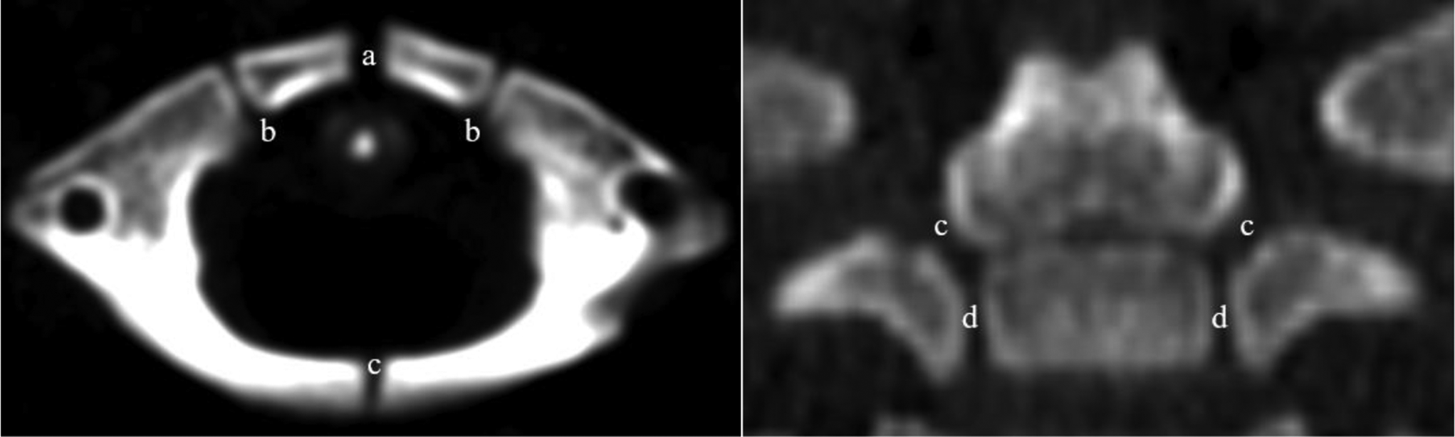
A retrospective collection of CT imaging of 160 cases of normal cervical spine in children aged 1 to 6 years old was conducted. The cases were divided into six age groups, with each group representing a one-year age range. Measure the morphological anatomical indicators and anatomical positional indicators of the atlantoaxial synchondroses. Record and statistically analyze the measurements of each indicator.
Results
Measurements were taken on various parameters of the atlantoaxial synchondroses. TD, SD, height, area, and perimeter all gradually decreased among the groups. Distance between bilateral atlantal anterolateral synchondroses increased gradually from Group A to Group F, while the angle formed along the long axis in the cross-section showed a decreasing trend. Distance between the axoid dentolateral synchondroses and between the neurocentral synchondroses increased gradually from Group A to Group F, with the angle value in the cross-section showing a gradual decrease, and distance from the odontoid apex increasing from Group A to Group F.
Conclusions
The atlantoaxial synchondroses gradually decrease in size with age, and ossification levels increase with age, with faster ossification occurring during a 1–2 years-old period. The anterolateral synchondroses, dentolateral synchondroses, and neurocentral synchondroses all gradually ossify towards the lateral direction with increasing age.


留言 (0)