
記住我


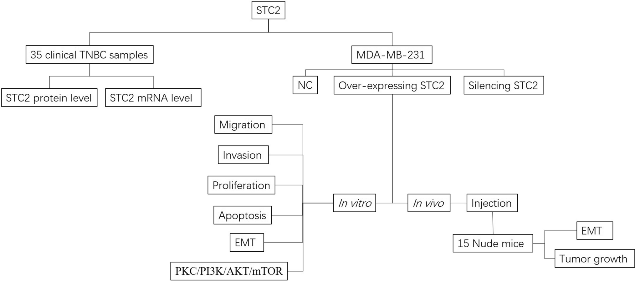
35 pairs of TNBC tissues and the para-carcinoma tissues were collected from the breast cancer patients in Yuebei People’s Hospital (Shaoguan, China) from December of 2019 to December 2022. All the patients received no preoperational antitumor therapy including radiotherapy and chemotherapy. All the patients were without lymph node metastasis and distant metastasis. Patients were undergone radical resection, and the cancer tissues and para-carcinoma tissues were collected avoiding of necrosis. Informed consents were obtained from every patient. The experiments were approved by the Ethics Committee of Yuebei People’s Hospital. This study was approved by the Yuebei People’s Hospital Ethics Committee with approval number KY-2021-299 (Fig. 1).
Fig. 1 2.2 STC2 levels in breast cancer tissues
STC2 levels in TNBC tissues and para-carcinoma tissues were measured by IHC. Tissues were separated from human body and then fixed in 4% polyformaldehyde (PFA) (Solarbio Sciences & Technology Co., Ltd., Beijing, China) for more than 48 h. Tissues were dehydrated by gradient ethanol (70%, 80%, 90%, 100%, 100%) for 30 min of each, and then cleared by twice of xylene (Bioss Antibodiess Co., Ltd. Beijing, China) for 30 min of each. Waxed tissued twice for 30 min of each, with the temperature between 58–62 °C. Paraffin embedded tissues were cut into 5 μm of slices and dried at 60 °C for 1 h for fixation. For STC2 detection, slices were dried at 60 °C for 1 h and dewaxed in twice of xylene for 30 min of each. Hydrated slices in gradient ethanol (100% for 2 min, 100% for 2 min, 90% for 2 min, 80% for 5 min, 70% for 10 min) and twice of ddH2O for 2 min. Washed slices twice with 1 × phosphate buffer solution (PBS) (Solarbio). Antigen repaired with 1 × Tris/EDTA (pH 9.0) (Bioss) in microwave oven for 10 min at high heat. Replenished with ddH2O and heated in microwave oven for 40 min at moderate heat. Cooled to room temperature, and then washed with 1 × PBS for 5 min, three times. Inactivated in 3% hydrogen peroxide solution (Bioss), sealed with 10% goat serum (Beyotime Biotechnology, Inc. Shanghai, China) for 10 min, and then incubated with rabbit anti-STC2 (ab255610,1:200) (Abcam, Shanghai, China) containing SignalUp™ immunostaining enhancer (Beyotime) at 37 °C for 1–2 h. Washed with PBST (PBS with 0.1% tween-20, Solarbio) twice for 5 min and incubated with Horseradish peroxidase (HRP) Polymer (Bioss) secondary antibody for 30 min. Washed with PBST twice for 5 min and stained with diaminobenzidine (DAB) (Bioss) for 10 min. Washed with water and re-stained with hematoxylin (Bioss). Dehydrated with graduated ethanol (70% for 10 s, 80% for 10 s, 90% for 10 s, 100% for 10 s) and cleared with twice of xylene for 5 min. Sealed slices with neutral balsam (Bioss) and observed under a microscope.
2.3 Quantitative real-time PCR measuring STC2 levels2.3.1 RNA extractionTotal RNA was extracted from the cancer tissues and the para-carcinoma tissues. Tissues were ground in liquid nitrogen as powder. Mixed with 1 ml Trizol (Beyotime) and let stand for 5 min. Mixed with 200 μl chloroform (Guangzhou Chemical Reagent Factory, Guangzhou, China) on vortex for 30 s, and let stand for 2 min. Centrifugated at 4 °C, 12,000×g for 10 min. Separated the top level (RNA), added equal volume of Isopropanol (Chemical Reagent Factory) mixed gently, and then let stand for 10 min. Centrifugated at 4 Centrifugated at 4 °C, 12,000×g for 10 min and collected the precipitation. Washed with 75% ethanol twice and dried in laminar flow cabinet. Dissolved with 20–60 μl diethyl pyrocarbonate (DEPC) water (Aladdin Biochemical Technology Co., Ltd. Shanghai, China).
2.3.2 RNA purity and integrity detection50 times diluted RNA was detected with BioPhotometer plus (Eppendorf China) to measure the OD260/OD280 > 1.8, representing pollution-free from proteins. 1 μl RNA was analyzed by 1% agarose (Biosharp Life Sciences, Hefei, China) gel with 80 V for 20 min, and the observed by ultraviolet (UV) cross link apparatus (Thermo Fisher Scientific, Inc. Shanghai, China) to confirm the RNA integrity, representing as clearing 5 s rRNA, 18 s rRNA and 28 s rRNA.
2.3.3 STC2 reverse transcription and quantitative PCR (qPCR)The STC2 reverse transcription (TransGen Biotech Co., Ltd. Beijing, China) was carried on according to the instruction of the kit. The qPCR reaction system includes SYBR Green Mix, primers (STC2 F: GGGTGTGGCGTGTTTGAATG, R: TTTCCAGCGTTGTGCAGAAAA; β-actin F: ATGGATGTAGAAAATGAGCAG, R: TAGTCGCCTTTTTGCCTTGG), cDNA template, and water, with a total volume of 20 μl. The reaction conditions are 95 °C pre-denaturation for 5 min, followed by 40 cycles of 95 °C denaturation for 15 s and 60 °C annealing and extension for 32 s.
2.4 Cell culturingMDA-MB-231 cell line obtained from China Center for Type Culture Collection (CCTCC). MDA-MB-231 cells were cultured with Leibovitz’s L-15 (Thermo) containing 10% fetal bovine serum (FBS) (Thermo), 10,000 U/ml penicillin and 10,000 μg/ml streptomycin, and maintained at 37 °C, 5% CO2 and saturated humidity. Cultured medium was changed every 2 to 3 days. Passage culturation was assayed when MDA-MB-231 cells reached 90% density. MDA-MB-231 cells in logarithmic growth phase were used for the following experiments.
2.5 Overexpressing or silencing STC2 in MDA-MB-231 cellsOverexpressed STC2 plasmid and siRNA-STC2 were constructed and synthesized by Shanghai Genechem Co., Ltd. (Shanghai, China). MDA-MB-231 were transfected with overexpressed STC2 plasmid (OE-STC2 group) or siRNA-STC2 (si-STC2 group) using Lipo.3000 (Thermo) according to the instruction. 48 h later, the cells were for measuring the related proteins expressions, cell apoptosis, metastatic and invasive abilities comparing to the negative control (NC group).
2.6 STC2 expression and EMT levels in MDA-MB-231 cellsThe transfected MDA-MB-231, mentioned before, were lysed with moderate radio immunoprecipitation assay (RIPA) buffer (Beyotime) containing 1 mM of Phenylmethanesulfonyl fluoride (PMSF) (KeyGEN BioTECH, Nanjing, China) on ice for 30 min. Centrifugated at 12,000×g, 4 °C for 10 min and collected the supernatant. Mixed with 5 × loading buffer and heated at 100 °C for 10 min, and then stored at − 80 °C or continued for proteins expressions measurement. Quantitated the total protein by BCA assay (KeyGEN) before western blotting (WB). 40 μg of total protein was analyzed by sodium dodecyl sulfate-polyacrylamide gel electrophoresis (SDS-PAGE) with 10% separating gel and 5% storing gel. Proteins were transferred to polyvinylidene fluoride (PVDF) (Merck Millipore cooperation, Germany) membrane and then sealed with 5% skim milk containing 2% bovine serum albumin (BSA) (Solarbio). Rabbit anti-STC2 (ab255610, 1:1000, Abcam), rabbit anti-E-cadherin (20874-1-AP, 1:10,000) (Proteintech Group, Inc. Wuhan, China), rabbit anti-N-cadherin (22018-1-AP, 1:5000, Proteintech), rabbit anti-Lamiin γ1 (ab233389, 1:1000, Abcam), rabbit anti-Fibronectin (15613-1-AP, 1:10,000, Proteintech), rabbit anti-β-catenin (51067-2-AP, 1:10,000, Proteintech), rabbit anti-Vimentin (10366-1-AP, 1:5000, Proteintech), rabbit anti-Claudin-1 (28674-1-AP, 1:2000, Proteintech), rabbit anti-FSP1 (20886-1-AP, 1:3000, Proteintech), rabbit anti-Snail 1 (13099-1-AP, 1:500, Proteintech), rabbit anti-ZEB1 (21544-1-AP, 1:500, Proteintech), mouse anti-α-SMA (ab7817, 1:1000, Abcam) and mouse anti-GAPDH (RM2002, 1:3000, Beijing Ray Antibody Biotech, Beijing, China) antibodies were used to incubate the corresponding protein bands at 4 °C, overnight. After washing with PBST (PBS containing 0.1% Tween-20) (Sangon Biotech Co., Ltd. Shanghai, China) for 3 times, peroxidase affinipure goat anti-rabbit IgG (H+L) (111-035-003, Jackson ImmunoResearch Inc. USA) or peroxidase affinipure goat anti-mouse (115-035-003, Jackson) antibodies were incubated the corresponding protein bands at 25 °C for 1 h. After washing with PBST for three times, protein bands were analyzed with ECL system (P0018FS, Beyotime).
2.7 Cell cycle and apoptosis assayThe transfected MDA-MB-231 mentioned before, were digested with trypsin without EDTA (Thermo) and suspended in L-15 with 10% FBS. Washed and suspended with 1 × PBS to prepare as single cell suspension with concentration of 1–5 × 10⁶ cells/ml. 100 μl of cell suspension was collected in the flow cytometry tube. 5 μl of Annexin V-FITC (Beyotime) was added into the cells, and then mixed with 5 μl of propidium iodide (PI) (Beyotime). After 10 min incubation at 25 °C in dark, the cell cycle and apoptotic cells were analyzed by flow cytometer (Attune CytPix, Thermo).
2.8 Cell proliferation measurement by cell counting kit-8 (CCK-8)The transfected MDA-MB-231 mentioned before, were digested with trypsin without EDTA (Thermo) and suspended in L-15 with 10% FBS. Washed and suspended with 1 × PBS to prepare as single cell suspension with concentration of 1–5 × 10⁶ cells/ml. 2 × 10³ cells were resuspended in 100 μl of L-15 with 10% FBS, and then seeded in a 96-well plate. The cell proliferation was measure at 0 h, 24 h, 48 h, 72 h and 96 h by using CCK-8 (Beyotime, C0038) according to the specification, separately.
2.9 Cell migration measurement by wound healing assayThe transfected MDA-MB-231 were digested with trypsin without EDTA (Thermo) and suspended in L-15 containing 10% FBS. 1–5 × 10⁵ cells were seeded in a 6-well plate (Corning Inc. Shanghai, China). Use a pipette tip to make a straight line. Continue to culture for 48 h. Observed the scratch healing under a microscope (Leica Microsystems, Danaher Life Sciences, Germany). Randomly selected 8 fields to measure the average width of the scratch healing at 0 h, 6 h, 24 h and 48 h, separately.
2.10 Cell invasion measurement by transwell systemThe transfected MDA-MB-231 were digested with trypsin without EDTA (Thermo) and suspended in L-15 without FBS to the appropriate concentration. For invasion assay, 1–5 × 10⁴ cells (no more than 300 μl) were seed to the upper chambers of the Transwell system with 24-well insert (8 μm pore size) (Corning). A upper chamber was coated with 5 μl Matrigel (Becton, Dickinson and Company, USA) mixed with 95 μl L-15. The lower chambers were loaded with 600 μl L-15 containing 20% FBS. After incubation for 24 h, removed the medium, fixed with methanol for 20 min, and stained with 0.1% Crystal Violet (Beyotime) in 20% methanol. Cleaned the cells on the upper surface of the upper chamber. Observed the transmembrane cells under a microscope (Leica). Selected 8 fields randomly and calculated the average number of the cells stained as purple.
2.11 PKC/PI3K/AKT/mTOR signaling pathway measurementsOn one hand, pAKT Ser473, pPKCδ Ser645 and pPKCδ Thr507 were measured in the transfected MDA-MB-231 with silencing STC2 or over-expressing STC-2, comparing to the NC. On the other hand, the transfected MDA-MB-231 were treated with rapamycin (25 nM, 24 h), LY294002 (10 μM, 24 h) or Rottlerin (3 μM, 24 h), separately. PCR and WB assays were for determining the STC2 level, and WB was for pAKT Ser473, pPKCδ Ser645 and pPKCδ Thr507 levels. Rabbit anti-pAKT Ser473 (9273, 1:1000, Cell Signaling Technology, Inc. Shanghai, China), rabbit anti-pPKCδ Ser645 (9376, 1:1000, CST), rabbit anti-pPKCδ Thr507 (bs-3727r, 1:1000, Bioss Antibodies, Inc. Beijing, China), rabbit anti-STC2 (ab80590,1:1000, Abcam) and mouse anti-GAPDH (RM2002, 1:3000, Ray) antibodies were used for primary antibodies. The following steps were mentioned as before.
2.12 Tumor formation in nude mouseAll animal care conditions and experimental protocols were approved by the Animal Ethics Committee of Yuebei People’s Hospital with approval number G2024014. Animal experiments were carried out according to Institutional Animal Care and Use Committee (IACUC) Guidelines. 15 BALB/c-nude mice (male, age 5 to 6 weeks, were obtained from SPF. All the nude mice were housed and kept in a specific pathogen-free (SPF) animal facility in 12 h light/dark cycles, with controlled humidity and temperature and free access to food and water. Nude mice were divided into 3 groups, randomly, including si-STC2 group, OE-STC2 group and the NC group, 5 in each group. Collected the transfected MDA-MB-231 with digestion by trypsin without EDTA (Thermo), washed with PBS 3 times, and suspended in 1 ml of PBS to the concentration of 107 cells/0.3 ml. Injected the cells into the mammary fat pad of nude mice through a 22-gauge needle 1 to 2 cm deep to prevent leakage of the inoculum, according to the grouping. 1 week before injection, each mouse received subcutaneous injection of 5 μg of 17β-estradiol valerate (Aladdin Biochemical Technology Co., Ltd, Shanghai, China), which was dissolved in 0.5 ml of sesame oil (Solarbio) as repository vehicle. Estrogen was injected every week until sacrifice in order to sustain tumor growth. When solid tumors reached palpable size, sacrificed the nude mice and separated the tumors. Cleaned the non-tumorous tissues, recorded the tumor size, and then fixed in 4% polyformaldehyde (PFA) (Solarbio) more than 24 h until cutting into sections.
2.13 ImmunohistochemistryThe tumor tissues removed from nude mice were dehydrated and waxed mentioned before. 5 μm of slices were obtained for dewaxing and hydrating. After antigen repairing, inactivating and sealing, primary antibodies including STC2 (1:100), N-Cadherin (1:100), E-Cadherin (1:100), β-catenin (1:100) and Claudin-1 (1:100) were added at 4 °C overnight. HRP Polymer secondary antibody were incubated for 30 min, and then stained by DAB and hematoxylin. Dehydrated and cleared the sections and observed under a microscope.
2.14 Statistical analysisStatistical analysis was performed by SPSS 19.0 (SPSS Inc., Chicago, IL). Experiments were repeated at least 3 times and the data were presented as mean. Student’s t test was used for comparisons between groups, while ONE-WAY ANOVA was used among groups more than two. A value of P < 0.05 was considered as significant difference.
留言 (0)