
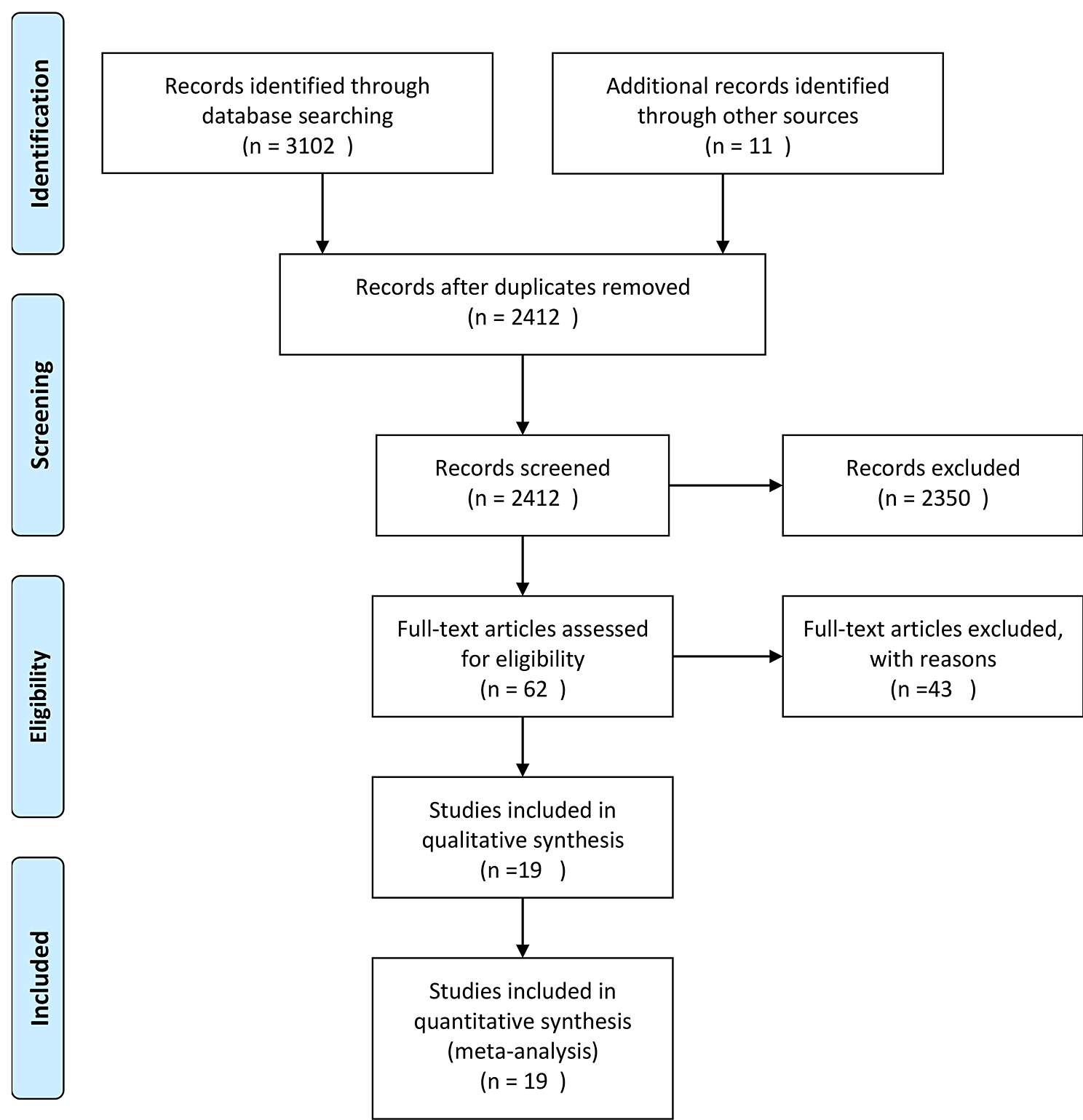
記住我

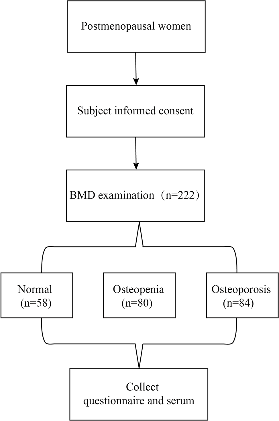

A total of 10,393 euthyroid Chinese adults, including 4,171 females and 6,222 males, were ultimately included for analysis, and their baseline characteristics by gender and TSH level were showed in Table 1. In female, there was a significantly difference in terms of age among the TSH tertile groups, however, no difference was observed in male. BMI was the lowest in the TSH_low group in both female and male, and there was significant difference between any group. BMD in both female and male was the highest in the TSH_low group, however, statistical difference was only observed in female. In female, there was a significantly difference in terms of osteoporosis and osteopenia among the TSH tertile groups, however, no difference was observed in male. TSH level was the lowest in the TSH_low group in both female and male, and there was significant difference between any group. fT3 and fT4 levels were the lowest in the TSH_low group in both female and male, however, statistical difference was only observed in male.
Table 1 Clinical characteristics of participants based on gender and TSH tertile groupsDistribution of BMD in normal TSH levelDue to the significant negative association between BMD and age, age was stratified by 60 years old (Supplemental Fig. 1). Fitting curves of BMD across normal TSH levels stratified by gender and age were illustrated in Fig. 2. In women aged less than 60 years, BMD decreased with the increase of TSH at normal level, while in women aged 60 years and older, BMD increased with the increase of TSH at normal level (Fig. 2A); besides, the BMD of women aged less than 60 years was higher than that of women aged 60 years and older (156.05 ± 39.34 mg/cm3 vs. 86.95 ± 29.51 mg/cm3, P < 0.001) (Fig. 2A). Both in men aged less than 60 years and aged 60 years and older, BMD decreased with the increase of TSH at normal levels (Fig. 2B); besides, the BMD of men aged less than 60 years was higher than that of men aged 60 years and older (143.08 ± 32.76 mg/cm3 vs. 108.13 ± 31.99 mg/cm3, P < 0.001).
Fig. 2
Fitting curves of BMD with normal TSH level stratified by age (A: female; B: male)
To further investigate the difference of BMD based on different serum TSH levels, these participants were equally divided into three groups according to tertiles of serum TSH levels. In women aged less than 60 years, the BMD of participants in TSH_low group was higher than that of participants in TSH_high group (161.55 ± 41.80 mg/cm3 vs. 150.86 ± 37.65 mg/cm3, P < 0.001). However, in women aged 60 years and older, these participants in the TSH_low group had lower BMD than those in the TSH_high group, but it did not reach statistically significant difference (82.52 ± 29.81 mg/cm3 vs. 87.73 ± 28.94 mg/cm3, P = 0.117) (Fig. 3).
In men aged less than 60 years, the BMD of participants in TSH_low group was higher than that of participants in TSH_high group, but it did not reach statistically significant difference (143.77 ± 33.33 mg/cm3 vs. 140.80 ± 31.62 mg/cm3, P = 0.077). Similarly, in men aged 60 years and older, participants in the TSH_low group had higher BMD than those in the TSH_high group, but it did not reach statistically significant difference (109.57 ± 33.29 mg/cm3 vs. 105.49 ± 29.09 mg/cm3, P = 0.381) (Fig. 3).
Fig. 3
Bone mineral density of three regions according to the TSH tertiles in female and male stratified by age
Linear regression resultsTable 2 showed the results of linear regression analysis for BMD and normal TSH level by gender and age. Negative associations of BMD and normal TSH level was indicated among total women (β=-4.01, 95% CI: -5.55 ~ -2.45, P < 0.001) and women with aged less than 60 years (β=-4.34, 95% CI: -5.82 ~ -2.86, P < 0.001), however, this inverse trend was found in women aged 60 years and older (β = 2.04, 95% CI: 0.08 ~ 3.99, P = 0.041). Besides, there was a significant negative association between BMD and normal TSH level in women aged 60 years and older after adjusting age (β=-2.02, 95% CI: -3.21 ~ -0.84, P = 0.001).
Table 2 Linear regression for BMD and normal TSH level by gender and ageIn male, BMD had a significant negative association with normal TSH level in men younger than 60 years (β=-1.05, 95% CI: -2.08 ~ -0.02, P = 0.047), but this association was not kept while adjusting for age (β=-0.71, 95% CI: -1.58 ~ 0.17, P = 0.116).
Multinomial logistic regression resultsWhile transferring BMD to three categories (normal BMD, osteopenia and osteoporosis), the association of osteoporosis and osteopenia with TSH tertile groups by gender and age was showed in Table 3. In female, whatever total participants or participants younger than 60 years, osteopenia was significantly higher in participants with TSH_high and TSH_mid groups than those with TSH_low group, but this significance was not observed after adjusting for age. Besides, it was only found that in women over 60 years old, women with TSH_mid group had a lower incidence of osteoporosis than those with TSH_low group.
Table 3 Multinomial logistic regression for osteopenia/osteoporosis and TSH tertile groups by gender and ageIn male with osteopenia, no significance among TSH tertile group was found, whatever total participants or participants aged less than 60 years, except for men aged 60 years and older. In men aged 60 years and older, osteopenia was higher in participants with TSH_high group than those with TSH_low group, and this significance was kept after adjusting for age. In male with osteoporosis, no significance among TSH tertile groups was found, whatever total participants, participants aged less than 60 years, or participants aged 60 years and older.
留言 (0)