
記住我


Due to a double-edged sword role of pericytes in the TME, imatinib treatment strategies can increase the effectiveness of antitumor therapies in the presence of excessive pericyte coverage, but inappropriate modulation of pericyte coverage has detrimental effects. Nevertheless, an effective imatinib regimen and a longitudinal monitoring technique for pericyte coverage are lacking. In this study, we employed mpMRI to monitor the effects of imatinib on vascular characterization and the TME in CRC with high pericyte coverage to determine for an optimized strategy. To generate tumor models with the highest level of pericyte coverage, subcutaneous tumors were generated in mice with four human CRC cell lines, and vascular characterization was conducted through IF analysis. Representative H&E-, CD31/PDGFRβ-, CD31/α-SMA-, CD31/dextran- and CD31-stained tumor tissue sections from the four model types are shown in Fig. 2A. HT-29 tumors had the highest PDGFRβ+ pericyte coverage, while HCT116 tumors displayed the lowest PDGFRβ+ pericyte coverage (Fig. 2B; Table 1). Similar distinctions in α-SMA+ pericyte coverage were observed between HT-29 and HCT116 tumors (Fig. 2C; Table 1). Compared to that in HT-29 tumors, PDGFRβ+ and α-SMA+ pericyte coverage in RKO and SW480 tumors was significantly lower (Fig. 2B, C). Notably, to exclude the possibility that α-SMA+ and PDGFRβ+ cell populations were vascular smooth muscle cells (VSMCs), colocalization analysis of αSMA and CD31 was performed.
Fig. 2
Vascular characteristics in the four CRC models. (A) Representative images of H&E staining at high magnification and CD31/PDGFRβ, CD31/α-SMA, CD31/dextran and CD31 staining in the four CRC models. (B)-(E) Quantification of PDGFRβ+ pericyte coverage, α-SMA+ pericyte coverage, relative vessel permeability and the MVD in the four CRC models. The data are shown as the mean ± standard deviation. *P < 0.05, **P < 0.01, ***P < 0.001; NS, nonsignificant
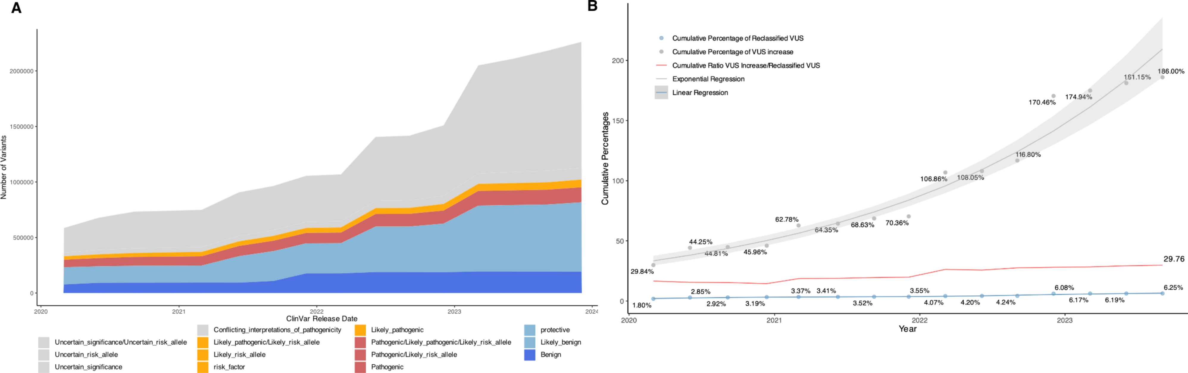
Table 1 Comparision of quantitative indicators of vascular characterization in four CRC modelsTwo other important vascular indicators, relative vessel permeability and Microvessel density (MVD), were also compared among the tumor models. HCT116 tumors were found to exhibit the highest vessel permeability, and HT-29 tumors showed lower vessel permeability (Fig. 2D; Table 1). Furthermore, HCT116 and HT-29 tumors showed the lowest MVD and a higher MVD, respectively (Fig. 2E; Table 1). Taken together, the results indicated that in HT-29 tumors with the highest pericyte coverage, lower vessel permeability and a higher MVD were observed. Conversely, in HCT116 tumors with the lowest pericyte coverage, the highest vessel permeability and lowest MVD were observed.
Monitoring CRC models with high and low pericyte coverage by mpMRIPrevious studies have demonstrated the value of mpMRI in evaluating hemodynamics, tumor cell characteristics, and pH changes [44, 49, 54]. To investigate the feasibility of distinguishing differences in tumors with the highest and lowest pericyte coverage by mpMRI and DCE-, DWI-, and APT CEST-MRI at 9.4T was utilized to monitor the entire tumor region of the HT-29 and HCT116 models. Representative multiparametric maps of HT-29 and HCT116 CRC models are shown in Fig. 3A. In HT-29 tumors with the highest pericyte coverage, lower Ktrans, Ve, and Kep values corresponded to lower relative vessel permeability, and a higher AUC value corresponded to a higher MVD (Fig. 3B). In HCT116 tumors with the lowest pericyte coverage, higher Ktrans, Ve, and Kep values corresponded to higher relative vessel permeability, and a lower AUC value corresponded to a lower MVD. Additionally, significant differences were shown in ADC values and MTRasym values that were calculated from the z-spectrum at B1 = 1.0 µT and 2.0 µT between HT-29 and HCT116 models, and the results of these differences were further investigated (Fig. 3B, C). Collectively, these results indicated that DCE- parameters on 9.4T MRI exhibited significant differences among the CRC models and were consistent with vascular characteristics.
Fig. 3
9.4T mpMRI maps of CRC models with high and low pericyte coverage. (A) Representative T2WI images and maps of DCE-derived Ktrans, Ve, and Kep and AUC, DWI-derived ADC, and APT-CEST-derived MTRasym (1 µT and 2 µT) at 3.5 ppm, of HT-29 and HCT116 tumors. (B) Quantification of multiple parameters, including the Ktrans, Ve, Kep, AUC, and ADC values, of HT-29 and HCT116 tumors. (C) Corrected-z-spectrum and MTRasym values at 1 µT and 2 µT.of HT-29 and HCT116 tumors The data are shown as the mean ± standard deviation. *P < 0.05, **P < 0.01 and ***P < 0.001
Antitumor effect of imatinib in CRC with high pericyte coverageTo explore the antitumor effect of imatinib in CRC with excessive pericyte coverage, the effect of imatinib on HT-29 tumors with the highest pericyte coverage was evaluated by measuring the changes in tumor volume using T2-weighted imaging (T2WI), and the tumor necrosis volume was assessed through DCE imaging. Tumor volumes and necrosis volumes at each time point during imatinib treatment are shown in Fig. 4A. As shown in Tables 2 and 3, the tumor volumes of the imatinib group on Day 0, Day 4, Day 7, and Day 10 were 182.91 ± 22.29, 229.75 ± 24.91, 328.29 ± 15.56, and 340.37 ± 19.67 mm3, respectively, while the tumor volumes of the control group were 186.83 ± 16.50, 293.46 ± 22.78, 451.09 ± 42.51, and 578.63 ± 47.87 mm3. Starting from Day 4, the imatinib group exhibited a trend of slower growth and smaller tumor volume than the control group (Fig. 4B). Additionally, the imatinib group exhibited a smaller tumor volume change over 10 days than the control group (Fig. 4C). Furthermore, the necrosis volumes of the imatinib group on Day 0, Day 4, Day 7, and Day 10 were 3.49 ± 0.45, 58.10 ± 10.83, 73.07 ± 9.61, and 89.72 ± 10.34 mm3, while the necrosis volumes of the control group were 3.44 ± 0.73, 4.66 ± 0.65, 8.15 ± 1.63, and 17.20 ± 2.56 mm3 (Tables 2 and 3). From Day 4, the imatinib group displayed a faster tumor necrosis and larger necrosis volume than the control group (Fig. 4D). In addition, the imatinib group exhibited a higher proportion of necrosis volume compared to the control group on Day 10 (Fig. 4E). During the treatment process, mice showed no obvious abnormal reactions, and none of the mice died; however, none of the mice showed a complete tumor response. Together, the findings showed that imatinib has significant antitumor effects on CRC with high pericyte coverage but also suggested that it might not be suitable for monotherapy.
Fig. 4
Antitumor effect to imatinib treatment in CRC with high pericyte coverage. (A) Representative T2WI and DCE images used for HT-29 tumor volume and necrosis volume measurement during imatinib treatment. (B) Curves of tumor volumes of the imatinib group and control group measured at different timepoints. (C) Quantification of tumor volume changes in the two groups. (D) Curves of necrosis volumes of the imatinib group and control group measured at different timepoints. (E) Proportion of necrosis volume in the two groups. The data are shown as the mean ± standard deviation. *P < 0.05, **P < 0.01 and ***P < 0.001
Table 2 Quantitative variations of tumor volume, histological and imaging results in control groupTable 3 Quantitative variations of tumor volume, histological and imaging results in imatinib groupImatinib alters vascular characteristicsTo identify a proper imatinib regimen, the pattern of the pericyte coverage decrease induced by imatinib over time must be assessed. To elucidate the trends in pericyte coverage and important vascular characteristics caused by imatinib treatment, histological assessment was performed at each time point during treatment. Representative sections used to assess PDGFRβ+ pericyte coverage, α-SMA+ pericyte coverage, MVD, and relative vessel permeability during treatment are shown in Fig. 5A. In the imatinib group, intratumor PDGFRβ+ pericyte coverage and α-SMA+ pericyte coverage both showed a decreasing trend (Fig. 5B; Tables 2 and 3). PDGFRβ+ pericyte coverage gradually declined from Day 0 to Day 7, reaching a plateau after Day 7 (F = 186.22, P < 0.001). Similarly, α-SMA+ pericyte coverage declined gradually in the first 4 days, subsequently showed a slower decrease and reached a plateau after Day 7 (F = 119.112, P < 0.001). Additionally, relative vessel permeability gradually increased from Day 0 to Day 7, reaching a plateau after Day 7 (F = 12.867, P = 0.002, Fig. 5B; Tables 2 and 3). Subsequently, the MVD showed a gradual decline from day 0 to day 4, a significant decline from Day 4 to Day 7, and then a slower gradual decline after Day 7 (F = 35.952, P < 0.001, Fig. 5B; Tables 2 and 3). There were no significant trends observed for any of the indicators among the control group (P > 0.05). Starting from Day 4, significant differences were observed in PDGFR-β+ pericyte coverage, α-SMA+ pericyte coverage, MVD and relative vessel permeability between the two groups. Taken together, these data demonstrated that the imatinib-induced decrease in pericyte coverage reached a plateau on Day 7 and was accompanied by increased vessel permeability and decreased MVD in CRC with high pericyte coverage.
Fig. 5
Histological assessment of vascular characteristics at different time points during imatinib treatment. (A) Representative images of H&E, CD31/PDGFRβ, CD31/α-SMA, CD31/dextran and CD31 staining during treatment. (B) Longitudinal assessments of vascular characteristics, including PDGFRβ+ pericyte coverage, α-SMA+ pericyte coverage, relative vessel permeability and the MVD, in the imatinib group and control group. The data are shown as the mean ± standard deviation. *P < 0.05, **P < 0.01, ***P < 0.001; NS, nonsignificant
Molecular and histological profiling of treatment-induced TME alterationsA previous study demonstrated that imatinib downregulates the expression of Bcl-w, a component of the Bcl-2/bax pathway, in tumor ECs [42]. However, the relationship between the Bcl-2/bax pathway and imatinib-mediated modulation of pericyte coverage remains unknown. To explore whether imatinib modulated the gene expression of Bcl-2/bax pathway components, the expression of key genes in the Bcl-2/bax pathway and the related gene AKT1 was compared between the imatinib and control groups on Day 10. In the imatinib group, the mRNA expression of antiapoptotic genes, such as MCL-1, BCL-2, BCL-W, BCL-XL, and AKT-1, was significantly downregulated, while that of proapoptotic genes, such as BAX, BAK, CASP3, and CASP8, was significantly upregulated (Fig. 6A). MCL-1 is a multidrug resistance gene, and Mcl-1 overexpression indicates inhibition of tumor apoptosis and resistance to multiple antitumor therapies [39,40,41]. More importantly, an inappropriate decrease in pericyte coverage leads to increased hypoxia-associated metastasis and decreased survival, highlighting that elucidating the impact of imatinib treatments on TME hypoxia is crucial for identifying a proper imatinib regimen. To elucidate the trends in tumor apoptosis and TME hypoxia induced by imatinib, the expression of the antiapoptotic protein Mcl-1, proliferation marker Ki-67, and TME hypoxia marker HIF-1α and apoptosis, which was assessed by TUNEL staining, during treatment were evaluated by IF staining; representative sections are shown in Fig. 6B. In the imatinib group, a significant decrease in Ki-67 expression was observed in tumors, indicating inhibition of tumor proliferation (F = 109.248, P < 0.001, Fig. 6C; Tables 2 and 3). Similarly, a significant increase in the intensity of TUNEL staining, which was used to assess apoptosis, and a significant decrease in the expression of the anti-apoptotic protein Mcl-1 indicated increased tumor apoptosis (F = 460.606, P < 0.001 for TUNEL and F = 130.236, P < 0.001 for Mcl-1, Fig. 6C; Tables 2 and 3). Notably, HIF-1α expression showed a slow decline from Day 0 to Day 4, followed by a gradual increase after Day 4, suggesting alleviation followed by aggravation of TME hypoxia (F = 37.043, P < 0.001, Fig. 6C; Tables 2 and 3). Among the control group, no significant trends were observed in Ki-67, HIF-1α, or Mcl-1 expression, but there was a mild upward trend in the TUNEL staining intensity. Starting from Day 4, Ki-67, TUNEL, HIF-1α, and Mcl-1 expression was significantly different between the two groups. According to hematoxylin and eosin (HE) staining, there was a large area of tumor necrosis from Day 4 in the imatinib group, while only a small area of necrosis was observed on Day 10 in the control group. Together, these data indicated that imatinib induces tumor apoptosis through the Bcl-2/bax pathway and that 4 days of imatinib treatment alleviates hypoxia in CRC with high pericyte coverage.
Fig. 6
qPCR analysis of the gene expression of Bcl-2/Bax pathway components and histological assessment of TME indicators during imatinib treatment. (A) qPCR analysis of the gene expression of Bcl-2/Bax pathway components in the imatinib group and control group on Day 10. (B) Representative images of H&E, Ki-67, TUNEL, HIF-1α and Mcl-1 staining during treatment. (C) Longitudinal assessment of TME indicators, including Ki-67 expression, the TUNEL staining intensity, HIF-1α expression and Mcl-1 expression, in the two groups. The data are shown as the mean ± standard deviation. *P < 0.05, **P < 0.01, ***P < 0.001; NS, nonsignificant
Alterations of mpMRI parameters during imatinib treatmentmpMRI has the potential to overcome the limitations of histological for longitudinal monitoring of pericyte coverage, and is thus a potential technique for evaluating of the effects of imatinib treatment. To prove this hypothesis, DCE-, DWI-, and APT CEST-MRI were conducted to monitor the effects of 10-day imatinib treatment. Representative multiparametric maps obtained during imatinib treatment are shown in Fig. 7A. The influence of imatinib treatment on tumor hemodynamics was reflected by changes in DCE-MRI parameters, including Ktrans, Ve, Kep, and the AUC. In the imatinib group, increasing trends in the Ktrans, Ve, and Kep values were observed in the tumor region, suggesting increased vessel permeability (Fig. 7B; Tables 2 and 3). First, the Ktrans value significantly increased from Day 0 to Day 4 of treatment, followed by a gradual increase (F = 70.465, P < 0.001). Second, the Ve value increased significantly from Day 0 to Day 4, followed by a gradual increase, and increased significantly again after Day 7 (F = 24.004, P < 0.001). Third, the Kep value exhibited a significant increase from Day 0 to Day 7, reaching a plateau after Day 7 (F = 18.626, P < 0.001). Moreover, a decline in the AUC in the tumor region was noticed in the imatinib group, indicating a reduction in vascular volume (Fig. 7B; Tables 2 and 3). The AUC significantly decreased from Day 0 to Day 4, followed by a gradual decline, and reached a plateau after Day 7 in the imatinib group (F = 44.470, P < 0.001). The control group showed no significant trend in any of the parameters (P > 0.05). Starting from Day 4, a significant difference was observed in the Ktrans, Ve, Kep, values and AUC between the two groups. Together, the results indicated that the DCE-derived Ktrans, Ve and Kep values were sensitive indicators of increased vessel permeability induced by a decrease in pericyte coverage and that the AUC reflected the reduced accumulation of the contrast agent.
Fig. 7
mpMRI monitoring of the CRC model with high pericyte coverage during imatinib treatment. (A) Representative T2WI images and maps of DCE-derived Ktrans, Ve, and Kep and AUC, DWI-derived ADC, and APT-CEST-derived MTRasym (1 µT and 2 µT) at 3.5 ppm during treatment. (B) Longitudinal assessment of multiple parameters, including the Ktrans, Ve, and Kep, AUC, ADC, and MTRasym (1 µT and 2 µT) values, in the imatinib group and control group. The data are shown as the mean ± standard deviation. *P < 0.05, **P < 0.01, ***P < 0.001; NS, nonsignificant
To evaluate the feasibility of using DWI- and APT CEST-MRI to quantify TME alterations, DWI-derived ADC and APT CEST-derived MTRasym values were longitudinally monitored during imatinib treatment. In the imatinib group, there was a moderate increase in the intratumor ADC value (F = 45.103, P < 0.001), indicating increased diffusion of water molecules and increased tumor necrosis (Fig. 7B; Tables 2 and 3). Moreover, the MTRasym value increased initially, followed by a subsequent decrease, indicating a pH change in the TME (Fig. 7B; Tables 2 and 3). At B1 = 1 µT and 2 µT, the MTRasym value showed a gradual increase from Day 0 to Day 4, followed by a gradual decline after Day 4 (F = 12.701, P < 0.001 for B1 = 1 µT and F = 4.904, P = 0.013 for B1 = 2 µT). The trend in the MTRasym value was more pronounced at B1 = 1 µT than at B1 = 2 µT. There were no significant trends observed for any of the parameters among the control group (P > 0.05). From Day 4, there were significant differences observed in the ADC value between the two groups. Significant differences in only the MTRasym value were observed on Day 4 at B1 = 1 µT and 2 µT and on Day 7 at B1 = 1.0 µT. Collectively, the findings indicated that the DWI-derived ADC value was sensitive reflecting increased diffusion of water molecules due to imatinib-induced tumor necrosis, and the APT CEST-derived MTRasym (1 µT) value for reflecting pH level was experiencing a 4-day increase followed by a subsequent decline.
Correlation analysis of the changes induced by imatinib treatmentTo investigate the correlation between pericyte coverage and TME indicators after imatinib treatment, correlation analysis among the histological data was conducted, and the results are shown in Fig. 8A. Among vascular features, PDGFRβ+ pericyte coverage showed the strongest positive correlation with α-SMA+ pericyte coverage (r = 0.90, P < 0.01). Moreover, relative vessel permeability was negatively correlated with PDGFRβ+ pericyte coverage (r= -0.71, P < 0.05). Furthermore, the MVD exhibited a positive correlation with PDGFRβ+ pericyte coverage (r = 0.80, P < 0.05). Regarding the correlation between vascular and TME indicators, Mcl-1 expression was positively correlated with PDGFRβ+ pericyte coverage (r = 0.90, P < 0.01) and α-SMA+ pericyte coverage (r = 0.86, P < 0.01). Additionally, HIF-1α expression was negatively correlated with PDGFRβ+ pericyte coverage (r = -0.76, P < 0.05). PDGFRβ+ pericyte coverage displayed a significant correlation with Ki-67 expression and the TUNEL staining intensity (Ki-67: r = 0.72 (P < 0.05) and TUNEL: r = -0.76 (P < 0.05)), indicating that imatinib had an antitumor effect. Taken together, the results showed that PDGFRβ+ pericyte coverage is not only correlated with vessel-related α-SMA+ pericyte coverage, vessel permeability and the MVD but also correlated with Ki-67, TUNEL, HIF-1α and Mcl-1 expression in the TME during imatinib treatment.
Fig. 8
Correlation analysis among the histological results and between the imaging and histological results
To identify MRI parameters for histological indicators that maybe useful for monitoring the treatment effect of imatinib, correlation analysis between MRI and histological data was conducted, and the results are shown in Fig. 8B and C. In terms of vascular characteristics, the permeability-related parameter Ve showed a very strong correlation with PDGFRβ+ pericyte coverage (r = -0.89, P < 0.01). Moreover, Ktrans exhibited the strongest correlation with α-SMA+ pericyte coverage (r = -0.82, P < 0.05). The parameter that was the most strongly correlated with Ve was relative vessel permeability (r = 0.92, P < 0.01). The perfusion-related parameter AUC exhibited a positive correlation with the MVD (r = 0.92, P < 0.01). In terms of TME indicators, the ADC value strongly correlated with Ki-67 and Mcl-1 expression and the TUNEL staining intensity (Ki-67: r = -0.73 (P < 0.05), TUNEL: r = 0.79 (P < 0.05), and Mcl-1: r = -0.81 (P < 0.05)). Tumor volume was most strongly correlated with TUNEL (r = -0.88, P < 0.01). Moreover, the MTRasym (1 µT) value showed a negative correlation with HIF-1α expression (MTRasym (1 µT): r = -0.80 (P < 0.05)). Collectively, the findings showed that MRI and histological data were well correlated, indicating that Ve and Ktrans are useful for evaluating pericyte coverage, Ve for vessel permeability, the AUC for MVD, the ADC for tumor apoptosis and the MTRasym value at 1 µT for HIF-1α expression during imatinib treatment.
留言 (0)