
記住我

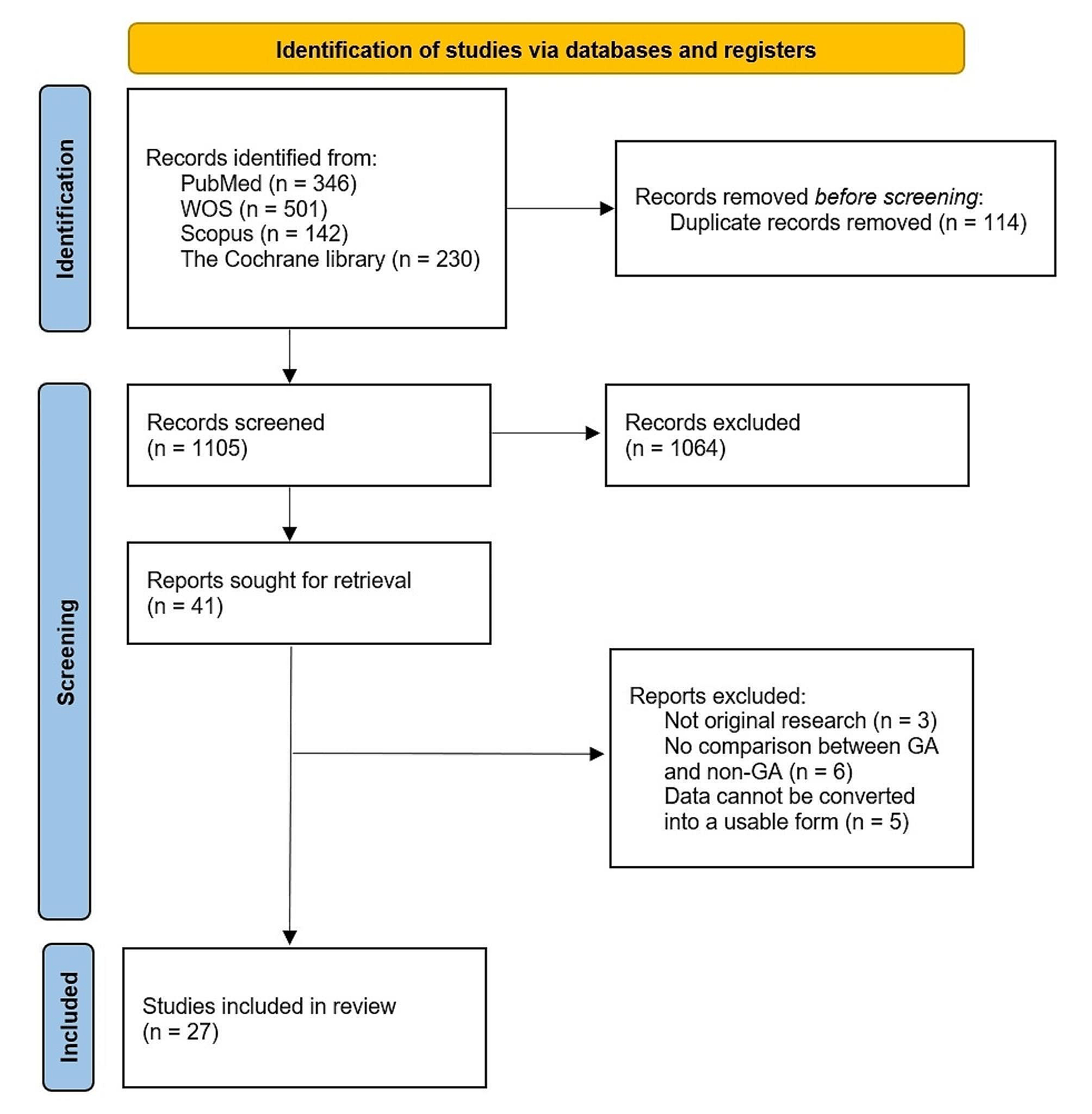


A 42-year-old man, in good health, standing at 168 cm and weighing 79 kg, was hospitalized due to multiple deformed steel bars piercing his left lower waist and hip. The injuries occurred as a result of an accidental fall and impact on his back and waist. The patient was promptly transferred to the central operating theater in a prone position through the designated “green channel” (Fig. 1).
Fig. 1
The patient in prone position
Upon arrival at the central operating theater, the patient was conscious but displayed signs of pain, such as a distressed facial expression, pale eyelids, and cold, clammy limbs. He was breathing spontaneously, although decreased breath sounds were noted in the left lower lung lobe. His oxygen saturation level (SpO2) was 95%, and there were no signs of airway obstruction or abnormal head and neck movements. The patient’s hemodynamics appeared stable, with a blood pressure reading of 135/65 mmHg, a heart rate of 113 bpm, and a regular cardiac rhythm.
A computerized tomography (CT) scan revealed the presence of metallic foreign bodies in the abdominal and pelvic cavities, multiple fractures involving the ribs and the pubic symphysis, as well as indications of possible contusion in the liver, blood vessels, scrotum, and muscle groups. In light of these findings, a multidisciplinary team consultation was convened, and the patient was diagnosed with hemorrhagic shock and severe multiple trauma. The preliminary plan involved extracting the deformed steel bars and performing an exploratory laparotomy.
One of the main challenges in the perioperative period was performing emergency endotracheal intubation while the patient was in the prone position. This procedure needed to be carried out carefully to avoid further damage to organs and prevent exacerbation of the hemorrhagic shock during the extraction of the deformed steel bars.
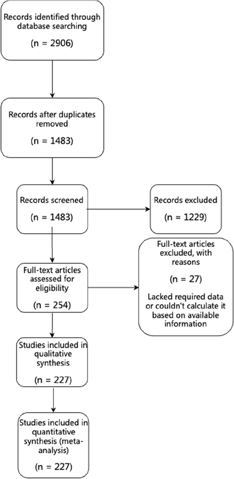
Since the patient was in a prone position and central venous catheterization was not feasible, two peripheral venous channels were established on the patient’s right upper and lower limbs respectively, upon transfer to the central operating theater. Another venous channel had been previously established in the emergency room. Considering the need to extract the deformed steel bars without causing pain or potential damage to abdominal and pelvic organs before anesthesia induction, our anesthesia team opted to establish the airway (tracheal intubation) while the patient remained in the prone position on operating table. The patient’s head was tilted to the right and extended beyond the operating table, with an anesthetist supporting it manually. Successful cricothyroid membrane puncture and spray with 1% tetracaine 1 ml were performed. Superficial anesthesia was achieved by spraying 1% tetracaine 1 ml on the floor of the mouth and the root of the tongue, while ephedrine 5 mg and 1% tetracaine 1 ml were carefully administered into both nasal cavities separately. Topicalisation was established and tested by an anesthetist, and another anesthetist helped him performing the fiberoptic intubation. After preoxygenation via nasal cannula with 6 L/min of oxygen for 5 min, the patient received sedation through an intravenous infusion of 5 µg of sulfentanyl and 1 mg of midazolam, aiming for a Ramesay score of 2–3. Fiberoptic intubation using a reinforced endotracheal tube (size 7.0) was successfully carried out through the right nasal cavity. The tube was first inserted blindly for a few centimeters and then the endoscope inserted over it in a second step. Once the flexible bronchoscope was in the trachea, and the carina was identified, the endotracheal tube was advanced continuously. Oxygen insufflation was performed via the working channel of the endoscope during the intubation process. Following confirmation of the proper placement of the endotracheal tube, additional doses of 2 mg of midazolam, 50 mg of propofol, 20 µg of sulfentanyl, and 50 mg of rocuronium were administered intravenously. The endotracheal tube was then held firmly in position and connected to the anesthetic breathing circuit, and mechanical ventilation was initiated. Anaesthesia was maintained using 2% sevoflurane in air/oxygen mixture with a flow rate of 1 L/min, while remifentanil and propofol were infused at rates of 0.1–0.15 µg·kg− 1·min− 1 and 50–150 µg·kg− 1·h− 1, respectively. Firstly, the deformed steel bars were extracted, and pledgets were used to pack the wound and control bleeding. Subsequently, the patient was repositioned into a supine position. A median abdominal incision was made for exploratory laparotomy following rapid disinfection and draping of the abdomen with a surgical sheet. Then nasopharyngeal temperature was monitored continuously, and a warm air blower was utilized to maintain the patient’s body temperature at 36–37℃. Since arterial blood gas analysis performed at the beginning of the operation revealed metabolic acidosis for HCO3−act 18mmol/L presented, a subsequent infusion of 100 ml of 5% sodium bicarbonate solution was administered. During the exploratory laparotomy, it was found that three deformed steel bars had penetrated the patient’s body to a depth of 12–15 cm (Fig. 2), causing damage to the liver, pancreas, stomach, spleen, and kidney. The surgical procedures included left nephrectomy and repair of the liver, pancreas, and stomach. Internal fixation was performed on the eighth, ninth, and tenth ribs. The duration of the surgery was 7 h, and the intraoperative blood loss amounted to 3000 ml. The fluid input consisted of crystalloid fluid, colloid fluid, calcium gluconate and blood components. Continuous infusion of norepinephrine was performed to prevent intraoperative hypotension. At the conclusion of the surgery, the urine volume was measured at 1500 ml, and another arterial blood gas analysis revealed normal acid-base balance and electrolytes.Despite of stabilization, considering the severity of the multiple trauma, the patient was transferred to the intensive care unit (ICU) without extubation immediately after the surgery. Extubation of the tracheal tube was performed on the second day after surgery when the patient regained consciousness and demonstrated stable respiratory and circulatory functions. The patient made a successful recovery and was discharged from the hospital one month post-surgery without any functional disorders in key organs.
Fig. 2
Three deformed steel bars penetrateted in the patient’s body
留言 (0)