DGCT accounts for approximately 0.28% to 0.38% of all odontogenic tumors and is thus an extremely uncommon benign odontogenic tumor [1, 2, 6, 7]. DGCT can occur in all age groups but shows higher frequencies in middle-aged to elderly patients [2, 7]. A male predilection has been noted in the literature [1, 2]. DGCT is more commonly found in the mandible than maxilla [2]. More cases of a central/intraosseous pattern than peripheral/extraosseous pattern have been reported [1, 2].
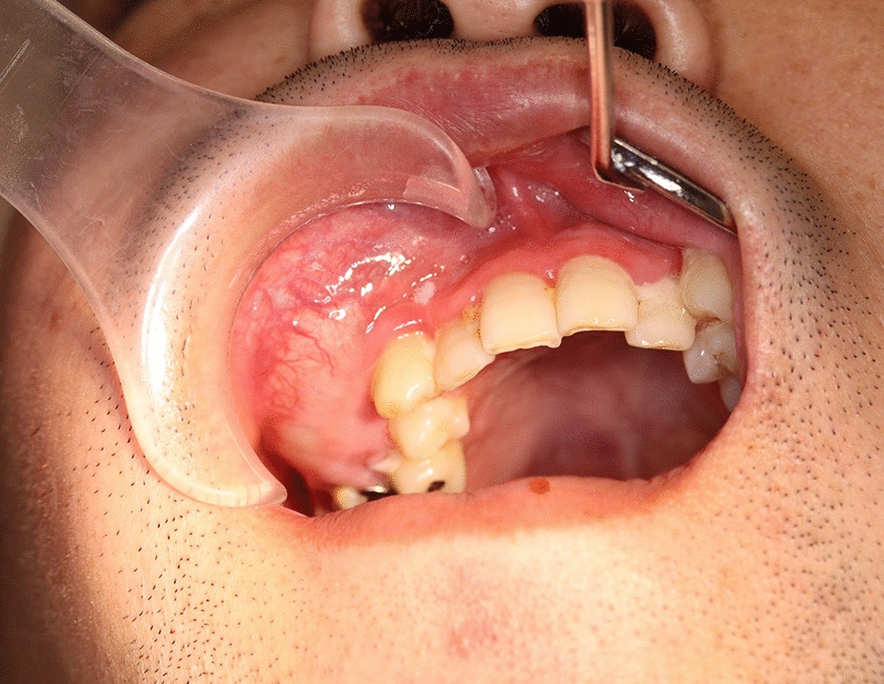
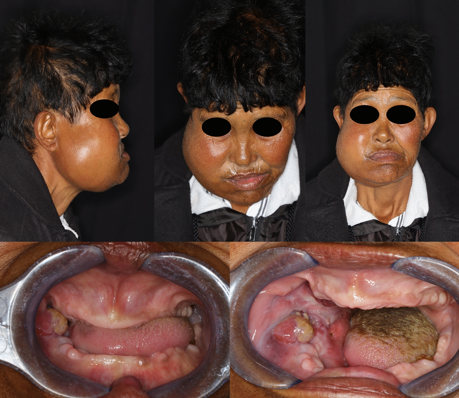
Our patient presented with a mass in the right maxilla but did not complain of pain at the site of origin. Instead, he had difficult in mouth opening and developed swelling of the right buccal region and cervical lymph nodes. We considered that the swelling was the result of bone expansion caused by the mass, and the patient’s history of autism spectrum disorder and intellectual disability might have resulted in his late presentation to the hospital.
DGCT presents certain characteristic findings on imaging examinations. We searched PubMed for reports of DGCT that included imaging findings written in English beginning in 2017, the time point at which DGCT began to be continuously classified as a benign odontogenic tumor. Fifteen reports describing 16 cases of DGCT with imaging findings were reviewed [1, 3, 7,8,9,10,11,12,13,14,15,16,17,18,19] (Table 1). The patients’ ages ranged from 11 to 80 years. Six cases occurred in the maxilla [3, 7, 8, 12, 13, 18], and 10 occurred in the mandible [1, 9,10,11, 13,14,15,16,17, 19]. Panoramic images were obtained in 12 cases [1, 3, 8,9,10,11, 13, 14, 16, 17, 19], a Waters image was obtained in 1 case [18], and CT images were obtained in 11 cases [1, 3, 7,8,9, 11,12,13,14, 17, 19].
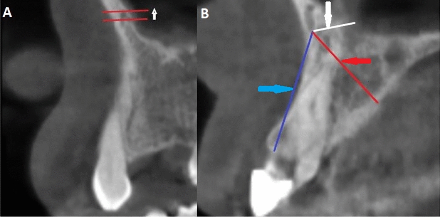
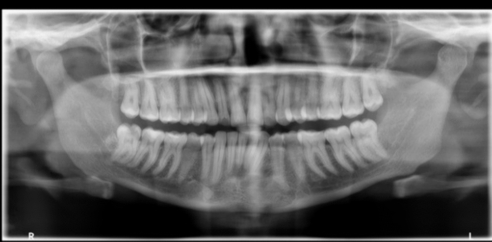
Table 1 List of reports of dentinogenic ghost cell tumor with image findings written in English from 2017The panoramic images and the Waters image in these previous reports showed a well-defined uni- or multilocular radiolucent region, radiopaque calcification, and root resorption [1, 3, 8,9,10,11, 13, 14, 16,17,18,19]. In our case, the panoramic image showed a relatively well-defined and unilocular radiolucent region in the molar area of the right maxilla. The lesion occupied the right maxillary sinus, and root resorption of tooth 16 was suspected. Our findings of a well-defined border, unilocular radiolucent region, and root resorption were consistent with these previous reports. However, tooth displacement and calcification were not observed.
On CT images in these previous reports, the tumor generally appeared as a well-defined, uni- or multilocular, low-density region with high-density structures and cortical bone expansion [1, 3, 7,8,9, 11,12,13,14, 17, 19]. In our case, CT images showed a well-defined, multilocular low-density region with cortical bone expansion, consistent with previous reports. However, although we had suspected that the minute high-density region around the molar teeth represented alveolar bone change, it instead represented dentin formation. This resulted in difficultly diagnosing the lesion.
MRI was subsequently performed, the mass showed homogeneous isointensity on T1WI, and heterogeneous hypointensity to hyperintensity around the alveolar region but most of the mass showed homogeneous strong hyperintensity on STIR images. In addition, heterogeneous enhancement was observed around the alveolar region at the lower half site of the lesion, and enhancement was present along the edge of the upper region on contrast-enhanced T1WI. We judged that the mass was divided into a solid region and cystic region. The ADC was at 1.5 × 10⁻³ mm2/s around the alveolar region at the lower site of the lesion, and high at 2.8 × 10⁻³ mm2/s at the upper site of the lesion. The CI curve rapidly increased and reached a plateau at approximately 30 s, and the plateau was sustained to approximately 400 s. We previously reported the CI curves of ameloblastoma calculated from dynamic contrast-enhanced MRI parameters [20,21,22,23,24]. These curves could be divided into two patterns. In one pattern, the curve increased and reached a plateau at 100 to 300 s, and the plateau then either remained unchanged or gradually decreased to 600 to 900 s. In the other pattern, the curve increased relatively rapidly and reached a plateau at 90 to 120 s, decreased relatively rapidly to 300 s, and then decreased gradually thereafter. The CI pattern in the present case was similar to the former pattern, although the plateau was reached earlier. We considered that a benign tumor could be suspected and that a malignant tumor could be differentiated at least from the pattern of the CI curve. To the best of our knowledge, no study to date has focused on the MRI findings of DGCT. Our MRI findings in this case report are the first such findings reported worldwide, and they show the difference in intensity between the solid region and cystic region of the DGCT. Therefore, we believe that this case is extremely valuable and meaningful in a clinical context.
Histopathologically, DGCT is a benign odontogenic tumor consisting of epithelial neoplastic islands that resemble ameloblastoma and are accompanied by ghost cells and dentin [1,2,3]. According to the WHO, a proportion of ghost cells and dentin exceeding 1% to 2% is useful for the diagnosis of DGCT [2, 5]. In the present case, the tumor comprised epithelial neoplastic islands resembling ameloblastoma inside tight fibroconnective tissue. Masses of ghost cells and formation of dentin were also observed. As a result, the minute high-density area around the molar teeth that we suspected to represent alveolar bone change on the CT images was actually the formation of dentin. These findings were consistent with the typical pathologic findings of DGCT [1,2,3]. However, at the boundary between the solid area and cystic area, epithelial neoplastic islands resembling ameloblastoma were present whereas ghost cells were absent. The epithelium relining the cystic area was thin stratified squamous epithelium similar to that seen in a dentigerous cyst. To the best of our knowledge, no reports to date have described the pathologic findings of the cystic area of DGCT, making the present findings very valuable.
In conclusion, we have presented a rare case of DGCT that occurred in the maxilla with bone expansion, and we focused particularly on new imaging findings (especially MRI). An accurate imaging diagnosis of DGCT is difficult because of its low frequency and often ambiguous findings, such as the presence or absence of an impacted tooth without obvious calcification. The minute high-density area around the molar teeth that we suspected to be alveolar bone change on the CT images was subsequently found to be the formation of dentin in the histopathologic examination. These findings made it difficult to determine the differential diagnoses of lesions with calcification. The present case suggests that we should consider the possibility of an odontogenic tumor with calcification when high-density structures are observed inside the lesion.

留言 (0)