Venous malformations in children: comparison between magnetic resonance imaging and histopathological findings
Background
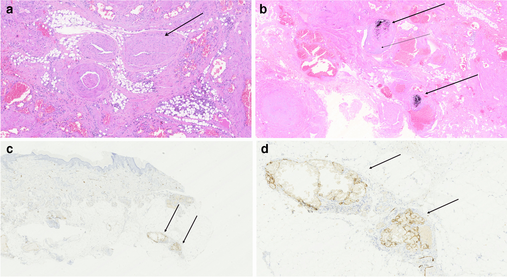
Among low-flow vascular malformations, venous malformations are relatively frequent. The pathological patterns vary in severity and are generally characterized by dilated vessels and low-flow blood that over time can organize into phleboliths. Sometimes small capillary and/or lymphatic vessels may be associated, micro- and/or macro-shunts may form alone or in different combinations, and finally adipose tissue may be interposed between the malformed vessels. Magnetic resonance imaging (MRI) is a crucial examination for confirming venous malformations because it can accurately identify different features of the lesions.
Objective
The aim of our study was to compare MRI and histopathological findings of venous malformations in children to assess the possibilities and limitations of MRI.
Materials and methods
In a retrospective study, two observers independently evaluated the contrast-enhanced MRI of 26 children with venous malformations. Several radiological parameters were considered and compared with histopathological findings. The agreement between the interobserver radiological evaluation and between histopathological and radiological diagnosis was verified using Cohen’s kappa.
Results
MRI interobserver agreement was excellent for micro-shunts and good for the remaining findings. The radiological-pathological agreement was perfect for the presence/absence of phleboliths and of macro-shunts and almost perfect for the presence of intralesional adipose tissue, lymphatic component, and micro-shunts.
Conclusion
MRI in venous malformations can detect the presence of phleboliths, adipose tissue, and lymphatic components with excellent accuracy and good to excellent interobserver agreement. Furthermore, MR angiography can detect micro-shunts in simple and combined venous malformations with substantial agreement with histopathological findings.

留言 (0)