A case of pancreatic body cancer with disappearance of the dilated pancreatic duct on the tail side during preoperative treatment
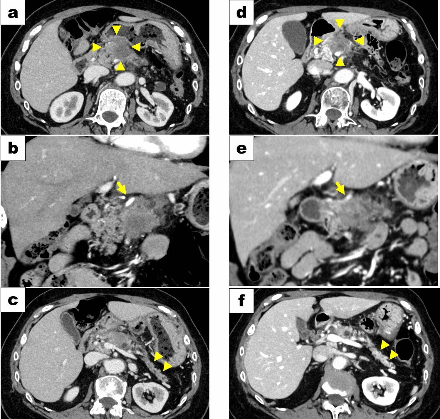
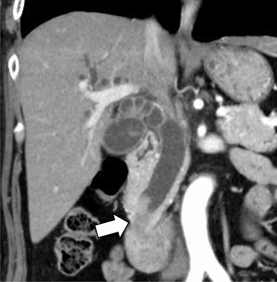
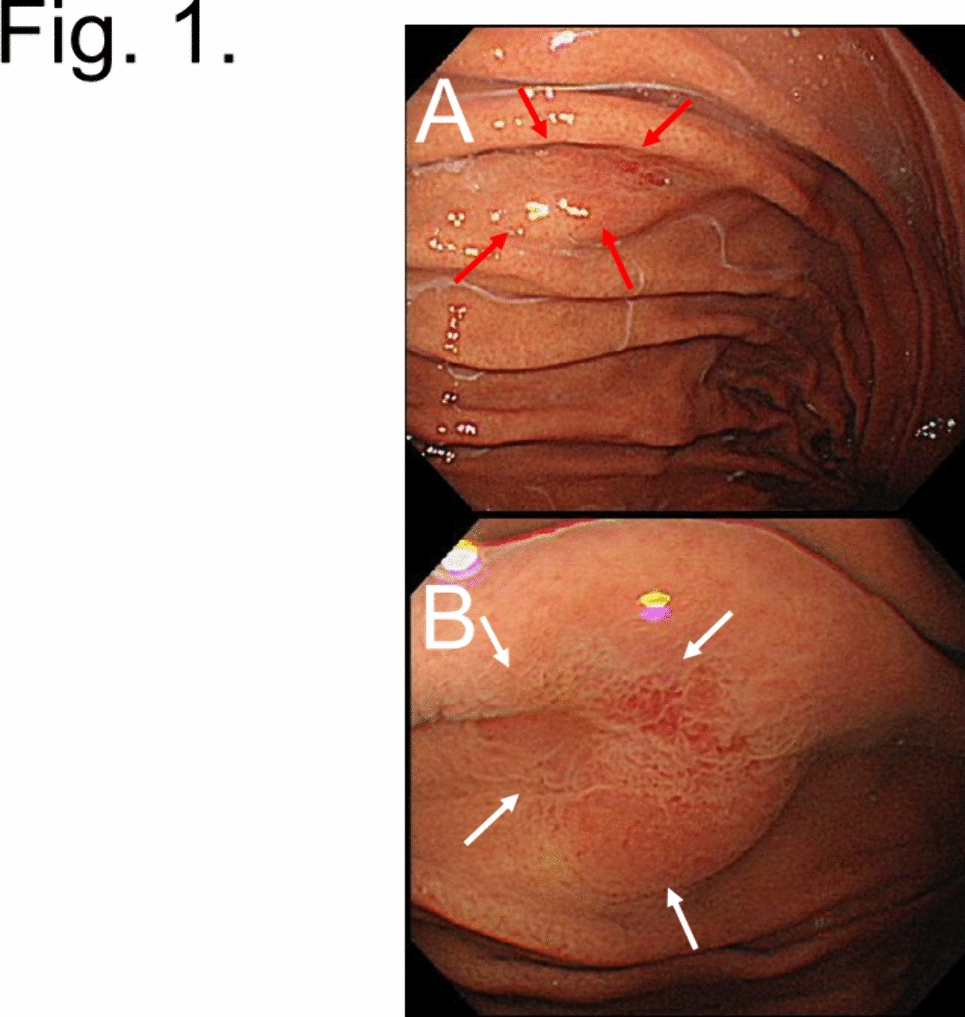
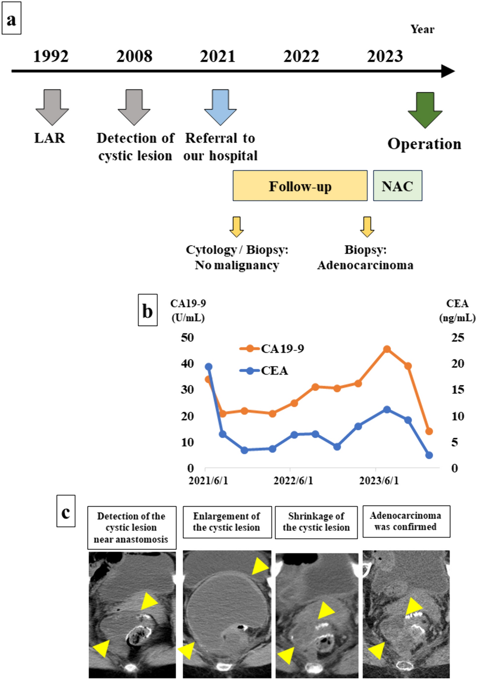
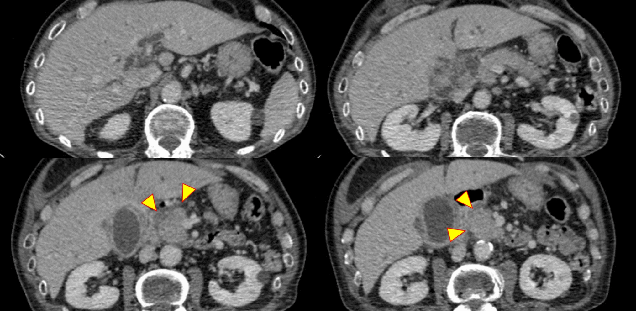
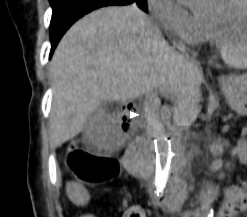
This is a case of a 67-year-old woman diagnosed with a 35-mm pancreatic body cancer with a chief complaint of epigastric discomfort. Computed tomography demonstrated invasion of the common hepatic artery, portal vein, and stomach, and chemotherapy was initiated for locally advanced pancreatic cancer. After 9 months of chemotherapy, the tumor remained stable on imaging, and the tumor markers were within the normal range. After additional chemoradiotherapy, the patient underwent a conversion surgery, a pancreaticoduodenectomy. Magnetic resonance cholangiopancreatography (MRCP) at the time of diagnosis demonstrated main pancreatic duct (MPD) dilatation on the tail side of the tumor; however, most of the MPD signal disappeared on MRCP after chemotherapy. Surgical findings failed to identify MPD on the first pancreatic resection plane, and additional resection was conducted; however, no MPD was found. As a pancreatic duct anastomosis was not available, pancreatic reconstruction was selected for pancreaticogastric anastomosis using the invagination method. Pathologically, the pancreatic tissue on the tail side of the tumor was replaced by fibrotic tissue, and MPD could not be identified. To the best of our knowledge, this is the first case report of the disappearance of a dilated pancreatic duct on the tail side accompanied by exocrine tissue loss during preoperative treatment for pancreatic cancer.


留言 (0)