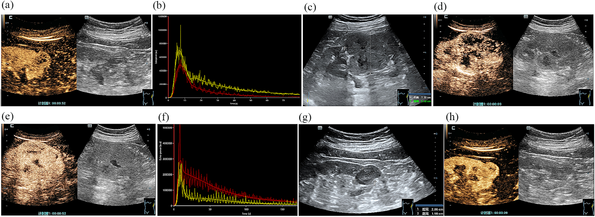
Hypospadias is a congenital condition that refers to the abnormal positioning of the urethral opening in the male reproductive organ. Surgery for hypospadias serves as the primary approach for managing this condition. Despite surgery being the primary treatment, postoperative complications such as urethral stricture, urethral fistula, and infection can still occur [13]. Postoperative ultrasound examination is crucial in the management of hypospadias as it provides essential information to help physicians evaluate the effectiveness of the surgical repair, detect complications, and follow up on the treatment outcome. Ultrasonography can reveal urethral conditions in real time [14]. The injection of saline into the urethra not only discloses the urethra’s normal condition but also simulates urination, thereby underscoring the importance of ultrasonography in assessing urethral conditions. The discharge of urine is an important observation index after urethral reconstruction surgery [15]. Via ultrasound examination, physicians can observe the flow of urine in the urethra and evaluate the smoothness of urine discharge. Should difficulties in urination or abnormal urine reflux be detected, physicians can implement timely interventions to ensure normal urine discharge. Postoperative urethral stricture, a common complication of hypospadias surgery, often manifests as difficulty with local dilation or dilation challenges associated with a local bulge in the anterior region. Patients might experience difficulties in urinating. Moreover, the accumulation of a substantial volume of urine in the bladder can stimulate the bladder’s nerves, resulting in symptoms such as frequent urination and urgency [16]. Isolated clinical manifestations may not accurately reflect the actual condition of urethral strictures, while ultrasonography, when combined with intraurethral liquid injection, can unveil the specific location and degree of stenosis. Manifestations of urethral diverticula encompass hematuria, urethral infections, urethral calculi, dysuria, and potentially carcinoma. Retrograde urography or MRI can be utilized for the diagnosis of urethral diverticula [17, 18]. Compared to the aforementioned methods, ultrasound, being economical and convenient, and lacking radiation, offers real-time insights into urethral conditions. This assists doctors in estimating the size and location of diverticula, serving as a reference for subsequent treatment steps. Postoperative intraurethral hair growth presents another challenging issue, leading to dysuria and penile pain. Traditionally, intraurethral hair has been treated with surgical resection; however, recent findings suggest lasers can also effectively remove hair from the urethra [19, 20]. Ultrasonography distinctly reveals intraurethral hair-like structures, aiding doctors in making initial assessments and guiding further treatment. Furthermore, given the necessity for long-term follow-up post-hair removal, ultrasonography offers a straightforward examination method. Persistent dysuria, residual urine, and infections may lead to urethral calcification and calculi formation. Calcification lesions exhibit a strong echo on ultrasound, whereas calculi present a strong echo accompanied by a posterior shadow, attributed to their high tissue density. Furthermore, it is advisable for doctors to instruct patients to void their bladder before undergoing urethral ultrasonography and to measure the residual urine during the procedure, thereby assessing their urinary conditions. The results of urethral ultrasonography should be interpreted in conjunction with patients’ clinical manifestations and medical history, as some may report dysuria or other symptoms even when ultrasonic findings indicate no anomalies. Ultrasound can further assist doctors in evaluating the urethral wall by observing its structure, such as thickness and smoothness, to assess the healing status of the surgical site [21]. A well-healed urethral wall will exhibit a uniform and continuous structure, whereas poor healing or fistulas may result in interrupted or irregular continuity of the urethra, clearly discernible through ultrasound examination. Finally, given that ultrasound does not expose patients to radiation and is convenient to perform and repeat [22], it plays a pivotal role in the follow-up treatment after urethral repair surgery. Through regular ultrasound examinations, doctors can evaluate the durability of treatment effects and promptly identify and manage potential complications.


留言 (0)