Cell culture
Several cell lines (5637, MRC-5, MDA-MB-231, MCF-7, U87, SW1116, Jurkat, HT29, EJ [MGH-U1], AGS, Raji, and HEK293) were obtained from the National Cell Bank (Pasture Institute, Iran). JAM-ICR, a new cell line isolated from a 64-year-old patient with high-grade (grade IV) BLCA, was established at the Shiraz Institute for Cancer Research [13]. Adipose-derived mesenchymal stem cells (MSCs) were derived from breast adipose tissues of a healthy donor who underwent cosmetic mammoplasty by the explant method. Cell lines were cultured in RPMI-1640 medium (Gibco, USA) supplemented with 10% fetal bovine serum (FBS; Gibco) and 1% penicillin‒streptomycin (P/S; Sigma‒Aldrich, Germany). HEK293 and MSCs were cultured in Dulbecco’s modified Eagle medium (DMEM). The selection medium was composed of RPMI-1640 supplemented with hypoxanthine-aminopterin-thymidine (HAT) medium (2X, Sigma‒Aldrich), 20% FBS, nonessential amino acids (Gibco), 1 mM sodium pyruvate (Gibco), and conditioned medium obtained from cultured peritoneal feeder cells. HAT medium was replaced with hypoxanthine-thymidine (HT) medium (Sigma‒Aldrich) and complete culture medium on the 14th and 21st days of fusion, respectively. All cells were grown in a humidified 5% CO2 incubator at 37 °C.
Immunization of BALB/c mice with the JAM-ICR cell line
Inbred 6-week-old BALB/c mice were obtained from the Pasteur Institute of Iran (Tehran, Iran). When the JAM-ICR cell line reached the logarithmic phase of growth, the cells were scraped and collected. Then, 500 µl of cell suspension with a density of 1 × 10⁷ cells/ml was adjusted and injected intraperitoneally. The subsequent boosts were performed at three-week intervals and repeated until a sufficient serum titer was obtained. The last boost was injected three days before the fusion.
ELISA
An indirect ELISA was used to titrate the concentration of antibodies in the mouse serum. Blood samples from the tail veins of inoculated mice were taken one day before and one week after each injection. JAM-ICR cell lysate was obtained by RIPA lysis buffer and diluted in carbonate/bicarbonate buffer (5 µg/well: Merck, Germany). Then, 100 µL JAM-ICR lysate was coated in MaxiSorp flat-bottom 96-well plates (Nunc, Denmark). After overnight incubation, 250 µL blocking buffer [1% bovine serum albumin (BSA); Biosera, France] was added and incubated at room temperature (RT) for 2 h. Following the addition of mouse sera (1:500) to wells and washing, 100 µl of HRP-conjugated goat anti-mouse antibody (1:1500, BD Biosciences, USA) was incubated for 1 h at 37 °C. Then, 100 µl tetramethylbenzidine (TMB) substrate solution (Invitrogen, USA) was added for 15 min at RT, and the reaction was stopped with 100 µl H2SO4 (0.16 M). The optical density was measured at 450 nm using a microplate reader (Anthos 2020, Austria). All washes were performed with 1X PBS buffer containing 0.05% Tween-20 (Bio-Rad, USA).
Production of hybridoma
The fusion procedure was done according to previous work in our laboratory [14, 15]. Briefly, splenocytes and SP2/0-Ag14 cells at a 5:1 ratio in serum-free RPMI-1640 were mixed and centrifuged at 300×g for 5 min. Then, 1000 µl PEG (Sigma‒Aldrich) was added to the cell pellet (12 × 107) dropwise over 1 min while agitating the tube. The fusion mixture was incubated for an additional 3 min, and then 10 ml of serum-free RPMI-1640 was added over the course of 4 min. The cell suspension was centrifuged and then adjusted to 1.5 × 10⁶ cells/ml in the selection medium.
Flow cytometry
The surface expression of the target antigen was evaluated with flow cytometry [16]. For this purpose, 100 µl of supernatant from hybridoma cells was added to cell suspensions (2 × 10⁵ cells/tube) and incubated for 60 min at 4 °C. Following washing cells with 2 ml staining buffer, the cells were incubated for 30 min at 4 °C with 50 µl diluted FITC-conjugated sheep anti-mouse Ig secondary antibody (1 µl antibody in 49 µl staining buffer; SINA BIOTECH, Iran). After washing, data were acquired on a 4-color flow cytometer instrument (BD Biosciences) and analyzed by FlowJo software (version X.0.7, USA). Each test was done three times separately, and the frequencies of positive cells are shown.
Isotype determination of mAb
A limiting assay was performed three times to isolate single clone-producing antibodies. The isotype of antibodies was then evaluated with an eBioscience™ Mouse Ig Isotyping ELISA Kit (Invitrogen, Austria) according to the manufacturer’s instructions.
Expansion and purification of mAb
Female BALB/c mice (6 weeks old) were primed by intraperitoneal injection of 500 µl/mouse Pristane (Sigma‒Aldrich). After 7 days, hybridoma cells were harvested, and 500 µl of cell suspension (7 × 10⁶ cells/ml) was administered. The ascitic fluid was collected and purified by a Hi-Trap protein G column (GE Healthcare, Sweden) and fast protein liquid chromatography (FPLC) instrument (GE Healthcare). The sample was diluted with binding buffer (20 mM phosphate) and applied to the column. Then, the column was washed with binding buffer to remove the unbound proteins until the absorbance reached a steady baseline of 0.1 milliabsorbance unit (mAU). Attached antibodies were eluted with elution buffer (100 mM glycine, pH = 2.7) at a flow rate of 1 ml/min. Following dialysis with 1X PBS, the concentration of the mAb was determined using a NanoDrop 2000c Spectrophotometer (Thermo Fisher Scientific, USA).
Western blot
Purified mAb (20 µg) and target antigen (40 µg) were loaded on a 12.5% polyacrylamide gel under reduced conditions. The gel was either transferred to a PVDF membrane or stained with colloidal Coomassie Brilliant Blue G-250 (Bio-Rad). The transfer was done using 25 constant voltage and 2.5 limited ampers for 80 min. The blot was placed in blocking buffer (3% BSA in PBS containing 0.15% Tween-20) for 2 h, and then 20 µg/ml primary antibodies were added and incubated at RT with shaking for 1 h. After washing, the blot was incubated for 1 h at RT with HRP-conjugated goat anti-mouse Ig (BD Biosciences, 1:3000 in blocking buffer). After soaking the PVDF membrane in enhanced chemiluminescence substrate (Bio-Rad) for 5 min in the dark, the protein was detected using a ChemiDoc imaging system (Bio-Rad).
Immunoprecipitation (IP)
Target antigen was isolated with a Pierce Crosslink Immunoprecipitation Kit (Thermo Fisher Scientific). According to the company’s instructions, the lysate of JAM-ICR was obtained by using cold IP lysis buffer and coincubated with protein A/G agarose. After cross-linking the selected antibody to agarose with disuccinimidyl suberate (DSS), unbound proteins were washed, and the target protein was collected by elution buffer. The output was verified by western blotting, and the specific target was excised from the polyacrylamide gel and analyzed by liquid chromatography with mass spectrometry (LC/MS). Accordingly, the beta-actin (ACTB) protein was introduced as the target antigen of 6D6.
Immunohistochemistry (IHC)
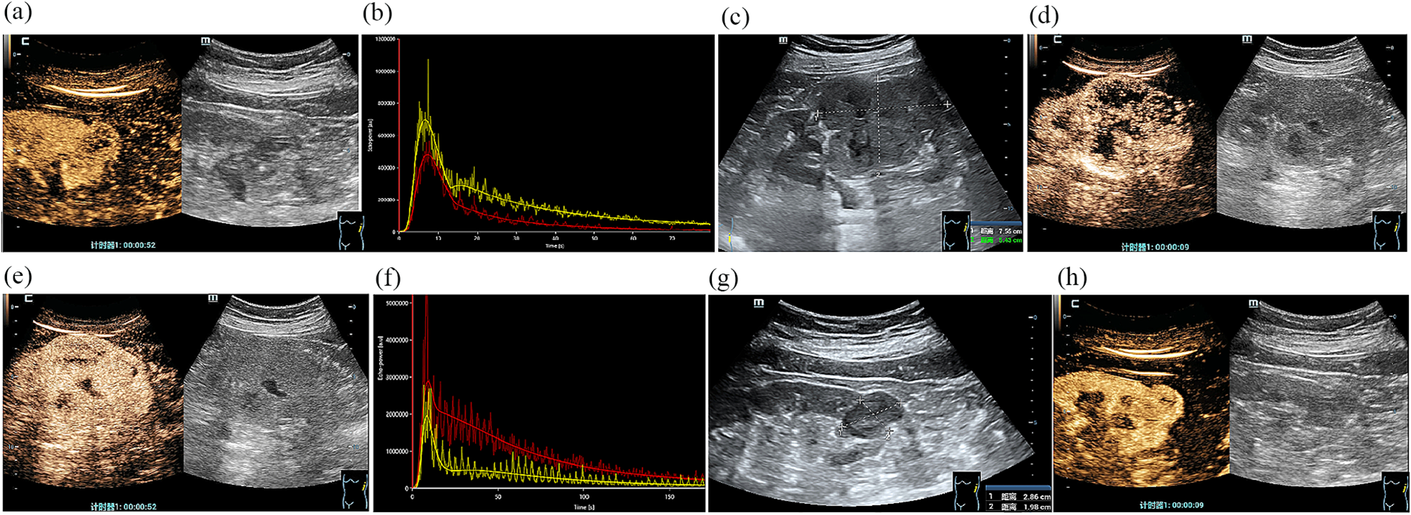
Expression of the target antigen in different bladder tumor tissues was assessed by immunohistochemical staining (IHC). In this connection, resected specimens from 35 patients (29 males and 6 females) with a mean age of 66 ± 2 years old were collected. Five samples from von Brunn’s nests (proliferating epithelial cells; benign tumors) were also assessed. Furthermore, nearby normal tissues were used for benign BLCA situations. The IHC procedure was carried out in accordance with our lab’s setup [13]. The intensity of expression was qualified by scoring from 0 to 2 (0: negative, 1: low, 2: high). The relationship between ACTB intensity and prognostic factors, including T- and N-stages, histological grade, tumor necrosis, lymph node involvement, carcinoma in situ, perivesical fat, invasion of the tumor to the adjacent muscle, perineural, lamina propria, and lymphovascular invasion, was also evaluated.
In silico evaluation
Gene Expression Profiling Interactive Analysis-2 (http://gepia2.cancer-pku.cn/), STRING (https://string-db.org/), cBioPortal (cBioPortal for Cancer Genomics, http://www.cbioportal.org), Catalog of Somatic Mutations in Cancer (COSMIC) database (https://cancer.sanger.ac.uk/cosmic), and the University of California Santa Cruz (UCSC) Genome Browser (https://genome.ucsc.edu/) web servers were used to study ACTB in BLCA and other cancers. First, the expression of ACTB in tumor and normal tissue and then in different stages and survival were obtained in BLCA. Then, the correlation between ACTB and the overexpressed gene and BLCA tumor biomarkers [17] was evaluated. Furthermore, the GEPIA2 server recommended 10 genes that have similar expression patterns with ACTB in BLCA. The expression, survival, and interaction of ACTB were evaluated in various cancers. The interaction network of ACTB with other proteins and genes was obtained from STRING and UCSC servers, respectively.
Statistical analysis
SPSS (version 25.0, USA) and GraphPad Prism (version 6.01, USA) software were used to conduct the statistical analysis and depict figures, respectively. The expression of the target antigen in different cell lines were compared using Mann-Whitney U test. The relationship between the intensity of ACTB and prognostic factors was analyzed with the chi-square (x2) test. Additionally, the survival of patients with different expression levels of ACTB was compared with the Kaplan–Meier estimator and the log-rank test. A P-value of less than 0.05 was regarded as statistically significant, and the data are reported as the mean ± SD.


留言 (0)