Cell culture
The primary cells of rabbit and human ureter smooth muscle cells were procured from Shanghai Saibaikang Biotechnology Co., Ltd. (Shanghai, China). The cells were cultivated in accordance with the recommended protocol using complete medium (10% fetal bovine serum (FBS), 100 U/mL penicillin, and 100 µg/mL streptomycin) in T25 culture flasks. Incubation was carried out in a 5% carbon dioxide environment at 37 °C with saturated humidity. Cells from passages 3 to 6 were employed in the experimental procedures.
Animals
In our previous study, we constructed a rabbit model of ureteral calculi using flowable resin [23]. In this study, we used a sample of 25 New Zealand rabbits, comprising both males and females, with an average weight of 3.0 ± 0.5 kg and an average age of 4 ± 0.5 months. The rabbits were obtained from the Laboratory Animal Center of Guizhou Medical University, and certified under license number SCXK-2018-001. The rabbits were housed separately in cages that were kept at a temperature between 17 and 19℃. The humidity level was maintained at 40–60%. The rabbits followed a 12-hour light/dark cycle and were given a regular chow diet. The current investigation obtained approval from the Medical Ethics Committee of Guizhou Provincial People’s Hospital (Ethical Approval No: 2,017,059).
Construction of ureteral calculi model
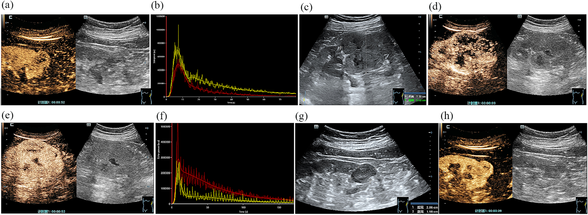
A total of twenty-five rabbits were assigned at random to five groups, with each group consisting of five rabbits. The groups were named as follows: normal group, 1st-day group, 3rd-day group, 5th-day group, and 7th-day group. The control group did not receive any treatment. We constructed a ureteral calculi model with flowable resin (FiltelKTM Z350 XT Flowable Restorative). The flowable resin is a good choice as calculi material due to its characteristics of rapid setting time, light-cured and low viscosity. The experimental groups underwent left ureteral calculus shaping according to our established procedure [23]. The rabbits in the first, third, fifth, and seventh groups were euthanized at 1, 3, 5, and 7 days after the operation, respectively. The rabbits in the control group were euthanized after 7 days. The rabbits were euthanized by administering a lethal dosage of sodium pentobarbital (30 mg/kg) through an intravenous injection. After removing the blocked tissues from the left upper ureter, the specimens were immediately placed in a pre-cooled PBS buffer. The tissues were washed with cold PBS, dried by blotting, transported to liquid nitrogen, and kept at a temperature of -80 °C. Figure 1 displays visual representations of the surgical procedure and specific urological dissections performed after the operation.
Cell and tissue protein extraction
Cellular proteins were extracted by gently washing the cells in the T25 bottle with pre-cooled PBS, adding 200 µL of RIPA containing 1% PMSF, placing on ice, and reacting for 30 min. The mixture was then centrifuged at 4 °C at 12,000 x g for 30 min, and the supernatant was transferred to an EP tube. Tissue proteins were extracted by adding 500µL of RIPA containing 1% PMSF and placing on ice for 30 min, followed by centrifugation at 4 °C for 30 min at 12,000 x g. The resulting supernatant was transferred to an EP tube. Extracted cell and tissue proteins were assayed for protein content according to the BCA reagent instructions. The proteins were then denatured by adding the appropriate amount of loading buffer to the proteins, boiled for 10 min, cooled and stored in portions for use.
Western blot
Equivalent proteins from different samples were isolated by 10% gel electrophoresis, after which the proteins were transferred to a PVDF membrane. The Genefist fast closure solution was applied for a duration of 10 min. EP1 (PTGER1), EP2 (PTGER2), EP3 (PTGER3), EP4 (PTGER4), mPGES-1, PKA, and p-PKA antibodies were diluted at 1:2000, while the internal reference antibodies GAPDH and β-Tubulin were diluted at 1:2000 and 1:10,000, respectively. Incubating different primary antibodies after cutting on a whole membrane results in the absence of images of adequate length. All antibodies were sourced from Affinity. Separate addition of corresponding antibodies was performed, followed by an overnight incubation at 4 °C. The membrane underwent TBST washing thrice, each for 10 min. Subsequently, goat Anti-rabbit IgG H&L (HRP) was added, and the incubation took place at room temperature for 1 h. Chemiluminescence assay kits (NCM Biotech, China) were used to visualize the protein bands in Genegenome XRQ System (Syngene, Britain). ImageJ software (v1.46r) was used to measure the density of protein bands and to convert the results into quantitative data.
Cell immunofluorescence
Fourth-generation rabbit ureteral smooth muscle cells, exhibiting robust growth, were utilized to create a cell suspension. A total of 5 × 10⁴ cells were evenly distributed per well in a 12-well plate with a coverslip. After 24 h, once the cells had adhered to the well walls and displayed vigorous growth, the medium was discarded. A PBS rinse was performed, followed by fixation with 4% paraformaldehyde for 15 min. Subsequently, a 0.5% TritonX-100 treatment was carried out at room temperature for 20 min. Subsequently, the cells were cultured with 10% goat serum at ambient temperature for a duration of 30 min. Drops of EP1, EP2, EP3, and EP4 antibodies (diluted at 1:300, Affinity) were added to a humidified chamber and left to incubate overnight at 4°C. Following the rinse with TBST, sections were incubated with anti-rabbit IgG (Alexa Fluor 488, Servicebio, China) in the dark at room temperature for 1 h.4,6-diamino-2-phenyl indole (DAPI) (Servicebio, China) was added dropwise for 10 min to stain the nuclei. Subsequent observation and image acquisition were performed under a fluorescent microscope (Olympus, USA).
Ureteral in vitro tension test
The rabbits were euthanized by administering a lethal dose of sodium pentobarbital through an intravenous injection. The ureter was promptly and entirely extracted into a 4 °C Krebs solution (pH7.4) pre-saturated with a gas mixture of oxygen (95% O2 and 5% CO2) with the following composition: NaCl, 118 mM; KCl, 4.7 mM; CaCl2, 2.5 mM; MgSO4, 1.2 mM; NaHCO3, 25.2 mM; NaH2PO4, 1.2 mM; Glucose, 11.1 mM. The surrounding fat, blood vessels, and excess tissue were microscopically removed, and the ureter was cut into small 1 cm sections. The ureter mounted horizontally on an isometric force transducer (DMT620, Denmark), filled with Krebs solution at 37 °C, and aerated with 95% O2 and 5% CO2 (Supplementary Fig. 1 in Supplementary material). The Lab Chart software, specifically the Data Monitoring Tool (DMT), was utilized to continuously record the stress in the ureter. Prior to the start of the experiment, the ureter was subjected to a static strain of 3 mN for 40 min to allow it to adapt.
(1)
Normal: No intervention was applied, and the tension of the ureter was examined for a duration of 1 h to monitor any alterations in its tone.
(2)
Measured concentration-diastolic rate curve: High potassium solution was gradually added to reach 80 mM, inducing ureteral constriction. After stabilization for 5 min, cumulative concentrations of PGE2 (ranging from 10 nM to 30 µM) were introduced. The control group was administered a comparable dosage of DMSO, and changes in ureteral tone were recorded.
(3)
Control: The contraction of the ureter was induced by progressively increasing the concentration of potassium solution to 80 mM, and then allowing it to stabilize for 5 min. Subsequently, 1 µM PGE2 was introduced, and the change in tension was recorded. The ureteral diastolic rate was then calculated.
(4)
The effect of EP receptor antagonists: Pre-incubation with 10 µM EP2 antagonist (PF-04418948) or 10 µM EP4 antagonist (L-161,982) for 10 min preceded the gradual addition of high potassium solution to 80 mM, inducing ureteral contraction. After stabilization for 5 min, 1 µM PGE2 was added, and no PGE2 was introduced to the control group. Tension changes were recorded, and the ureteral diastolic rate was calculated.
(5)
The effect of EP receptor agonist: Ureteral contraction was initiated by progressively introducing a high potassium solution up to a concentration of 80 mM, which was then followed by a 5-minute period overnight of stability. Afterwards, a concentration of 10 µM of the EP2 agonist (Butaprost) or the EP4 agonist (CAY10598) was introduced. Changes in tension were recorded, and the ureteral diastolic rate was calculated using the equation below.
Ureteral diastolic rate calculation: Dosed ureteral diastolic rate = (High Potassium Induced Maximum Tension - Recorded Tension After Dosing)/(High potassium Induced Maximum Tension - Eluted Tension).
cAMP measurement
Ureteral smooth muscle cells were seeded overnight in a 96-well plate at 37 °C and subjected to the following treatments:1.Left untreated (control). 2.Cells were exposed to PGE2 (10 µM), EP2 agonist (10 µM), and EP4 agonist (10 µM), followed by an additional 2-hour incubation. 3.Cells were incubated with EP2 antagonist (10 µM) and EP4 antagonist (10 µM) for 30 min, followed by treatment with PGE2 for 2 h. Subsequently, the cells were lysed.The cAMP levels in the smooth muscle were quantified using an ELISA kit (Elabscience Biotechnology Co., Ltd, China). The standards in the ELISA kit were used to make a standard curve. Concisely, 50 µL of samples and 50 µL of biotinylated detection antibody were introduced to cAMP pre-coated wells and incubated at 37 °C for 45 min. Following the plate washing, HRP-conjugated antibodies (100 µL per well) were introduced and incubated at 37 °C for 30 min. After performing another wash, a solution of substrate (90 µL per well) was added and incubated at 37 °C for 15 min. Subsequently, a stop solution of 50 µL per well was introduced, and the optical density (OD) values at 450 nm were assessed in order to determine the cAMP levels.
Data analysis and statistics
The experimental data were represented as the mean value plus or minus the standard deviation (Mean ± SD). Data analysis and visualization were performed using the statistical analysis program GraphPad Prism 8.0. The Student’s t-test was used to compare two groups. Multiple groups were compared by ANOVA, and post hoc comparisons were carried out by LSD method. A significance level of p < 0.05 denotes the presence of statistically significant differences.


留言 (0)