
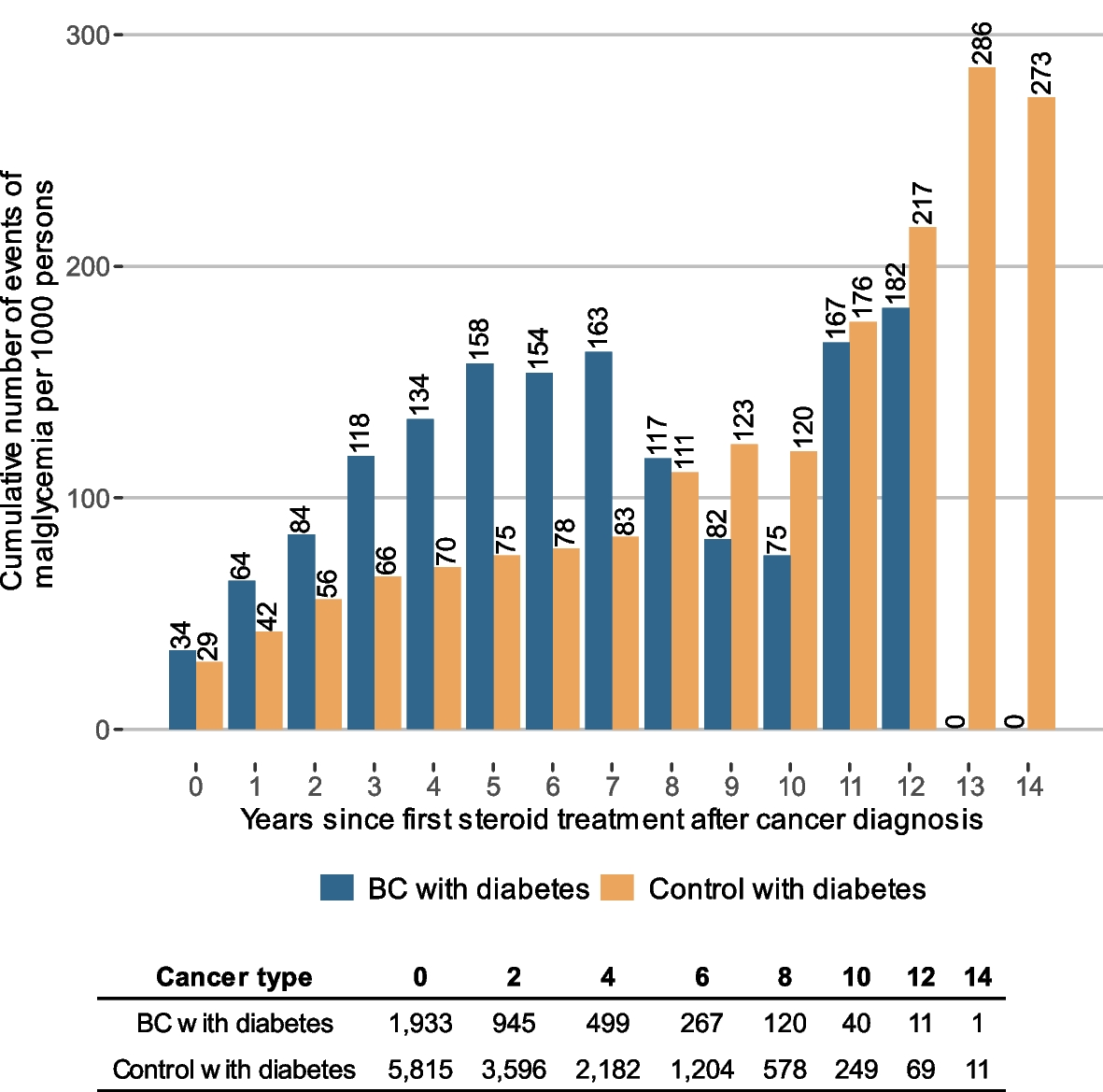
記住我


The system consisted of a perfusion and suction platform (Fig. 1) and a PC-UAS (Fig. 2) (PC-UAS, Inventor Technology, Jiangxi, China). The perfusion and suction platform comprised a main control unit, perfusion device, suction device, and pressure feedback device. Users can set the perfusion flow rate, control pressure value, warning pressure value, and limit value on the platform display. The main control unit of the platform adjusts the suction pressure using pressure feedback. The platform offers two modes: automatic (perfusion, suction, pressure monitoring, and pressure feedback control) and simple. It can display the actual suction and cavity pressures in real-time.
Fig. 1
Perfusion and suction platform
Fig. 2
PC-UAS; 1 pressure channel, 2 suctioning channel, 3 working channel, and 4, pressure measuring hole
The PC-UAS had an inner diameter of 12 Fr, an outer diameter of 14 Fr, and a length of 35 cm. Its transparent material allows for the direct observation of mucosal conditions through the sheath. The cavity pressure is measured using four pressure holes at the tip. The end of the sheath had two connection channels for suction and pressure monitoring. The suction channel can automatically suck out stones and adjust the pressure, whereas the pressure monitoring channel can monitor the cavity pressure.
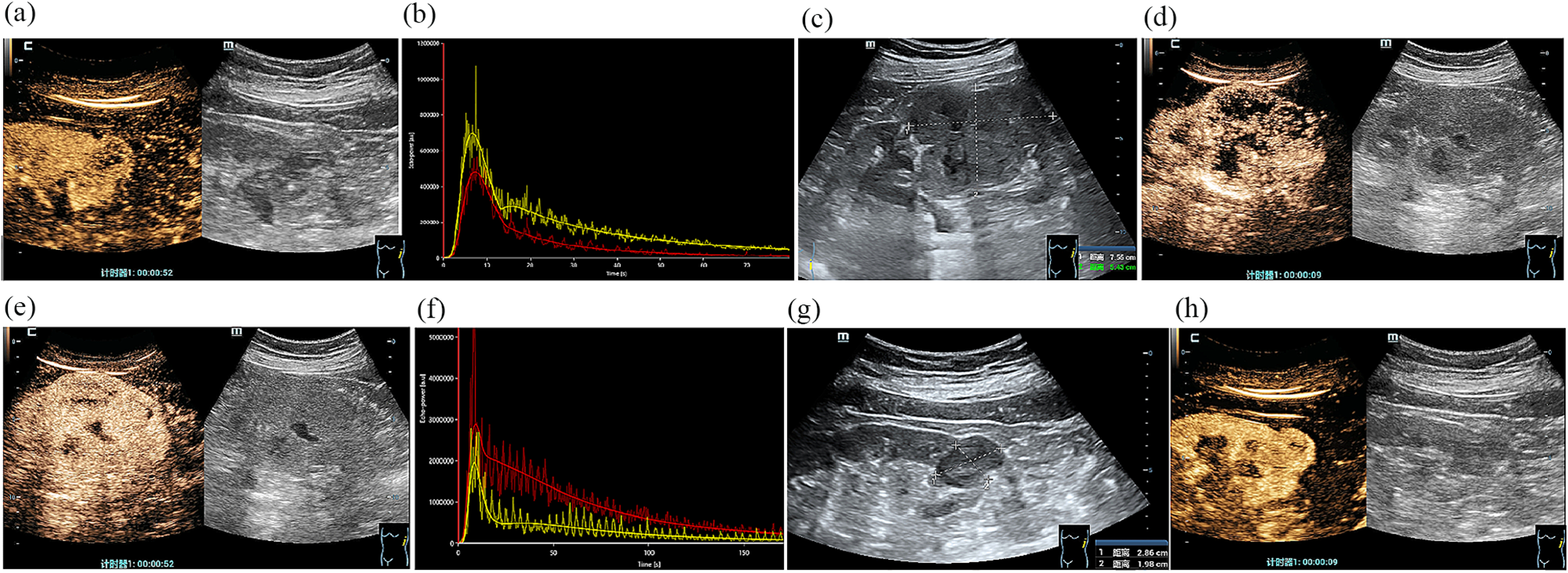
PatientsWe retrospectively analyzed data from consecutive patients with steinstrasse between June 2018 and October 2023 at Ganzhou People’s Hospital. The patient inclusion criteria were age 18–80 years and complex steinstrasse containing ≥ 4 stones or with an aggregate length ≥ 1.5 cm [7]. The exclusion criteria were pregnancy, urinary tract abnormalities, kidney malrotation, and positive urine cultures. Steinstrasse was confirmed by imaging, including urinary tract ultrasonography, intravenous urography, and computed tomography (CT). Stone size was calculated using CT. The study was approved by institutional ethics committee of Ganzhou People’s Hospital(TY-HKY2021-012). All patients provided written informed consent prior to surgery.
Procedural methodsRigid ureteroscopic lithotripsy was performed under general anesthesia with the patient in the oblique supine lithotomy position [8]. After the rigid ureteroscope was connected to the irrigation tube, the platform mode was switched to the simple irrigation mode, in which the platform operated similarly to a conventional irrigation pump with an irrigation flow of 50 ml/min. Under the guidance of a guidewire, a rigid 7/8.4 Fr ureteroscope (KARL Storz, Tuttlingen, Germany) was used for ureteroscopy. The rigid ureteroscope had an irrigation channel (2.4 Fr) and a working channel (3.4 Fr). After verifying the absence of ureteral stricture, a guidewire was inserted. The 12/14Fr PC-UAS was placed along the guidewire at the distal end of the stone. The pressure-measuring and suction channels of the sheath were connected to the perfusion suction platform (Fig. 3), and the pressure measuring channel was filled with water and zeroed. The perfusion suction platform was set to fully automatic mode, with a perfusion flow rate of 150 ml/min, control pressure of -15 to -2 mmHg, pressure warning value of 20 mmHg, and limit value of 30 mmHg. During the surgery, a 550 μm diameter fiber was used for rigid lithotripsy with a power of 2.0–2.5 J × 20–30HZ. The broken stone particles were automatically sucked out through the gap between the sheath and the scope, while particles larger than the gap but smaller than the sheath diameter were removed by withdrawing the scope. After surgery, a 4.6 Fr double J was routinely placed for 4 weeks. For patients with ureteral stenosis detected during ureteroscopy, the surgery was changed to ordinary ureteroscopy, and the stones were pushed into the kidneys for PCNL. Laparoscopy was performed for middle and lower ureteral stones that could not be pushed.
Fig. 3
Pipe connection; 1, pressure measuring tube, 2, working channel, and 3, suctioning tube
After surgery, the patients’ vital signs and blood tests, such as routine blood count, electrolytes, and procalcitonin, were closely monitored. A kidney, ureter, and bladder radiograph (KUB) was performed on postoperative days 1 and 30 to evaluate residual stones. CT scans were performed on patients with X-ray-negative stones. Ultrasonography was performed at 3 months to assess hydronephrosis. Stone-free status was defined as the absence of stone fragments or residual stones < 2mme in CT or < 4 mm on KUB in size. Procedure duration was defined as the time from insertion of the controlled-pressure ureteral suction sheath to the end of the procedure. Surgical complications were graded according to the Clavien-Dindo classification [9].
留言 (0)