Intraoperative Fluorescent Ureter Visualization for Transvaginal High Uterosacral Ligament Suspension for Severe Pelvic Organ Prolapse
Introduction and hypothesis
We aimed to demonstrate the feasibility of ureteral navigation using intra-ureteric indocyanine green (ICG) and near-infrared fluorescence (NIRF) imaging during transvaginal high uterosacral ligament suspension for prolapse repair to reduce the risk of iatrogenic ureteral injury.
Methods
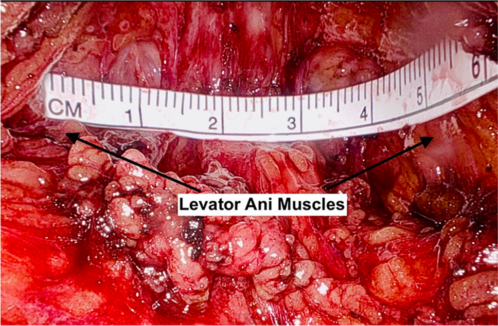
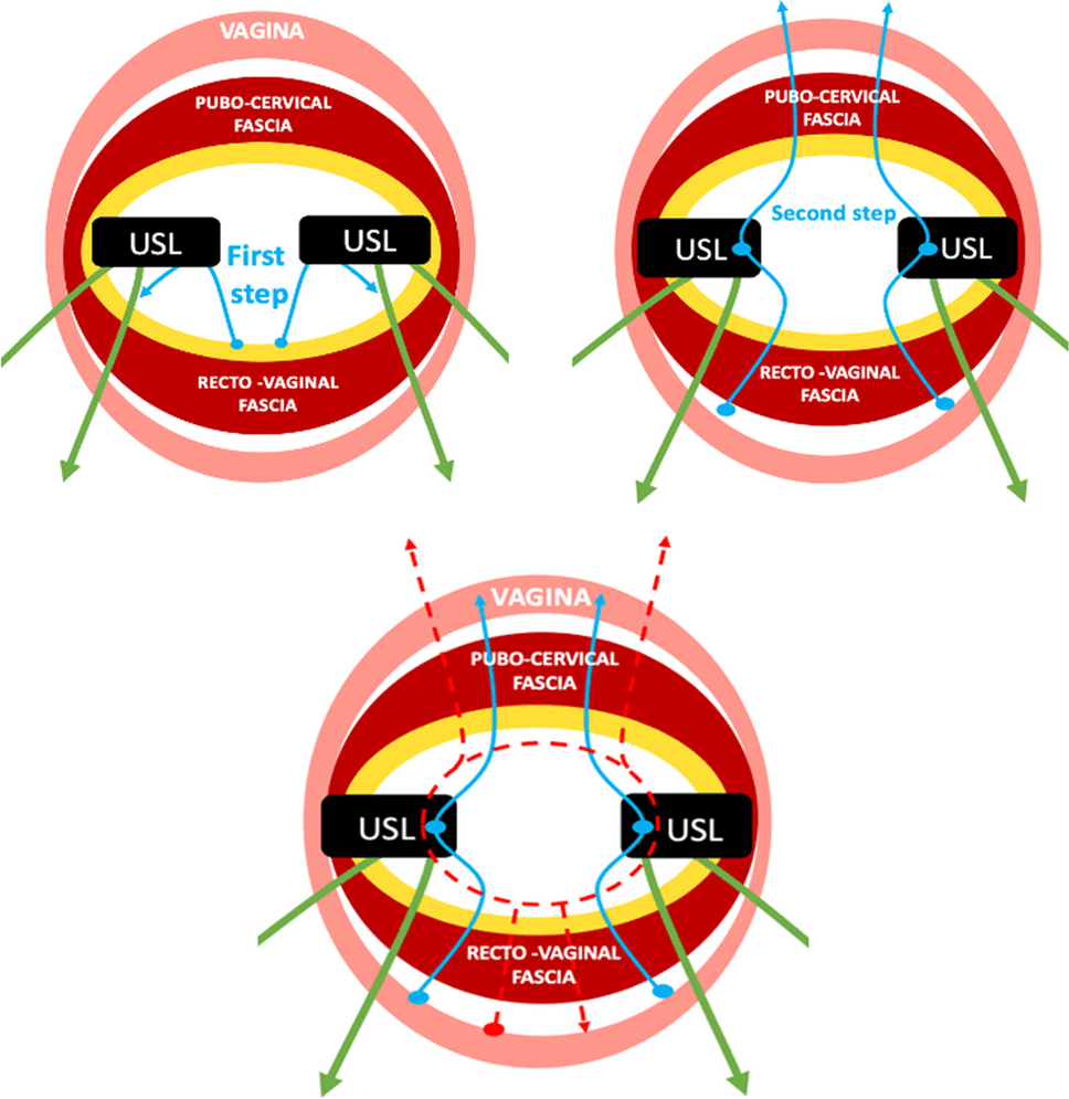
A cystoscope was inserted into the bladder, the tip of a 6-F open-end ureteral catheter was inserted into the ureteral orifices, and ICG was instilled into the ureters. The ureteral path was then clearly identified using NIRF imaging. Sutures were safely placed in the uterosacral ligaments at the level of the ischial spine, taking advantage of direct ureteral visualization.
Results
At the end of the procedure, diagnostic cystoscopy was performed to confirm ureteral patency. No intraoperative or postoperative complications were observed.
Conclusions
Intra-ureteric ICG-NIRF imaging represents a simple, inexpensive, and reproducible trick for intraoperative ureteral detection, and could reassure surgeons during difficult operations, for instance, in the case of severe prolapse and/or when ureteral course abnormalities are expected.


留言 (0)