
記住我


Acanthamoeba keratitis (AK) is a rare but potentially sight-threatening infection of the cornea caused by the free-living protozoan,1,2 mostly related to contact lens misuse. The disease is often misdiagnosed and can progress rapidly, causing severe pain, corneal perforation, or scarring. Usually, the infection last for months causing epithelial abnormalities, ring-shaped corneal infiltration, and stromal necrosis.3 This causes significant worsening of patient's quality of life, limitation in daily life activities and choices for years and in some case permanent loss of vision. Effective and rapid treatment is mostly related to a prompt diagnosis based on the physician clinical suspicion in early stage, confirmed by microbiological instrumental tests, and an early intense topical compounded eye drops protocol regimen.4
However, inspite of all the efforts, still AK infection drastically impact patient's real life, inducing the patients to change their life choices, lifestyles, and influencing psychological growth particularly in such young age groups. In fact, late diagnosis and no standardized treatments (only compounded and based on empirical or anecdotal practice) cause 8 to 12 months of painful progressive course until eradication, and usually ends up in a severe corneal scarring, poor visual outcomes, and need for high-risk corneal transplantation surgery.5
Late diagnosis, widespread use of topical steroids before AK diagnosis, a lack of current anti-amoebic therapies, and limited evidence of efficacy and safety and the absence of an evidence-based treatment delivery protocol are primary causes of such unsuccessful management.
MATERIALS AND METHODSTwo patients with confirmed diagnosis of Acanthamoeba keratitis was advised for compassionate administration of topical polihexanide 0.08% monotherapy as per novel protocol6,7 because the patients were severely worsening despite 4 weeks of treatment with a combined therapy of PHMB 0.02% and propamidine 0.1% hourly.
The treatment protocol used was one drop of each every hour for the first 72 hr, then one drop six times a day for 4 to 6 weeks, then 2 weeks off.
The novel protocol consists polihexanide 0.08% every hour daytime only (16 drops a day) for five days. No overnight treatment is recommended. Then every 2 hr (8 times per day) for seven days, then every 3 hr (6 times per day) for seven days, finally every 4 hr (4 times per day) until the disease is resolved.7 Polihexanide 0.08% is manufactured under Good Manufacturing Practice (GMP) conditions. The active ingredient is polihexanide 0.8 mg/mL manufactured in unidose preservative-free vials. All experimental procedures were performed in accordance with guidelines established by the Association for Research in Vision and Ophthalmology, adhered to the tenets of the Declaration of Helsinki, and approved by the Intramural Ethical Committee (University Campus Bio-Medico, Rome, Italy). All participants provided written informed consent. Patients' follow-up was at least 7 months after treatment begun. No patients were treated with topical or systemic steroids or NSAIDs for all follow-up duration.
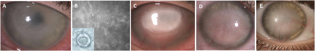
CASE 1A 27-year-old female contact lens wearer presented with severe pain and vision loss in her right eye. The best-corrected visual acuity (BCVA) of the right eye was 20/400, and there was a central corneal ulcer compromised by stromal and perineural infiltrates (Fig. 1A). Acanthamoeba keratitis was confirmed by in vivo corneal microscopy and scraping (Fig. 1B). Hourly administration of topical PHMB 0.02% and propamidine 0.1% was initiated and tapered weekly. A clinical deterioration was observed up to 4 weeks (Fig. 1C), with worsening of visual acuity and increase of corneal melting and thinning. Then, polihexanide 0.08% eyedrops were recommended worldwide for the first time. No other adjunctive topical therapy was used, and systemic pain relievers were not used. Within 15 days, the infection had cleared, and the cornea had healed. At 1 month, corneal opacities and neovascularization were reduced, vision improved at 20/200, and there was no recurrence of infection (Fig. 1D); therefore, treatments were stopped. No further treatments were advised for the further follow-up.
 FIG. 1.:
 Case 1: (A) Central corneal ulcer compromised by stromal and perineural infiltrates. (B) Acanthamoeba cyst in corneal in vivo microscopy. (C) Clinical deterioration after standard protocol. (D) Cornea healed within 15 days. (E) Corneal picture at 9 months.
At 9 months of follow-up, no recurrence was detected and clinical opacity slowly improved (Fig. 1E).
CASE 2A 17-year-old boy wearing contact lenses while swimming, came to our attention with severe pain and increasing vision loss in his left eye. Hypopyon, stromal infiltrates, and a central corneal ulcer were observed (Fig. 2A). BCVA of 20/200 was noted in his left eye.
 FIG. 2.:
 Case 2: (A) Hypopyon, multifocal stromal infiltrates, and a central corneal ulcer. (B) In vivo corneal microscopy. (C) Clinical deterioration after standard protocol. (D) Cornea healed within 30 days. (E) Corneal state at 7 months.
Acanthamoeba was identified after contact lens culture. In vivo microscopy and scraping then confirmed our suspicion of Acanthamoeba keratitis also (Fig. 2B). Prompt starting of hourly application of topical PHMB 0.02% and propamidine 0.1% was initiated. For 1 month, no clinical improvement was observed (Fig. 2C). Patient was suffering of persistent and severe pain with reduction of visual acuity, at clinical examination an increase of stromal infiltration and hypopyon were assessed. Then, polihexanide 0.08% eyedrops were suggested. No other adjunctive topical therapy was used, and systemic pain relievers were not used. The infection cleared up and the cornea healed within 30 days (Fig. 2D) At 1 month, corneal opacities and neovascularization had diminished, BCVA had improved at 20/125, and there had been no recurrence of infection. Hyaluronic acid artificial eye drops were administered in follow-up. At 7 months of follow-up, no recurrence was detected and clinical opacity slowly improved (Fig. 2E).
DISCUSSIONTopical polihexanide 0.08% monotherapy rapidly improved clinical outcomes in the first two AK patients treated. Corneal healing and parasite eradication was successfully achieved within 30 days since therapy begin. Slow and progressive resolution of secondary fibrosis and deep and superficial neovascularization was assessed at 3 and 7 months.
This is the first reported use of such novel concentration of preservative-free PHMB ophthalmic solution with a standardized protocol.
In our practice primary medical treatment for AK involves a combination of compounded antiamoebic agents such as PHMB 0.02%8 or chlorhexidine 0.02% to 0.06%,9 along with propamidine isethionate 0.1%.10 However, this regimen is not standardized and is frequently empirical, which means it is mostly directed by clinician observation and experience and is not consistent across various clinicians, even within the same hospital.
In severe cases AK may worsen in a corneal melting or perforation, despite medical therapy. In such cases, prompt surgical intervention is necessary including corneal transplantation with high risk of rejection and recurrence of primary infection.11
Then, early diagnosis and prompt initiation of treatment are essential for a good and fast visual recovery. Several studies have found that PHMB monotherapy is an effective treatment in AK. Lim et al.9 in randomized comparative study reported 55 AK patients successfully treated with PHMB 0.02% or chlorhexidine 0.02% monotherapy. However, Lim et al.9 had 23 subjects using PHMB 0.02% monotherapy, of whom 18 (78.3%) met the primary outcome and of whom 15 (65.2%) were cured without surgery. Their findings for chlorhexidine 0.02% were, respectively, 28 subjects, of whom 24 (85.7%) met the primary outcome and 22 (78.6%) were cured without surgery, although there were no statistically significant differences in final acuity scarring or the keratoplasty rate.
Papa et al., in a retrospective cohort study of 227 patients treated with various combinations of PHMB, diamidines, and chlorhexidine, reported that 47 patients were started on PHMB 0.02% monotherapy, with an additional three on PHMB 0.06%. These were baseline therapies; unfortunately, it was not specified how many of these patients later switched therapy.
Despite this, the study provided evidence that PHMB 0.02% monotherapy can be an effective initial treatment in AK and is easier to administer and less costly than other combinations.8
Polihexanide has been demonstrated (1) to have a positive safety profile, with low toxicity and high tolerance, and (2) an effective amoebicidal agent against both trophozoites and cysts1 (3) dose-dependent efficacy.7
In a prospective randomized clinical trial,7 0.08% preservative-free PHMB ophthalmic solution showed higher levels of tolerance and toxicity in AK subjects to justify the increased concentration in treatment trials. In our series, 0.08% may be the ideal concentration for a proper tolerability and safety and a prompt efficacy of the treatment. Moreover, the novel suggested protocol of PHMB 0.08% monotherapy for AK has been clearly effective in Acanthamoeba eradication. However, the application of high dose PHMB as a monotherapy in AK is not without its challenges, and further studies are needed to confirm such insights.
Moreover, it may not be suitable for all AK cases, especially in severe or advanced infections where a combination therapy might be required. In conclusion, polihexanide 0.08% monotherapy seems to be a promising therapeutic choice for AK because it clearly impacts on treatment duration and outcomes leading to not only in a clinical improvement of the local infection but actually more importantly to a improvement of the patients' quality of life, by reducing social, personal, and working-life consequences, which AK has on such usually young patients.
REFERENCES 1. Dart JK, Saw VP, Kilvington S. Acanthamoeba keratitis: Diagnosis and treatment update 2009. Am J Ophthalmol 2009;148:487–499.e2. 2. Alkharashi M, Lindsley K, Law HA, et al. Medical interventions for acanthamoeba keratitis. Cochrane Database Syst Rev 2015;2015:CD010792. 3. Lorenzo-Morales J, Khan NA, Walochnik J. An update on Aacanthamoeba keratitis: Diagnosis, pathogenesis and treatment. Parasite 2015;22:10. 4. Varacalli G, Di Zazzo A, Mori T, et al. Challenges in acanthamoeba keratitis: A review. J Clin Med 2021;10:942. 5. Hammersmith KM. Diagnosis and management of Acanthamoeba keratitis. Curr Opin Ophthalmol 2006;17:327–331. 6. Dart J, Papa V, Rama P, et al. The orphan drug for acanthamoeba keratitis (ODAK) trial: PHMB 0.08% (polihexanide) and placebo versus PHMB 0.02% and propamidine 0.1%. Ophthalmology 2024;131:277–287. 7. Papa V, Van der Meulen I, Rottey S, et al. Safety and tolerability of topical polyhexamethylene biguanide: A randomised clinical trial in healthy adult volunteers. Br J Ophthalmol 2022;106:190–196. 8. Papa V, Rama P, Radford C, et al. Acanthamoeba keratitis therapy: Time to cure and visual outcome analysis for different antiamoebic therapies in 227 cases. Br J Ophthalmol 2020;104:575–581. 9. Lim N, Goh D, Bunce C, et al. Comparison of polyhexamethylene biguanide and chlorhexidine as monotherapy agents in the treatment of acanthamoeba keratitis. Am J Ophthalmol 2008;145:130–135. 10. Hargrave SL, McCulley JP, Husseini Z. Results of a trial of combined propamidine isethionate and neomycin therapy for Acanthamoeba keratitis. Brolene Study Group. Ophthalmology 1999;106:952–957. 11. Di Zazzo A, Varacalli G, De Gregorio C, et al. Therapeutic corneal transplantation in acanthamoeba keratitis: Penetrating versus lamellar keratoplasty. Cornea 2022;41:396–401.
留言 (0)