A New Computed Tomography–Based Measure that Provides Insight Into Hip Stability in Patients With Posterior Wall Acetabular Fractures
OBJECTIVES:
To describe the technique and results of a new sagittal plane computed tomography (CT)-based angular measure for predicting stability after posterior wall acetabular fractures (PWF).
METHODS:
Design:
Retrospective review.
Setting:
Academic Level II trauma center.
Patient Selection Criteria:
Fifty-eight consecutive patients with PWF (AO/OTA class 62A.1), 98% were high-energy injuries.
Intervention:
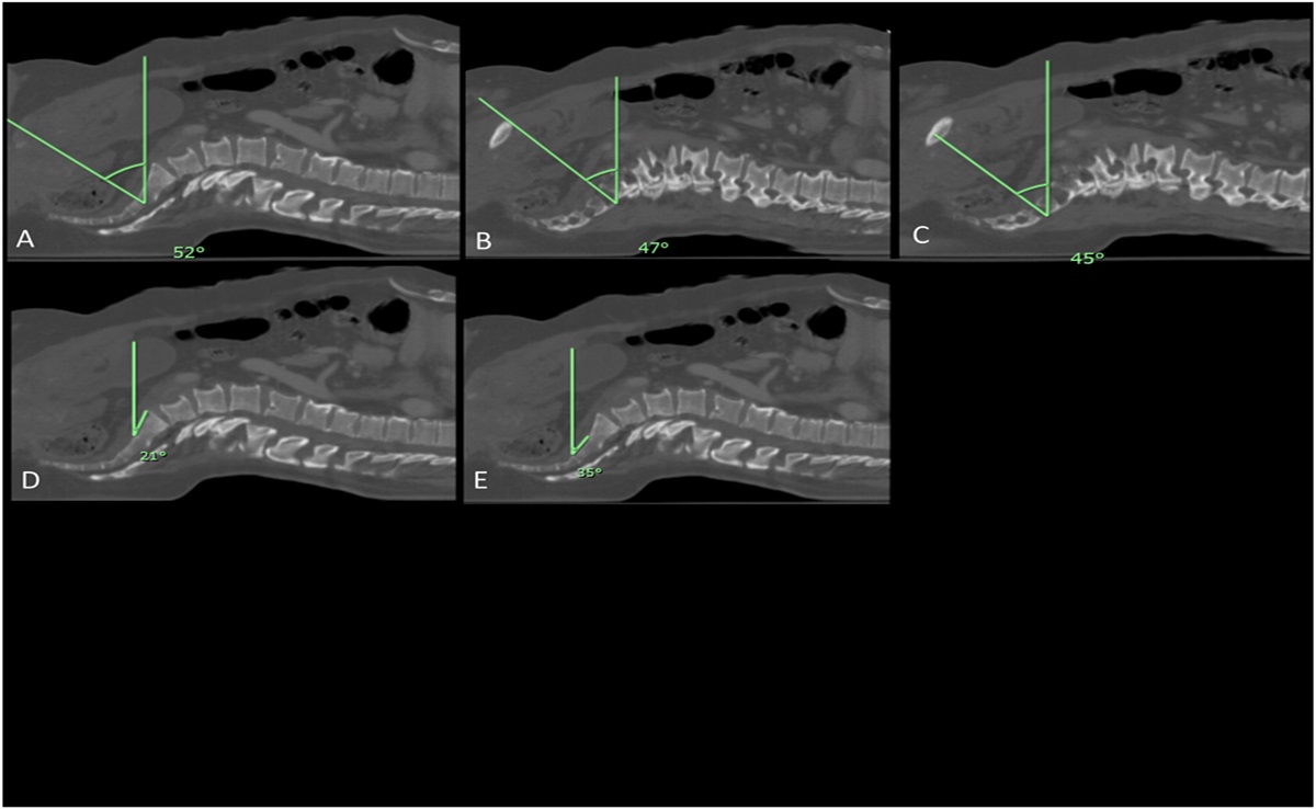
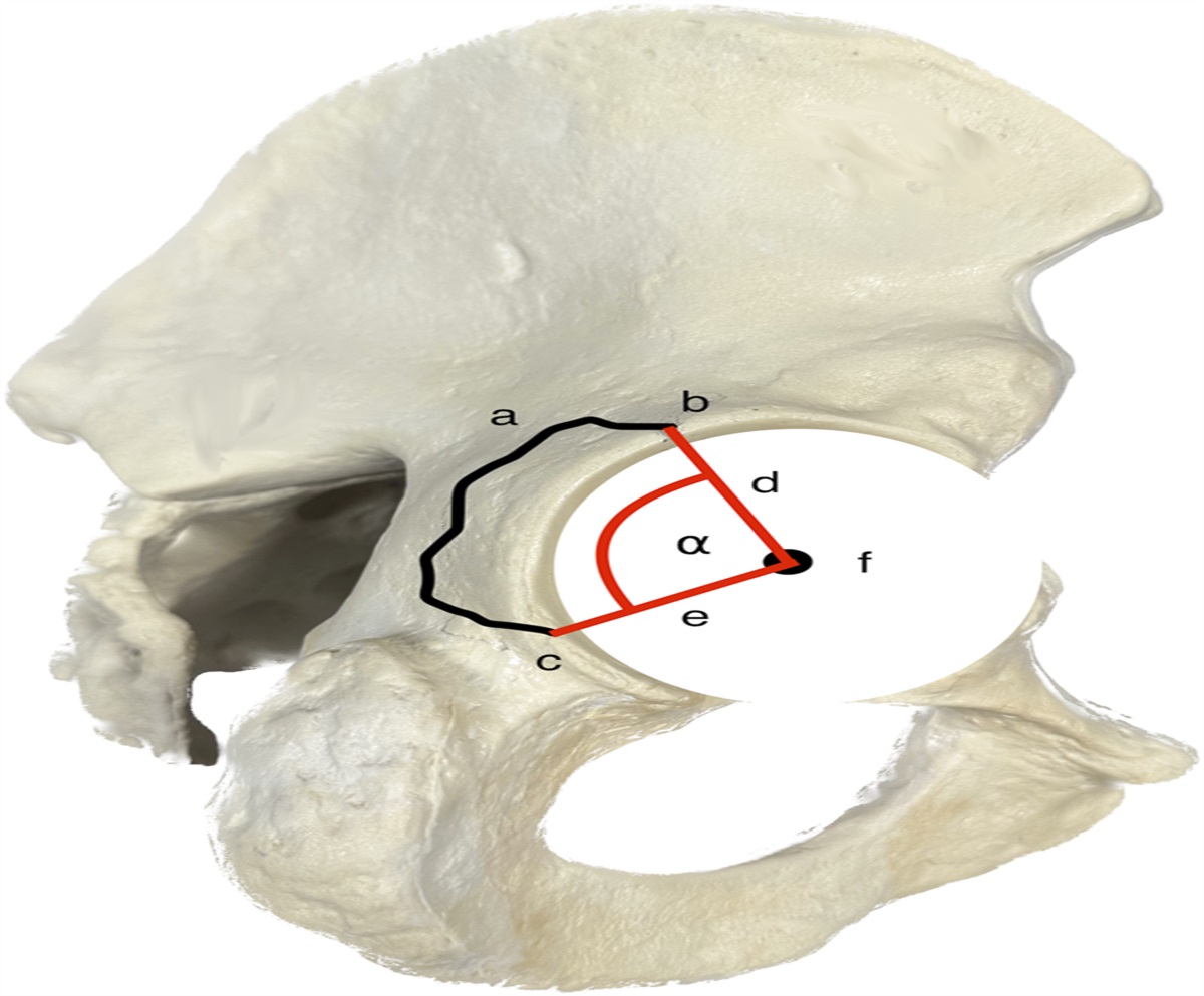
A new sagittal CT measure of PWF based on the angle subtending the joint center, cranial and caudal fracture exits.
Outcome Measures and Comparisons:
Hip incongruity or dislocation demonstrated using gold standard test, examination under anesthesia (EUA), or instability on static images. Prediction of hip instability using a sagittal CT angular measure based on cranial and caudal fracture exits was compared with previous axial CT measures suggestive of increased risk for instability including posterior wall size >50%, and those with cranial exit within 5.0 mm of the acetabular dome.
RESULTS:
There were 32 operative and 26 nonoperatively treated fractures. Thirty fractures were determined to be unstable, and 28 were stable after EUA. Measurements of >70 degrees using the sagittal CT angular measure predicted instability in 28 of 28 patients, and ≤70 degrees predicted stability in 30 of 30 patients (sensitivity 100% and specificity 100%). Prevalence of EUA confirmed instability for subgroups with PWF based on prior axial CT measures were as follows: ≥50% wall involvement (11/16; sensitivity 67% and specificity 60%; 95% CI, 45%–89%/45%–75%), fracture within 5.0 mm of dome (5/18; sensitivity 86% and specificity 73%; 95% CI, 71%–100%/59%–87%), fracture within 5.0 mm of dome and ≥50% involvement (1/9; sensitivity 89% and specificity 56%; 95% CI, 69%–100%/24%–88%).
CONCLUSIONS:
In a sample of 58 mostly high energy posterior wall fractures all having had an EUA, a new sagittal angular CT measurement of ≤70 degrees predicted hip stability and >70 degrees predicted instability with 100% sensitivity and specificity.
LEVEL OF EVIDENCE:
Diagnostic Level III. See Instructions for Authors for a complete description of levels of evidence.


留言 (0)