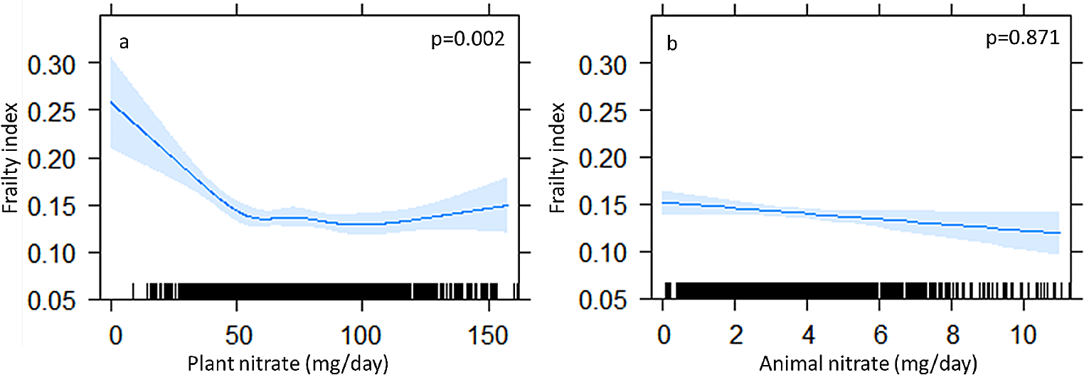
Study participants
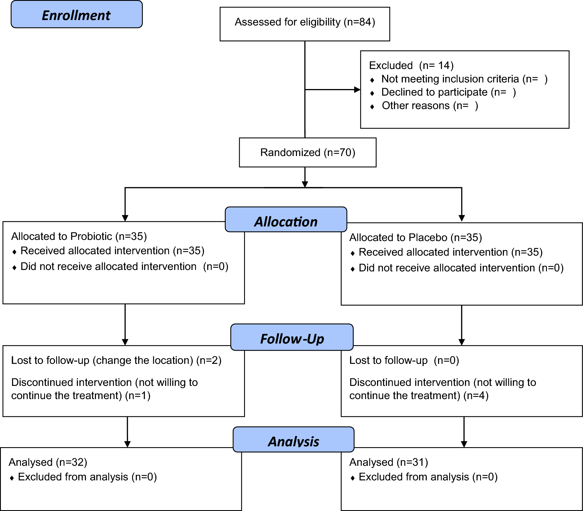
Forty-four older men and women aged between 60 and 75 with elevated SCF were recruited using online advertisements, posters in public buildings within Maastricht, and among participants who had taken part in prior studies. Interested candidates were invited for a screening visit to the university if they indicated that they had a BMI between 25 and 35 kg/m2 (overweight or obese), were right-handed, did not have any chronic medical conditions such as diabetes, hypertension, and active cardiovascular disease (CVD), and noticed that they were experiencing symptoms of cognitive decline. Anthropometrics, blood pressure, and fasting blood samples were taken during the screening visit. We utilized the Cognitive Failure Questionnaire (CFQ) [1, 26] to assess the presence of elevated SCF. This 25-item questionnaire evaluates the frequency of everyday cognitive errors based on a score of 1 (very often) to 5 (never). A cut-off value of ≥ 40 was used to define elevated SCF, which corresponds to approximately 23% of the older population [26]. In addition to the foregoing criteria, included participants needed to have a fasting plasma glucose < 7.0 mmol/L, serum total cholesterol < 8 mmol/L, serum triacylglycerol < 4.5 mmol/L, systolic blood pressure < 160 mmHg, diastolic blood pressure < 100 mmHg and a stable body weight. They also had to be willing to give up being a blood donor 8 weeks before and during the study, and 4 weeks after completion. Based on a questionnaire, they also needed to indicate that they did not possess any contraindications for an MRI scan. All study participants provided written informed consent before the start of the screening visit, and the study protocol was approved by the Medical Ethical Review Committee of the Maastricht University Hospital and Maastricht University (METC azM/UM) (NL75618.068.20) and registered at ClinicalTrials.gov in January 2021 as NCT04831203. This study has been performed in accordance with the ethical standards laid down in the 1964 Declaration of Helsinki and its later amendments and was conducted from May 2021 until October 2022.
Study design
A 36-week double-blind, randomized, controlled parallel study was performed. The randomization was performed by an independent researcher using WinPepi Etcetera software, stratified for sex. The sachets were packaged in numbered boxes, aligning with a randomization process of which the investigators were unaware. These boxes were then dispensed by M.S.A. All study outcomes were performed at two baseline test days and identical follow-up visits. These measurements included anthropometrics (height, weight, body mass index [BMI], waist and hip circumferences, and skinfold measurements to determine body fat percentage [27]), cognitive performance (CANTAB) and brain vascular function (CBF). At weeks 9, 18, and 27, the participants revisited the university to obtain additional supplies and to return the empty sachets in order to check compliance. Moreover, food frequency questionnaires (FFQ) were filled out during baseline and follow-up visits to evaluate dietary intake during the previous 4 weeks [28]. They were instructed not to perform any strenuous physical exercise or consume alcoholic beverages 48 h before the test days, and to fast for 12 h before blood sampling. Moreover, to standardize measurements, participants were asked to come to the university by public transport or car instead of walking or biking. All measurements were performed by the same investigator (M.S.A.), at the same time of day, and at the same location (Metabolic Research Unit [MRUM] and the Scannexus research facilities in Maastricht). Finally, study participants were requested to record in study diaries any protocol deviations or changes in their health status, medication use, and alcohol intake.
Study products
Participants either received 5.7 g of the egg-protein hydrolysate product (NWT-03; Newtricious R&D, ‘s-Hertogenbosch, The Netherlands) or 5.7 g of a maltodextrin placebo in dry powder sachets to mix with 200 mL of water and to consume every day in the morning before breakfast. In addition, the spray-dried egg-protein hydrolysate (NWT-03; Nizo Food Research, Ede, The Netherlands) contained citric acid, flavoring, acesulfame K, sucralose, and quinine HCL. The matching placebo consisted of maltodextrin from potato starch, flavoring, citric acid, cloudifier, tartaric acid, malic acid, acesulfame K, sucralose, caramel E150a, and quinine HCL. The products were similar in color and taste (lemon flavor) to ensure the double-blind research design and packaged in a box that was labeled according to Good Manufacturing Process (GMP) guidelines.
Cognitive performance
In line with our prior studies [6, 29], cognitive performance was assessed using the fully automated CANTAB software on a digital touchscreen tablet (iPad, 5th generation; Apple). Written and verbal instructions were provided by the investigator to familiarize the participants with the tablet before proceeding to the cognitive tasks. In addition, the software offered further guidance and practice trials to ensure their understanding and proficiency in using the tablet.
To investigate psychomotor speed, the reaction time task (RTI) was performed, and the resulting reaction time (ms) and movement time (ms) were evaluated as outcome parameters. The executive function domain was appraised using the multitasking task (MTT) with incongruency cost (ms), median reaction latency (ms), multitasking cost (ms), and total incorrect errors as the key outcomes. Furthermore, the spatial working memory (SWM) task additionally served to evaluate executive function by analyzing between errors, total errors, and strategy scores. Lastly, the memory domain was examined with a delayed matching to sample (DMS) and paired associates learning (PAL) task. The percentage of correctly answered trials for all delays specified DMS results, whereas PAL was evaluated based on total errors and the first attempt memory score. A more detailed description of these tests has previously been provided [6].
Brain vascular function
MRI with ASL scans were performed on a Siemens 3.0 Tesla Magnetom Prisma Fit scanner with a 64-channel head coil. Detailed information regarding the acquisition and processing of MRI data has formerly been published [6, 29]. In short, participants remained in the supine position for an approximately 20-min acclimatization period during which an MPRAGE (T1 image) structural scan was acquired, and a labeling plane was situated based on an angiogram perpendicular to the vertebral and carotid arteries. Afterwards, pseudo-continuous arterial spin labeling (pCASL) with spin echo readouts and background suppressed segmented 3-D gradients were performed. Over the course of the 9-min sequence, parameters included a TR 4050 (repetition time), TE 13.6 ms (echo time), GRAPPA 2, labelling duration 1750 ms, post-labelling delay 2000 ms, and ten label-control repetitions.
Motion correction was automatically performed on the Siemens scanner. To perform brain extraction and tissue segmentation for the anatomical MPRAGE image, Volbrain [30] was used. Quantitative CBF images were estimated from the ASL data using FSL software (Version 6.0) [31], and perfusion-weighted images were generated by pairwise subtraction of label and control images on the pCASL data. Based on the recommendations of the ASL White Paper [32], the BASIL tool (v 4.0.15) [33] quantified perfusion-weighted images. Using boundary-based registration to the brain extracted MPRAGE image by the FLIRT routine [34], the calibrated ASL images with absolute CBF values were co-registered. Gray matter, global brain, and cortical and subcortical CBF values were also determined using the MPRAGE scan. Subsequently, voxel-wise comparisons [35] were conducted by first registering the absolute CBF values to MNI (2 mm) space using a nonlinear algorithm (FNIRT). Due to a high signal-to-noise ratio (SNR), smoothing was not performed and nonparametric permutation testing with a threshold-free cluster enhancement within FSL was used to determine significantly different clusters. These clusters were determined from applying an unpaired t-task with a single-group a paired difference (FMRIB’s Local Analysis of Mixed Effects stage 1 (FLAME) [36]), voxel connectivity of 26, and a z-threshold of 2.3 (p value < 0.05). Based on the smoothness estimates, family-wise corrections for multiple comparisons were performed. The Harvard–Oxford subcortical structural atlas and Atlasquery function were used to determine the average probability of the cluster locations.
Brain-derived neurotrophic factor
At baseline and follow-up visits, fasting serum samples were collected using venipuncture on the forearm. Samples were centrifuged at 1300 × g for 10 min at 21 °C after clotting for approximately 60 min, aliquoted, frozen in liquid nitrogen, and stored at −80 °C. At the end of the study, all samples were analyzed for brain-derived neurotrophic factor (BDNF) concentrations by an enzyme-linked immunosorbent assay (Duo Kit ELISA, R&D Systems, Minneapolis, USA) according to manufacturer’s instructions. The results are reported as the sum of precursor BDNF and mature BDNF because the assay did not differentiate between them.
Statistical analyses
Cognitive performance and CBF were the primary study outcomes for this NWT-03 intervention. Power calculations were conducted for an analysis of covariance (ANCOVA) with baseline measures of the outcomes as a covariate. It was calculated that 18 participants per group were needed to detect clinically relevant changes of at least 5% in reaction latency within the executive function domain (MTT task). This power calculation was based on our prior trial using a between-subject variability of 10%, correlation coefficient of 0.75 [29], > 80% power, and two-sided alpha of 0.05. Moreover, a sample size of 18 participants per group was also sufficient to reach a power >80% to detect changes in CBF of at least 10%, using a between-subject variability of 18%, correlation coefficient of 0.68 [37,38,39], and two-sided alpha of 0.05.
Using the Shapiro–Wilk test, all variables were normally distributed. A one-way ANCOVA, using the baseline measurements of the outcome variables as covariates and intervention as a fixed factor was conducted to determine differences in responses between intervention and placebo groups. In preliminary analyses, we incorporated age as a potential effect-modifier into the ANCOVA model. However, this factor did not affect the outcomes and was, therefore, omitted from the final model. Furthermore, post-hoc analyses using a sex × intervention interaction as a fixed factor were also performed to test for differential effects between men and women. If the interaction was significant, then we separated the men and women and reran the analysis. If it was not significant, then we removed the interaction term from the final model. All statistical analyses were performed using SPSS (IBM Corp., IBM SPSS Statistics, V27, Armonk, NY, USA) and differences were deemed statistically significant using two-tailed tests at p ≤ 0.05.


留言 (0)