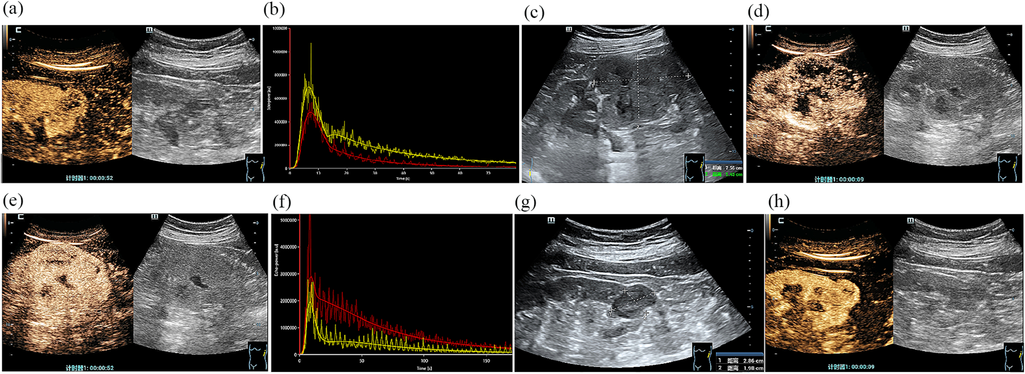
RSAML arises from renal sinus, accounting for quite a small part of renal AML. A growing tumor extends through renal hilum and outward into perinephric fat. RSAML is a typically complex renal tumor and often subjected to nephrectomy. With a high risk of long ischemia time, collecting system injury, and postoperative complications, NSS for centrally located renal tumors is technically demanding [18]. Even for experienced surgeons, it is quite a challenging work to perform NSS for these specially located tumors. Firstly, RSAML has a deep location, occupies renal sinus and compresses renal labium. There is little working space when handling the inner part. Secondly, renal vessels and collecting system are compressed by RSAML, resulting in abnormal hilar anatomy. Moreover, RSAML itself is enclosed by renal vessels and collecting system. Thirdly, due to RSAML’s fragility and thin tumor capsule, the tumor is easy to rupture and bleeding during the surgery procedure and leads to unclear operation field. In addition, spontaneous rupture results in giant retroperitoneal hematomas. Obvious tumor adhesion to adjacent organs is formed during the procedure of hematoma absorption, causing great difficulty in separating and exposing the tumor. In patients with highly complex renal tumors or AMLs, tumor complexity showed a good accuracy in predicting surgical outcomes and complications [19, 20]. Tumor size, the nearness to the urinary collecting system, and the involvement of renal sinus were the main predictors [19]. In our study, patients’ RENAL score was as high as 10, indicating that traditional partial nephrectomy may lead to poor surgical outcomes and high-grade complications. Therefore, a well-designed plan is critical in NSS for RSAML.
Here, we show modified robotic NSS as a safe, effective and feasible technique to treat RSAML. Our modified technique differs from traditional technique in two ways. Firstly, the inner part of tumor was grasped into pieces, to make curettage and aspiration more efficient and safer. Secondly, after removing the tumor, the parenchyma defect was not closed with sutures.
For AML, the goals of management are complete removal of the tumor, ameliorating symptoms, reducing risk of spontaneous rupture and renal function preservation as far as possible [21]. Robotic technology is being widely applied in renal surgery. Robotic surgery has the advantage of three-dimensional views, high quality images, flexible angle, and stable operation [22]. Compared with laparoscopic surgery, robotic partial nephrectomy may be a better option for treating large and complicated AML, as it can reduce warm ischemic time and preserve renal function more effectively [23]. Using minimally invasive technique, robotic NSS is performed successfully for malignant renal sinus tumors, but without more positive tumor margins or prolonged ischemia time [24]. Considering the complexity of RSAML, robotic NSS was planned. With clear view of operative field, renal vessels and tumor margins were easily determined. During dissection of the tumor, flexible angle and stable operation facilitated avoiding disruption of the tumor capsule, which could cause rupture and bleeding. During resection of the tumor, tumor among blood vessels and collecting system was accurately recognized and grasped into pieces.
The renal vessels and branches were dissected carefully. With sufficient exposure of them, the boundary between the tumor and the normal tissue was clearly identified, and the risk of injury and intraoperative blood loss were reduced and well-controlled. Injury to renal vessels leads to intraoperative and postoperative complications, such as bleeding and renal atrophy. Preoperative embolization could reduce blood loss and reduce tumor bulk during surgery [25, 26]. However, when confronted with AML arising from renal sinus, determining nutrient arteries of RSAML is difficult, as the direction of these vessels is parallel with that of renal hilar vessels. Therefore, we did not routinely perform a robotic NSS with preoperative embolization, but selectively clipped renal artery branch to reduce bleeding during the operation.
After blockage of renal artery and resection of the outer part of tumor, removal of the inner part in renal sinus was one the challenges of the operation. For skilled surgeons, a longitudinal incision is made along the Brodel line to remove malignant renal tumors. However, the total warm ischemia time is much longer [24]. AML is composed of varying proportions of fat tissues, smooth muscle, and blood vessels, which make it quite fragile. Previous studies indicate that laparoscopic curettage and aspiration could remove renal AML and avoid injury to the renal parenchyma and collecting system, especially for central renal AML [10, 27]. However, it was difficult to remove tumor among blood vessels and collecting system, in the condition of limited working space in renal sinus and abnormal hilar anatomy. Rough aspiration may cause mechanical injury. In our study, the inner part was bluntly grasped into pieces, to make curettage and aspiration more efficient and safer. These blunt procedures had little effect on renal artery and collecting system, as they had thick tube walls. However, venous wall was quite thin, and was prone to be injured. According to our experience, renal vein injury could be managed with sutures, to avoid excessive bleeding. In addition, this technique was an effective method of shortening the operation time, as it could fully expose small vessels at the base and stop bleeding points. A recent clinical study [28] reported that median warm ischemia time of robotic NSS in the treatment of central AML was only 21.50 min; this result is somewhat shorter than the warm ischemia time we reported, and we believe that this is mainly attributable to the much larger tumor size in our study(7.18 vs. 5.20 cm). Our single-center experience revealed the safety and effectiveness of the modified technique. We did not experience any conversion to open surgery or nephrectomy. One patient needed intraoperative blood transfusion. That surgery was performed at the beginning of our exploration of the modified robotic technique. No other intraoperative complications occurred.
Packing absorbable haemostat into the tumor bed and keep it tightly compressing parenchyma defect could achieve satisfied haemostasis [10]. In our study, several layers of absorbable gelatin sponges were applied and packed into the wound fossa, in order to reduce blood loss and drainage duration. The parenchyma defect was not closed with sutures, which may injure or obstruct renal vessels and collecting system. Compression and ischemic injuries to the renal parenchyma were avoided. The dosage of absorbable gelatin sponges depended on the size and depth of the wound fossa. Then total compression for a few minutes was carried out to ensure hemostasis. This procedure worked in case of small blood vessels injury or renal parenchymal hemorrhage. After the bulldog clamp was removed, renal hilum was inspected for hemostasis with low pneumoperitoneum pressure. Continued bleeding from sinus may be attributed to injury of tumor’s nutrient artery. Based on bleeding location, suspicious renal artery branch was clamped experimentally. When it did work, selective artery clipping was performed. The intraoperative blood loss was comparable to previous studies of surgery strategies for AML [6, 13] and no postoperative hemorrhage was detected, demonstrating that strategy for hemostasis was effective.
There are also some limitations in our study, including lack of control group, its retrospectively nature, small sample size and limited follow-up period. All the patients are treated in a single center, and a further prospective multi-center study is needed.


留言 (0)