Preservation of residual acoustic hearing following cochlear implantation is highly valuable but also highly variable, even with relatively short electrodes: post-implantation losses have been reported in up to ∼1/3 of cases (Lenarz et al., 2013; Roland et al., 2016). Some patients lose residual hearing acutely, presumably due to direct surgical trauma. These mechanisms include osseous spiral lamina fracture, spiral ligament perforation, disruption of the basilar membrane and organ of Corti, and, rarely, direct disruption of the modiolus (Eshraghi et al., 2003; Geerardyn et al., 2023; Nadol and Eddington, 2006). Of patients with initially preserved acoustic hearing, approximately 30-40% suffer delayed loss over weeks, months, or years (Gantz et al., 2018). Although acoustic-electric hearing rehabilitation is no longer possible, these patients typically still benefit from the electric hearing via the implant. Because the histopathology of the human inner ear can only be assessed at the cellular level using autopsy material, the reasons for post-implantation hearing loss, and especially delayed hearing loss, are poorly understood.
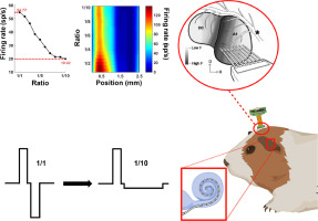
A particularly interesting case of post-implantation delayed hearing loss was described in a prior report (Quesnel et al., 2016). The patient had adult-onset bilateral, slowly progressive, down-sloping sensorineural hearing loss, likely due to genetic factors and occupational noise exposure history from factory work. He underwent left cochlear implantation with a 10 mm electrode (Iowa/ Nucleus Hybrid S8, Cochlear Corporation, CO, USA) at age 63. At the time of the implantation surgery, his audiogram showed sharply down-sloping thresholds bilaterally, ranging from normal at 250 Hz to profound loss at 3000 Hz and above, and his word recognition scores were 40% and 24% in the right and left ears, respectively (Figure 1). Over the course of the next 4.5 months, he experienced a near-total loss of acoustic hearing sensitivity in the implanted ear (Figure 1) and died of unrelated causes 7 years later. Utilizing classic and well-established quantification techniques (Merchant and Nadol, 2010), the prior study of the temporal bones in this case concluded that there were no remarkable interaural differences in the loss of inner or outer hair cells or the survival of spiral ganglion cells (SGCs). Since the most notable finding in the implanted ear was the deposition of the fibrous tissue in the scala tympani and vestibuli, obstruction of the round window, new bone growth in the scala tympani, and endolymphatic hydrops, speculation focused on changes in cochlear mechanics as an explanation for the loss of low frequency hearing, although the degree of hearing loss could not be fully accounted for.
Several years ago, we began a large-scale re-evaluation of prior pathological studies of human temporal bones in the Mass Eye and Ear collection, as a rigorous test of the existing dogmas surrounding the functionally important structural changes in presbycusis (Wu et al., 2020a). As part of that re-evaluation, we concluded, using high-power light microscopy, that prior studies of H&E stained celloidin sections, across many laboratories, failed to detect significant fractional losses of both inner and outer hair cells (Wu et al., 2020b). The difference in the resulting analyses were not subtle, as is illustrated by one example in Figure 2. The striking differences changed the view of the functionally important structural changes underlying sensorineural hearing loss in important ways.
There are two reasons why the classic approach to hair cell counting in sectioned temporal bones underestimated the amount of hair cell loss. First, the classic methods relied largely on evaluating cell nuclei (Merchant and Nadol, 2010), rather than cuticular plates and hair bundles (which can be clearly seen using differential interference contrast (DIC) imaging), and surviving supporting cell nuclei can easily be confused with hair cells, particularly in the inner hair cell area (Wu et al., 2020b). Second, the classic methods “binarized” the hair cell counts, considering the hair cell survival in each view of the sensory epithelium as either 100% or 0% (Merchant and Nadol, 2010), whereas, with high-power DIC, the observer can optically section the tissue and assess the fraction of hair cells surviving in each row (Wu et al., 2020b).
As part of this histopathological re-evaluation, we re-examined the two ears of this interesting hybrid implant case and concluded that the prior analysis failed to note a dramatic loss of inner hair cells on the implanted side that likely contributed to the delayed loss of residual hearing in this case. Current results also suggest that spiral ganglion cell survival was slightly reduced on the implanted side and that many surviving spiral ganglion cells in both ears were lacking peripheral axons.


留言 (0)