
記住我
Complex regional pain syndrome (CRPS) is a chronic pain condition that is debilitating and dramatically decreases the quality of life. CRPS is characterized by a constellation of sensory, motor, and autonomic dysfunction.1 CRPS occurs as type 1 (from the known noxious event that commonly involves immobilization with no identifiable nerve injury) or type 2 (distinct nerve injury). CRPS differs from other chronic pain conditions: neuropathic such as radicular pain or postherpetic neuralgia or generalized nonspecific pain conditions such as fibromyalgia, in which pain is associated with obvious and distinct alterations in sensory processing, sympathetic nervous involvement, and motor dysfunction. An approach to treating CRPS is through the induction of neuroplasticity via repetitive transcranial magnetic stimulation (rTMS). This form of stimulation has advantages over other forms of neuromodulation such as spinal cord stimulation, as rTMS is noninvasive and relatively low risk.2 High-frequency rTMS (10 Hz) applied over the primary motor cortex reduces pain from CRPS.3,4
CRPS is also associated with structural and functional changes in the brain areas responsible for motor control and sensory processing.5 Specifically, cortical representations of the affected limb in the somatosensory cortex are smaller and possess greater overlap with adjacent areas.6 This is speculated to be a result of a decrease in afferent input originating from the periphery and is reinforced by minimal usage of the affected limb to avoid pain.5 Therefore, a sensorimotor training (SMT) task that stimulates afferent input originating from the periphery may aid in reinstating somatosensory cortical territory and promote motor function. The beneficial effects of SMT may be enhanced using rTMS. Specifically, rTMS delivered to the primary motor cortex may create an environment within the sensorimotor cortex that promotes neuroplasticity through increased cortical excitability.7 This in turn promotes intraneuronal connectivity and reorganization achieved through sensorimotor integration from SMT.
The objective of this case report is to investigate the use of rTMS combined with SMT in a patient presenting with CRPS type 2. This article adheres to the applicable Enhancing the QUAlity and Transparency Of Health Research (EQUATOR) guidelines. This study was approved by the Hamilton Integrated Research Ethics Board (protocol number 14029) and written informed patient consent was obtained for the publication of this case report.
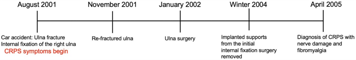
CASE DESCRIPTIONThe participant is a 42-year-old woman diagnosed with CRPS type 2 in the right wrist and hand due to a motor vehicle accident and subsequent ulna fracture that occurred in August 2001 (Figure 1).
 Figure 1.: Timeline of participant medical history. Diagnosis of CRPS type 2 was confirmed by a pain specialist and coauthor (H.S.) using the modified Budapest criteria.8 In addition, the patient’s diagnosis was confirmed based on previous medical notes made during her initial diagnosis of CRPS type 2 in 2005. Specifically, in this report, it was noted that the patient had a history of injury that involved her ulnar nerve. In this study, her clinical symptoms were noted to be in her ulnar nerve territory. CRPS indicates complex regional pain syndrome.
Past treatments included physiotherapy, opioids, anticonvulsants, and acupuncture, with no particular success with individual treatments. The participant was prescribed fentanyl from 2005 to 2019. Past interventions did not provide significant relief of CRPS-related symptoms. For the duration of the study, medications maintained included Pregabalin, Topiramate, Celecoxib, Duloxetine HCL, Oxycodone HCL, and Taro-Diclofenac 1.5% topical.
Diagnostic AssessmentThe participant presented with allodynia and hyperalgesia of the right forearm, wrist, and hand, changes to skin color (red and mottled), temperature (hot or cold to touch), edema, and trophic changes (nails on the right hand were more brittle and hair was darker on right arm). This was accompanied by motor changes including reduced range of motion of the wrist and digits, stiffness due to swelling, and pain during movement. Participant also presented with perceptual changes of limb position in space and dysesthesia (wet sensations and burning). Participant noted that they were unable to perform basic activities of daily living that required their right hand and arm. Pain was defined as neuropathic in nature based on the douleur neuropathique en 4 questions (DN4) and leeds assessment of neuropathic symptoms and signs (S-LANSS) criteria.
This was an open-label study involving an intervention comprised of an induction and tapering periods (Figure 2). rTMS is unlabeled for the use under discussion and is still investigational. The induction period consisted of 5 sessions per week of rTMS+SMT for 4 weeks (20 sessions total). The tapering period involved 5 sessions of rTMS alone delivered over 5 weeks (week 1: 2 sessions, weeks 2 and 3: 1 session, week 4: no session, and week 5: 1 session). There was no session in week 4 to extend the time between sessions. The goal of the tapering period is to prolong the effects achieved during the induction period. The outcomes of this study followed the Core Outcome Measures for Complex Regional Pain Syndrome Clinical sTudies (COMPACT) recommendations. Pain intensity, pain catastrophizing, wrist and hand function, and Patient-Reported Outcomes Measurement Information System 29-Item Profile (PROMIS-29 v2.0) were assessed at T0 to T4. Patient global impression of change questionnaire (PGIC) was assessed at T1, T2, T3, and T4. Allodynia using the rainbow pain scale was assessed at T0, T2, and T3.
rTMS was delivered over the hand representation of the primary motor cortex using a Magstim Rapid 2 stimulator (Magstim) with a 10 Hz protocol used elsewhere.9 This required ~11.5 minutes of stimulation and was delivered immediately in advance of the SMT. SMT used a custom-built hand device (Figure 3). The participant experienced a brief nerve stimulation to the skin of digits 2 to 5 of the right hand. This prompted the individual to move a slider that controlled their digit location to match a target on the custom-built user interface (Figure 3). Each training session required ~20 minutes in which 80 trials were performed with the CRPS-affected hand. Movement complexity was increased during the 4 weeks of training.
ResultsThe participant completely adhered to the intervention by attending all sessions outlined in the protocol and tolerated rTMS and SMT as there were no adverse effects. Figure 4 plots the pain intensity scale, pain catastrophizing, and wrist and hand evaluation. Figure 5 shows the changes in the PROMIS-29 v2.0 profile. The rainbow pain scale is presented in Figure 6. Using the PGIC, the participant indicated minimally improved at T1 and much improved at T2, T3, and T4.
 Figure 2.:
 Experimental timeline. *Indicates the number of experimental sessions per week.
 Figure 3.:
 SMT setup. The right hand was rested on the hand device such that each digit (2–5) was rested on a slider. These sliders allowed the participant to control the moving line on the user interface. SMT indicates sensory motor training.
 Figure 4.:
 Changes in pain intensity, pain catastrophizing, and hand and wrist evaluation across baseline. Higher scores are associated with worse outcomes in all 3 scales.
 Figure 5.:
 PROMIS-29 v2.0 profile. Outcomes included the PROMIS-29 v2.0 profile. Outcomes are reported relative to the reference population using T-scores. T-score values are rated as within normal limits, mild, moderate, and severe. PROMIS-29 v2.0 indicates Patient-Reported Outcomes Measurement Information System 29-Item Profile.
 Figure 6.:
 Rainbow pain scale and clinical interpretation. Changes in pain perception across the study. Colors represent the severity of allodynia-type pain in 4 locations measured on the dorsal side and 3 locations measured on the ventral side of the hand. Each area of the hand was divided based on its innervation by either the MN or UN. “P” indicates the smallest monofilament number that elicited pain. “S” indicates the smallest monofilament number that elicited touch sensation, but no pain. No color indicates no pain was elicited in that area (no allodynia). Only the affected hand (right hand) is illustrated. Rainbow pain scale colors and clinical interpretation are provided. MN indicates median nerve; UN, ulnar nerve.
DISCUSSIONThis case report describes the use of rTMS and SMT as a treatment for CRPS. 10 Hz rTMS applied over the hand representation of primary motor cortex combined with SMT of the right hand was effective in reducing the magnitude of pain intensity during the treatment period (~30% reduction) and at 3 months after intervention (~40% reduction). This effect is consistent with previous reports in CRPS which cite a change of 22% to 50% after 10 Hz rTMS.3,4,10,11 Importantly, pain intensity continued to improve at 3 months after intervention.
Pain reduction was associated with improvements in allodynia of the right hand and wrist such that there was an abolishment of allodynia on the ventral and dorsal surfaces of the hand and a reduction in the forearm from serious (2.44) to significant (4.56). This is the first report in CRPS to demonstrate an improvement in allodynia from rTMS and SMT.
This is the first study in CRPS to show that rTMS and SMT improve the physical function of the affected limb. Wrist and hand function improved such that there was a 22-point reduction in hand and wrist evaluation score immediately after the intervention and 12.5-point reduction from baseline to 3 months after intervention, which exceeds the minimal clinically important difference of 12 points for this measure.12 Although the mechanism for improvement in physical function are not fully understood, SMT may act to reinstate the sensorimotor cortical representation of the affected limb. This may have been further reinforced by preceding rTMS which increased the propensity for plasticity after SMT. rTMS and SMT may be more effective than graded motor imagery and multimodal physiotherapy, which has demonstrated low to very low-quality evidence for improving physical disability in CRPS.13
Similarly, improvements in pain catastrophizing revealed a reduction of 70% immediately after the intervention and a 20% reduction 3 months after intervention compared to baseline. Previous reports in CRPS have shown a 31% decrease in pain catastrophizing after graded motor imagery and transcranial direct current stimualtion.14 rTMS has also been shown to improve pain catastrophizing by 33% in individuals with fibromyalgia.15
PROMIS-29 v2.0 reported outcomes indicated that no change from the intervention reached normal levels however, there were improvements in pain interferences (moderate to mild), fatigue (moderate to moderate/mild), depression (moderate to mild), ability to participate in social roles (moderate to mild), and physical function (moderated to mild) from baseline to 3 months after intervention.
A limitation of this study is the ability to assess whether improvements in pain outcomes are equally contributed to by the rTMS and SMT. It is likely that each intervention provides benefits, and their summative effects are only examined herein. Further, a placebo was not included.
CONCLUSIONSThe lack of viable treatment options for patients with CRPS underscores the importance of identifying new effective options for this growing population. This case report demonstrates that CRPS type 2 of the upper limb may be treated using a combined approach of rTMS and SMT to alleviate pain and improve physical function, and quality of life. The participant in this study indicated an improvement from the intervention stating, “I have been able to pick up my 4-year-old son again,” “I can spend more time doing the things I enjoy,” and “emotionally, I don’t feel so helpless.” As such, this initial evidence is promising and suggests that rTMS combined with SMT for CRPS should be further explored on a larger scale.
DISCLOSURESName: Stevie D. Foglia.
Contribution: This author helped in conception of the study, acquisition, analysis, interpretation, drafting, and revising the article.
Name: Chloe C. Drapeau.
Contribution: This author helped in data acquisition and revising the article.
Name: Ravjot S. Rehsi.
Contribution: This author helped in data acquisition and revising the article.
Name: Karishma R. Ramdeo.
Contribution: This author helped in data acquisition and revising the article.
Name: Harsha Shanthanna.
Contribution: This author helped in conception of the study, interpretation, drafting, and revising the article.
Name: Aimee J. Nelson, PhD.
Contribution: This author helped in conception of the study, interpretation, drafting, and revising the article.
This manuscript was handled by: Christina L Jeng, MD, FASA.
REFERENCES 1. Gierthmühlen J, Binder A, Baron R. Mechanism-based treatment in complex regional pain syndromes. Nat Rev Neurol. 2014;10:518–528. 2. Sun L, Peng C, Joosten E, et al. Spinal cord stimulation and treatment of peripheral or central neuropathic pain: mechanisms and clinical application. Neural Plast. 2021;2021:1–9. 3. Pleger B, Janssen F, Schwenkreis P, Völker B, Maier C, Tegenthoff M. Repetitive transcranial magnetic stimulation of the motor cortex attenuates pain perception in complex regional pain syndrome type I. Neurosci Lett. 2004;356:87–90. 4. Picarelli H, Teixeira MJ, De Andrade DC, et al. Repetitive transcranial magnetic stimulation is efficacious as an add-on to pharmacological therapy in complex regional pain syndrome (CRPS) Type I. J Pain. 2010;11:1203–1210. 5. Echalier A, Borg C, Creac’h C, Laurent B, Michael GA. Spontaneous sensations reveal distorted body perception in complex regional pain syndrome (CRPS). Brain Cogn. 2020;142:105568. 6. (Karin) Swart CMA, Stins JF, Beek PJ. Cortical changes in complex regional pain syndrome (CRPS). Eur J Pain. 2009;13:902–907. 7. León Ruiz M, Rodríguez Sarasa ML, Sanjuán Rodríguez L, Benito-León J, García-Albea Ristol E, Arce S. Current evidence on transcranial magnetic stimulation and its potential usefulness in post-stroke neurorehabilitation: opening new doors to the treatment of cerebrovascular disease. Neurologia. 2018;33:459–472. 8. Harden RN, Oaklander AL, Burton AW, et al. Complex regional pain syndrome: Practical diagnostic and treatment guidelines, 4th edition. Pain Med. 2013;14:180–229. 9. Foglia SD, Rehsi RS, Turco CV, Shanthanna H, Nelson AJ. Case report: the feasibility of rTMS with intrathecal baclofen pump for the treatment of unresolved neuropathic pain following spinal cord injury. Front Rehabil Sci. 2022;3:893014. 10. Gaertner M, Kong JT, Scherrer KH, Foote A, Mackey S, Johnson KA. Advancing transcranial magnetic stimulation methods for complex regional pain syndrome: an open-label study of paired theta burst and high-frequency stimulation. Neuromodulation. 2018;21:409–416. 11. Delon-Martin C, Lefaucheur JP, Hodaj E, et al. Neural correlates of pain-autonomic coupling in patients with complex regional pain syndrome treated by repetitive transcranial magnetic stimulation of the motor cortex. Neuromodulation. 2023;27:188–199. 12. MacDermid JC, Tottenham V. Responsiveness of the Disability of the Arm, Shoulder, and Hand (DASH) and Patient-Rated Wrist/Hand Evaluation (PRWHE) in evaluating change after hand therapy. J Hand Ther. 2004;17:18–23. 13. Smart KM, Ferraro MC, Wand BM, O’Connell NE. Physiotherapy for pain and disability in adults with complex regional pain syndrome (CRPS) types I and II. Cochrane Database Syst Rev. 2022;5:1–169. 14. Lagueux E, Bernier M, Bourgault P, et al. The effectiveness of transcranial direct current stimulation as an add-on modality to graded motor imagery for treatment of complex regional pain syndrome a randomized proof of concept study. Clin J Pain. 2018;34:145–154. 15. Guinot M, Maindet C, Hodaj H, et al. Effects of repetitive transcranial magnetic stimulation and multicomponent therapy in patients with fibromyalgia: a randomized controlled trial. Arthritis Care Res (Hoboken). 2021;73:449–458.
留言 (0)