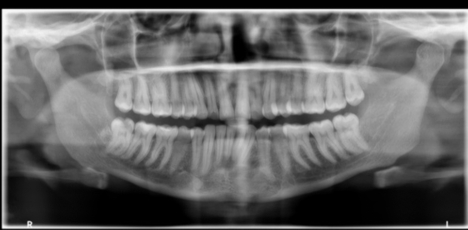
Caries is a common disease worldwide, and bitewing radiographs are most commonly used for caries screening. Nonetheless, bitewing technique is compromised by the fact that projection radiographs are used. Therefore, the novel BW + technique was developed that operates by means of tomosynthesis and enables clinicians to scroll through different layers and angles. The goal of the present study was to compare BW + with 2D- and 3D-alternatives in terms of sensitivity/specificity, reliability, perception of observers, and effective dose.
Analysis of sensitivity revealed that BW + radiographs were associated with a significantly higher sensitivity for C3 lesions, i.e., lesions of the outer dentin. This is of clinical relevance, as C3 caries is a clear indication for invasive treatment, i.e., caries excavation followed by a restauration. Specificity was relatively high as well, especially when compared to CBCT radiographs, which, however, are not frequently used for caries detection in dental practice.
Prior to the scoring, observers expected BW + to be useful for caries detection. After assessment, they reported that they would rate BW + to be more useful than CBCT. All observers stated that they would not prefer BW + over intraoral radiographs and digital sensors, to which they were more familiar. As a reason, lack of experience with the novel technique was frequently mentioned.
A possible reason why the observers did not believe BW + to be of added diagnostic value compared to digital sensors might owe to the discrepancy between their self-rated experience, and the actual caries detection rate. In fact, compared to the µCT reference images, around 70% of caries remained undetected by the observers. It remains unclear to what extent this impacted on the answers in the questionnaire and the caries detection rates. Future studies are needed to elucidate the perception of BW + once observers got familiar with the technique.
Inter-rater reliability showed fair agreement between the observers. Interestingly, clinic experience did not seem to be of relevance in caries detection, as the two observers with the greatest accuracy in caries detection ranged from 1.5 to 36 years of working experience.
Previous studies have shown that up to 90% of carious lesions are solely diagnosed through radiological imaging, while 50% of molars of 12–20 years present visually not detectable dentinal lesions [16,17,18]. In this context, bitewing imaging has been shown to provide extra diagnostic value compared to visual clinical examination [19] and has repeatedly been shown to be of significant diagnostic value [20,21,22].
Digital radiography in particular offers a quick and inexpensive method to evaluate both teeth and bone structure alike. It has also been shown to result in less radiation exposure than conventional radiographic imaging [23,24,25]. However, for every application the cost–benefit ratio of potential health hazards must be considered, especially in children and women during pregnancy [26, 27].
According to the European guidelines on radiation protection in dental radiology, the effective dose of intraoral bitewing radiography amounts to approximately 1–8.3 µSv, roughly a third of dental panoramic imaging (3.85–30 µSv). This translates to a risk of fatal cancer of 0.02–0.6 per million [28]. Two bitewing radiographs, i.e., one for each half of the jaw, therefore, account for approximately 1 day’s worth of background radiation, or a continental flight. For the novel BW + mode and the tested Pat 3/Pat 4 settings, doses were slightly higher than for conventional intraoral radiographs, which amounted to 15 µSv for a full intraoral examination [29]. According to the dose measurements specified in the handbook of the CBCT, effective dose values of BW + were slightly higher than the CBCT LD images (28.2 µSv compared to 4 µSv, Pat 3 setting), but lower compared to CBCT HD images (28.2 µSv compared to 69 µSv maxillary / 71 µSv mandibular, Pat 3 setting). It has to be noted that the values for CBCT are much lower compared to literature, and that no information is given in which region of the jaw they were reported. These higher values can be justified by the benefits of a local third dimension. Nevertheless, the European guidelines recommend no more than six-monthly intervals for posterior bitewing radiographs for high-risk, and annually for patients with moderate caries risk.
Furthermore, European guidelines, as to which intervals are adjusted for low-, moderate-, and high-risk patients, may differ from other international guidelines, as a review by Goodwin et al. indicated [30]. Therefore, careful consideration of individual age, risk, added diagnostic value, and likeliness of treatment alteration through radiography must be taken to justify each decision. As caries takes up to 4 years or longer to invade the dentin through the enamel layer, annual or even biannual radiographic monitoring may not be of additional diagnostic value in case that only initial C1 lesions are present [31, 32], and is, therefore, not indicated for these cohorts of patients.
Even though projection errors occur rarely in bitewing radiography [33], they can impair sensitivity and specificity. However, it must be mentioned that there are other factors impairing bitewing radiographs, such as burn-out artefacts, and whether they are also present in BW + was not investigated in the present study.
In dentistry, cone beam computer tomography (CBCT) has become gold standard for 3D imaging. It has significantly lower radiation exposure when compared to traditional computed tomography [34] and is usually very fast in regard to recording time and image reconstruction. Despite its broad range of indications, it is commonly used in addition to previous 2D-imaging in case that further information is required. Moreover, it requires the practicing dentist to obtain an extra qualification for CBCT in many countries.
Interestingly, the inter-rater reliability was only fair in the present study. Also, the intrarater reliability varied among observers, and only two doctors reached an agreement of more than 85% between day 2 and day 1. This emphasizes that many lesions were not seen by all the observers, as reported previously by another group [35]. Hence, specific trainings for dentists to improve skills in caries detection might be beneficial, as suggested in the field of radiation protection in the EURATOM guidelines [36]. In addition, combination of human and artificial intelligence might improve detection rates in the future [37].
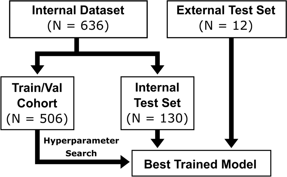
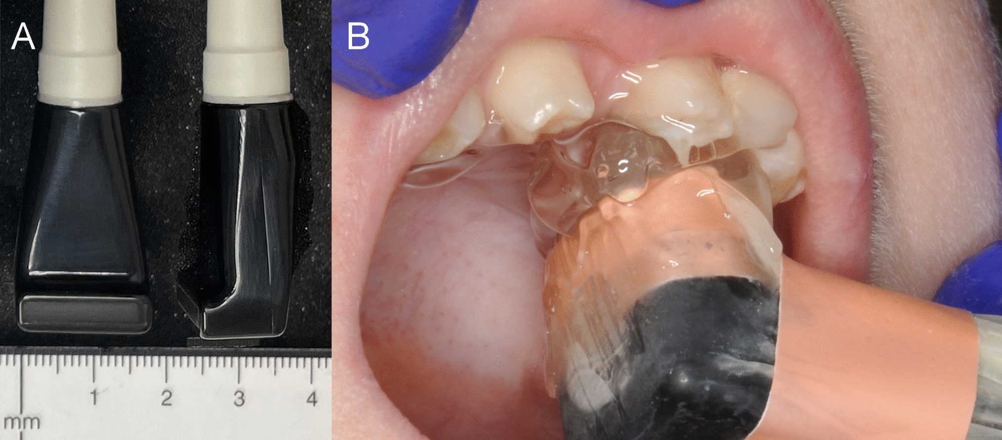
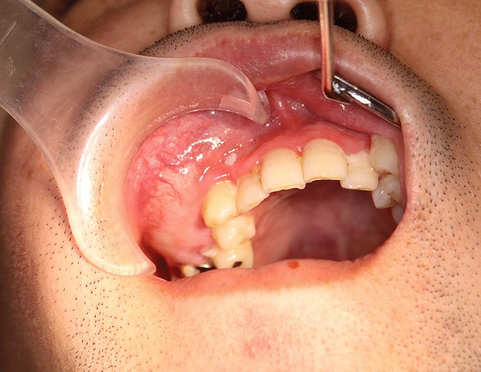
This design of the present study was chosen to simulate clinical reality as much as possible, without the need of exposing patients to hazardous radiation. Nevertheless, a limitation of this study is its ex vivo nature. As only two specimens were in possession of their natural dentition, teeth had to be surgically transplanted into the alveolar bone of three edentulous cadavers. As this procedure was extensive and complex, the respective pre-drilled holes were used for various insertions of tooth combinations. In addition, the high number of carious lesions may not reflect a typical individual, and might have contributed to lower detection rates, as lesions might have been easier overseen by the raters. For imaging plates, it has to be noted that recording was impaired by the low intraoral temperature of the cooled cadavers. Another limitation owes to the fact that not all potential voltage and current combinations could be evaluated. Around a decade ago, a study by Hellén-Halme and Nilsson has shown that a voltage of 70 kV leads to 40–50% higher absorbed dose in patients than 60 kV [38], and another study found that a voltage of 70 kV does not result in a significantly higher sensitivity than 60 kV [39]. This is in line with the sensor recordings of the present study, even though a slight reduction in specificity was found for 60 kV. Regarding the extraoral bite wings, only the most typical dose settings (Pat 3 and Pat 4) were evaluated. Therefore, future studies are needed to evaluate the applicability lower voltage protocols, specifically for extraoral BW and BW + .
It has to be noted that the present study was designed to evaluate whether there is a general benefit of BW + . However, no focus was put on more specific fields of indication, such as its advantages for caries diagnosis in disabled patients, or in patients with strong parapharyngeal reflex. From clinical practice it is known that intraoral radiology techniques are sometimes not applicable in these patients, so extraoral approaches might become a valuable alternative here. Another field in which BW + might be beneficial was mentioned in the questionnaire, where advantages of BW + were seen for patients with posterior crowding. Indeed, clinical reality sometimes requires separate intraoral radiographs for each interproximal contact, so the overall radiation dose would be lower with the BW + technology. Another limitation of the current study is that no differentiation was performed between interproximal and occlusal caries lesions. However, in future studies, it would be interesting to assess the specific benefits of BW + at the different localizations. Furthermore, it has to be noted that calibration only included a group discussion which lesions visible on exemplary radiographs would be represented by the different scores. Despite, additional test was run to validate consistent scoring among observers. However, since all observers were part of the same department and used the scores in clinical routine, a pre-calibration was assumed.
In conclusion, and within the limitations of the present study, BW + displayed increased sensitivity for detection of C3 caries lesions, and slightly higher specificity compared to other 2D- and 3D-imaging methods. These results prove that BW + is suitable for caries diagnostics and also has the potential to become a relevant tool for intraoral bitewing diagnostics in the future. Despite the diagnostic advantages, the risk–benefit ratio should be weighed up individually for each patient according to age and caries risk, as the 3D effect results in increased radiation exposure.

留言 (0)