Reagents
Asiaticoside was obtained from Chengdu Push Biotechnology Co., Ltd. (Sichuan, China) and prepared in dimethyl sulfoxide (DMSO) at a stock concentration of 10 mM. Anti-Semaphorin 4D/CD100, anti-CD72, and anti-GAPDH antibody were purchased from Abcam (Cambridge, USA). Anti-Bax, anti-Bcl-2, NF-κB p65 Rabbit Polyclonal Antibody, and HRP-Goat Anti-Rabbit-IgG (H+L) conjugate were purchased from Beyotime Biotechnology (Shanghai, China). Phospho-NF-κB p65 (Ser536) antibody and caspase 3 antibody were obtained from Cell Signaling Technology (Danvers, USA). DRP1 polyclonal antibody and FIS1 antibody were purchased from Signalway Antibody (Maryland, USA). β-Actin antibody was purchased from Boster (Wuhan, China). TRIzol reagent were gained from Invitrogen (Grand Island, USA). miRNA primer of miR-21, as well as U6, was purchased from Gene Pharma (Shanghai, China). HiScript III All-in-One RT SuperMix Perfect for qPCR and ChamQ Universal SYBR qPCR Master Mix were purchased from Vazyme (Jiangsu, China). IL-10 and IL-1β ELISA kits were obtained from Fankewei (Shanghai, China). Annexin V-FITC (fluorescein isothiocyanate) apoptosis detection kit, mitochondrial membrane potential assay kit with JC-1, reactive oxygen species assay kit, BCA protein assay kit, penicillin-streptomycin solution (100×) were purchased from Beyotime Biotechnology (Shanghai, China). Cell Counting K-8 (CCK8) reagent was purchased from ApexBio Technology (Houston, USA). Fetal bovine serum (FBS) was purchased from Tianhang Biotechnology (Hangzhou, China). Lipopolysaccharide (LPS 055: B5), DMEM high glucose medium, and other reagents were obtained from Sigma-Aldrich (St. Louis, USA).
Asiaticoside and lipopolysaccharide concentration
Asiaticoside’s molecular weight is 959.12, and chemical formula is C48H78O19 (Figure S1).The dose of Asiaticoside is used in our study as the reference (Qiu et al. 2015, Liu et al. 2023, Zhang et al. 2008).
1. Concentration preparation of Asiaticoside in cell experiment: 9.59 mg of Asiaticoside was accurately weighed and dissolved in 1 mL of DMSO; the final concentration was 10 mM, and frozen at 4 °C for later use. The final use concentration of DMSO is within 1‰.
Concentration preparation of Asiaticoside in animal experiment: accurately weigh 40 mg of Asiaticoside dissolved in 10 mL of ultrapure water to make a suspension (4 mg/mL). In the experiment, mice were given 0.1 mL/10 g by gavage, that is a dose of 40 mg/kg. The 10 and 20 mg/kg groups were diluted proportionally and then given to mice by gavage at 0.025 mL/10 g and 0.05 mL/10 g.
2. Concentration preparation of lipopolysaccharide in cell experiment: 10 mg LPS was dissolved in 10 mL of sterile ultrapure water, and the final concentration was 1 mg/mL.
Concentration preparation of lipopolysaccharide in cell experiment:10 mg LPS was dissolved in 1 mL of sterile ultrapure water at a final concentration of 10 mg/mL. Animals were given LPS at a dose of 0.1 mg/10 g (10 mg/kg), equal to 10 μL/10 g by intratracheal instillation.
Cell culture
RAW 264.7 cell is obtained from Procell Life Science & Technology Co. Ltd. (Hubei, China), and cultured in DMEM containing 10% FBS at 37 °C, 5% CO2, 1% 100U/mL penicillin, and 1% 100 μg/mL streptomycin. The operations required for the cells cultivation were operated on the Clean Bench (Shanghai Boxun Industry & Commerce Co., Ltd., Shanghai, China) in accordance to the standard procedures.
Cell Counting Kit-8
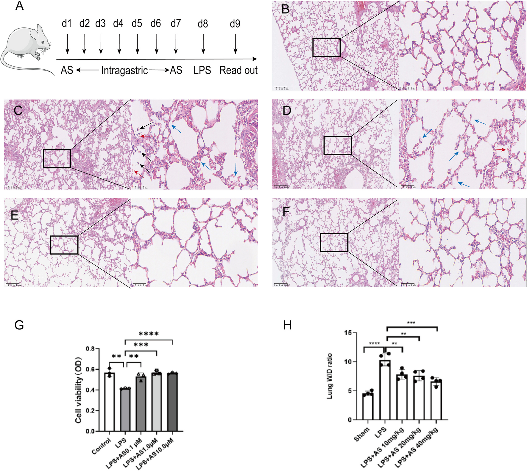
The cell viability of RAW264.7 after treatment with LPS (0.1, 1, 10, 20 mg/L) or AS (0.1, 1, 10 μM) was detected by CCK8. CCK8 reagent 10 μL of supplemental solution was added for 1 h at 37 °C humidity and 5% CO2. The optical density of each sample was measured using microplate reader at 450 nm (Bio-Rad, USA); all operations are performed according to the instruction.
Flow cytometry
RAW264.7 cells (1 × 10⁵) were seeded into 6-well plates. The cells were treated with LPS or AS, the spent medium was drained, and the cells were washed twice with phosphate-buffered solution (PBS). The washed cells were disintegrated with trypsin, and a gentle tap removed the cells from the bottom of the well. The cells were then resuspended in the pre-absorbed culture medium, washed with PBS, and counted. Annexin V-FITC and propidium iodide were added. A blank hole was set, an FITC hole was set only with Annexin V-FITC, and a PI hole was set only with PI. Samples were incubated in the dark for 20 min and then subjected to flow cytometry according to the manufacturer’s instructions.
Real-time fluorescence quantitative PCR
The total RNA of the cells or lung tissues was extracted with the TRIzol reagent according to the manufacturer’s instructions. RNA extraction protocol ensured the prevention of degradation of RNA and contamination with RNAases. Total RNA was finally dissolved in DEPC-treated water (Beyotime, China). The ratio of absorbance at 260 nm and 280 nm was used to assess the purity of and RNA. The extracted RNAs were used to prepare the reverse transcription reaction system. The thermal cycling conditions were 25 °C for 30 min, 42 °C for 30 min, 85 °C for 5 min, and conservation at 4 °C. Subsequent PCRs were performed on the ice. The PCR program included an initial pre-denaturation step at 95 °C for 3 min, followed by 40 cycles for denaturation at 95 °C for 12 s and annealing at 62 °C for 40 s. The fluorescence signals were then detected. RNA expression was normalized to that of U6. When the samples were added to the eight linked tubes, each gene for each sample was replicated in at least three repeated holes. The Ct values were suggested not to exceed 30.00. The differences in Ct values between the repeated holes were suggested not to exceed 0.50; otherwise, more repeated tests were recommended. The primer sequences were:
miRNA-21(F): 5′-AAGCGACCGTAGCTTATCAGA-3′
miRNA-21(R): 5′-GTCGTATCCAGTGCAGGGT-3′
U6(F): 5′-CTCGCTTCGGCAGCACA-3′
U6(R): 5′-AACGCTTCACGAATTTGCGT-3′
The final real-time PCR data were analyzed using the 2-ΔΔCt method.
Measurement of mitochondria membrane potential
JC-1 dye was used to detect changes in mitochondrial membrane potential (MMP). RAW264.7 cells were cultured in 6-well plates for 24 h and then treated with LPS or AS for 24 h. The treated cells were washed twice with PBS, and cell culture medium (1 mL) containing serum and phenol red was added to each well. Approximately 1 mL of JC-1 staining solution was added to each well, and the plates were incubated at 37 °C for 20 min. JC-1 staining buffer (1×) was prepared by adding 4 mL distilled water in 1 mL JC-1 staining buffer (5×) and placed in an ice bath. The cell supernatant was removed, cells were washed twice with JC-1 staining buffer (1×), and images were obtained using a fluorescent microscope (Nikon Corporation, Tokyo, Japan). The excitation and emission wavelengths were set to 490 nm and 530 nm, respectively.
Measurement of reactive oxygen species
An ROS assay kit was used to detect reactive oxygen species (ROS) production. The cells were cultured in 6-well plates for 24 h and then treated with LPS or LPS + AS for 24 h. Fluorescent probe DCFH-DA was diluted with serum-free culture medium (1:1000), and 1 mL of dilution was added to each well. The cells were incubated at 37 °C for 20 min. The supernatant was removed, and cells were washed with a serum-free culture medium and observed under a fluorescence microscope.
Animals
Eight-week-old male Balb/c mice (body weight 20 ± 2 g) were procured from Chengdu Dossy Experimental Animal Co., Ltd. (Chengdu, China; SYXK (Chuan) 2023-0017). Mice were kept at the Institute of Zoology of Southwest Medical University (Luzhou, China), and adaptive patterns of mice were labeled and grouped after a week. Mice were free from disease, and they were laid on clean bedding at any time, fed with sufficient water and food, and kept in a 12-h/12-h light/dark cycle. This study was conducted in accordance with the recommendations of the National Health Association Guidelines for the Care and Use of Laboratory Animals, and the protocol (SWMU20230043) was approved by the Animal Care and Utilization Committee of Southwest Medical University. Animals were randomly divided into 5 groups (sham, LPS, LPS + 10 mg/kg AS, LPS + 20 mg/kg AS, LPS + 40 mg/kg AS, N = 6 each group). The three AS dose groups were given 10, 20, and 40 mg/kg/day of AS by intragastric administration for 7 days before establishing the ALI model. All surgeries were performed under sodium pentobarbital anesthesia to minimize pain.
Establishing the ALI model
The ALI model was established according to the methods in previous reports (Zhu et al. 2018). The mice were anesthetized with 50 mg/kg sodium pentobarbital by intraperitoneal injection (Zhu et al. 2018). A nasal catheter was inserted through the mouth into the bronchus and connected to a syringe for infusion. The LPS and AS groups were administered LPS (10 mg/kg) by intratracheal instillation, and the sham group was treated with an equal amount of sterile ultrapure water. Mice were sacrificed 24 h later.
Bronchoalveolar lavage fluid collection
The mice were sacrificed under anesthesia with 50 mg/kg sodium pentobarbital, exposed mice on the operating table; after tracheal bronchial catheter inserted into the side, the other side of bronchus was ligated, 0.5 mL sterilized PBS lavage and perfusion each time, a total of 3 times, aspirate with a syringe pump for many times to obtain bronchoalveolar lavage fluid (BALF). BALF was collected in a 5-mL centrifuge tube and at 4 ℃, 1500 g × 10 min; the supernatant was collected and stored in the refrigerator at − 80 °C.
Hematoxylin and eosin staining
Mice were anesthetized with 1% pentobarbital sodium (50 mg/kg; i.p.), and the chest cavity was fully exposed. The right lower lobe of the lung was placed in 4% fixative (BioSharp, Hefei, China) for 48 h. The fixed lung tissue was wrapped in paraffin, sliced into 5-µm thick sections, stained with hematoxylin and eosin (H&E), and observed under a light microscope (200× magnification; BX-50 Olympus (Tokyo, Japan)).
Lung wet/dry weight ratio
After anesthetizing, the right lung of mice was obtained and blotted with absorbent paper and weighed on an electronic balance, and the lung wet weight (W) was recorded. The lung tissue was then placed in a 70 °C incubator to dry for 48 h and then weighed again, which was the dry weight (D). Lung wet/dry weight (W/D) ratio = W / D.
Western blotting
Western blotting was used to detect the concentration of Sema4D, CD72, Bcl-2, Bax, caspase 3, Drp1, Fis1, NF-κB p65, Phospho-NF-κB p65, and GAPDH of RAW264.7 cell or lung tissue. After obtaining these proteins, it was performed with the BCA test kit. Separation was performed on a 10% or 12% SDS polyacrylamide gel and transferred to PVDF membrane (Millipore, Ameica), which was mixed with Sema4D (1:1000), CD72 (1:500), NF-κB p65 (1:1000), Phospho-NF-κB p65 (1:1000), Drp1 (1:1000), Fis1 (1:1000), Bcl-2 (1:1000), Bax (1:1000), caspase 3 (1:1000), and GAPDH (1:10,000), respectively, at 4 °C overnight, and incubated with Goat anti-Rabbit IgG antibody HRP conjugated (1:1000) at room temperature for 1 h, then washed 3 times for 10 min, and visualized by ECL reagents, the bands were analyzed by ImageJ software 6.0.
Enzyme-linked immunosorbent assay
Supernatant was collected after cell treatment, and BALF was collected from anesthetized mice for detection; the concentrations of IL-1β and IL-10 of RAW264.7 cells and BALF were analyzed according to the manufacturer’s instructions.
Statistical analysis
All data were analyzed with Student’s t test or one-way ANOVA (SNK method was used for comparison between groups). The results were presented as mean ± SD. This paper uses SPSS22.0 and GraphPad Prism 9 software to analyze all the data. P < 0.05 was considered to be a statistically significant difference.


留言 (0)