
記住我
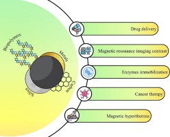


MOFs [[1], [2], [3]] are composed of organic ligands and metal centers (metal ions or metal oxide clusters). They are crystalline materials with nanoporous periodic network structures, also known as porous coordination polymers. By adjusting the topology, pore sizes, and/or chemical composition [[4], [5], [6], [7], [8], [9], [10], [11], [12], [13], [14]], MOFs can be designed into a large number of different architectures [15] and tailored for specific uses. For example, using ligands with similar structures but different lengths, it is possible to customize a framework MOF-5 structures [2,16] with different pore sizes and surface areas, as shown in Fig. 1A. MOF materials can also achieve robust reaction stability through proper modification and treatment, such as re-decomposition and oxidation after synthesis. In addition, the secondary building units (SBUs) [17] of MOFs can also be regulated by cation exchange, cation valence modification, defect formation, and new species introduction. Due to unique structural topological features [15,17] such as high specific surface area (SSA), ultra-high porosity, tunable architecture, and abundant functional groups, MOFs have been widely used in laboratory and industry. Examples includes adsorption [[18], [19], [20]], separation [21], sensing [22], catalysis [13,14,[23], [24], [25], [26], [27], [28], [29], [30], [31]], energy preservation and conversion [32,33], imaging and drug delivery [[34], [35], [36], [37], [38], [39]]. Some MOFs with special porous structures are shown in Fig. 1B.
An extremely important application of MOF materials is to prepare soft matter and bio-related materials. For examples, the biomimetic mineralization [[40], [41], [42], [43], [44], [45], [46], [47], [48], [49], [50], [51]] of biomolecules or drug molecules into MOFs attracted wide interests. Biomimetic multi-enzyme hybrids [41] was prepared using in situ encapsulated natural enzymes in iron‑cobalt bimetallic MOFs (FCM), and the performance study found that the hybrids had a multi-level porous structure and excellent catalytic activity. In the synthesis of GOx@FCM-tannic [41], tannins acid, as a post-etching agent, was further used to construct the mesoporous structure. The combination [40] of biomimetic mineralization of MOFs with Pickering interface system enhanced the catalysis and recycling of lipase in organic media, which successfully established a completely new biocatalytic system. A Zn-based MOF [50] with mixed ligands representing, respectively, DNA and protein residues was designed and successfully synthesized. MOF nanoparticles [45] were designed for protein delivery. A MOF-immobilized two-in-one engineered enzyme with enhanced cascade reaction activity for tumor therapy [4] was prepared. GOx-Fe0@ZIF-8 has been constructed, exhibiting higher enzyme cascades activity than MOF-immobilized two enzymes, GOx@ZIF-8 and Fe0@ZIF-8. In the MOF biological related materials, there must be an interface [52] between MOF and protein, polysaccharide, DNA, etc. Li et al. systematically reviewed the research progress in the field of MOF biological interface [46]. All the size of biomolecules, the MOF pore structures, and the interfacial region are in nanometer scale, being very suitable for SAS studies.
The miniaturization [53] of any material particles with uniform size and shape, regardless of chemical composition, has the potential to form colloidal particles and may open a door to new functions and applications. Therefore, when MOFs particles shrink to nanometer size, MOFs colloidal (CMOFs) [10,26,29,34,36,38,39,[53], [54], [55], [56], [57], [58], [59], [60]] materials were formed. The pioneering colloidal MOF material [53] is zeolitic imidazolate framework-8 (ZIF-8). Troyano et al. reviewed the growth process of ZIF-8, which provided useful guidance for the preparation of the colloidal form of ZIF-8. The results show that additives (or modulators), reagent concentrations, as well as the reaction time are the key parameters controlling the size [60,61] and monodispersity [62] of ZIF-8 colloidal crystals. Using colloidal MOF particles [59] of uniform size and shape as building blocks, colloidal materials can further provide additional functionality [3,10,[63], [64], [65], [66]] through self-assembly in solvents. Because colloidal MOF materials have the advantages of high porosity, large SSA, adjustable structure, heteroatoms, and uniform pore size, they have shown great application potential in many fields such as adsorption [12,13,18,67,68], separation [69,70], sensing [71,72], catalysis [7,20,29,73,74], energy storage [32,75,76] and imaging [58], and drug delivery [4,39,57], and so on.
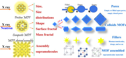
Up to now, >100,000 of MOF-related papers [1,16,46,77,78] can be retrieved. The topics covered are rich and diverse, mainly including MOF-derivation, coating, protection, doping, support, assistance, origin, regulation, encapsulation, trigger, transformation, base, template, MOF@MOF@others, MOF-on-MOF, supramolecular MOF, MOF (fabric) composite, MOF nanosheets, nanorods, nanoflakes, film, membranes, core-shell, fibers, MXene, COF, etc. In a broad sense, MOFs can be divided into three types: pristine MOFs [79,80], MOFs composite [17,19,22,37,38,73,[81], [82], [83]], and MOF-derived [10,24,35,[84], [85], [86], [87], [88], [89]] materials. Numerous synthesis approaches and self-assembled manners offer rich structural research connotations. Understanding the formation mechanism of MOF colloidal materials, as well as the structural evolution during synthesis and/or post-processing, is of great significance for the development of more efficient CMOF materials.
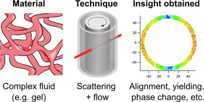
Scattering, microscopy, spectroscopy, and theoretical calculation are four types of commonly used techniques for structural characterization of MOF colloidal materials. Among them, scattering techniques, including small angle X-ray scattering (SAXS), small angle neutron scattering (SANS), dynamic light scattering (DLS), static light scattering (SLS), play an important role in extracting the particle size, shape, pore structure, SSA and internal structural information. Compared with the spatially localized microscopic techniques such as atomic force microscopy (AFM), scanning electron microscopy (SEM), transmission electron microscopy (TEM), scattering techniques have better sample compatibility, can obtain better statistical experimental signals, and are more helpful to quickly acquire the structure evolution process. Compared to visible light scattering (DLS and SLS) techniques with longer incident wavelengths, X-ray or neutron beam scattering (SAXS and SANS) techniques can analyze finer particle sizes and shapes, as well as internal structure information. Arguably, SAXS and SANS [23,81,86,[90], [91], [92], [93], [94], [95], [96], [97], [98], [99], [100], [101]], are powerful techniques that can truly be used for the dynamic characterization of nanostructures. In this paper, the recent advances of small angle scattering (SAXS and SANS) techniques in the structural study of MOF colloidal materials are introduced.
SAXS is commonly used to detect the X-ray scattering signals from nanoparticles with sizes ranging from a few nanometers to a few hundred nanometers. In this paper, wide-angle X-ray scattering (WAXS) and ultra-small-angle X-ray scattering (USAXS) are also classified as SAXS. The difference between the three is that SAXS is dedicated to detecting nanoparticle size and shape, USAXS extends the detectable particle information to micrometer scale, and WAXS extends the detectable particle information to below nanometer scale or is used for low-angle X-ray diffraction that is used to probe ordered structure with periodic structures on angstrom to nanometers. Usually, the detectable particle sizes are determined by the detectable scattering vector q-values (q = 4πsinθ/λ), where 2θ and λ are, respectively, the X-ray scattering angle and the incident X-ray wavelength. It is generally accepted that the maximum detectable particle size is determined by π/qmin and the minimum distinguished particle size is 2π/qmax. It is well known that the SAXS intensity (I(q) = F(q)S(q)) is the co-contribution of form factor F(q) and structure factor S(q). The surface characteristics, particle shape, size distribution, pores in MOFs [90,102], and so on can be extracted from the form factor. While the interactions between particles and even the network structure in MOFs [38,91] can be obtained from the structure factor. Thanks to the application of advanced synchrotron radiation source, high-brilliance SAXS technique can be used to pursue the in-situ dynamic reaction of MOFs in solution at the millisecond or even microsecond level. There already are some researches reporting the process of structural evolution of MOFs in nucleation, growth, self-assemble [85], aggregation [103], crystal conversion [104], and so on. SAXS can also be used to elucidate fractal structure and its evolution [20], giving surface fractal irregularities and roughness. Next, we will present the recent advances in SAXS applications for pristine MOFs, composite MOFs, and self-assembled MOFs, respectively.
The final products are closely related to the synthesis process. The ordered porous structure or pore array in MOFs is the foundation of all its applications. Evidently, pore structure information in MOFs is a general concern. Since the SAXS intensity is determined by the electron density difference between the scatter and its surrounding medium, both real nanoparticles and nanopores contribute to the SAXS intensity. For a monodisperse system with same particle shape and dimension, Pair-distance distribution function PDDF(R) in real space and the X-ray scattering intensity I(q) in reciprocal (q) space are Fourier transform relations with each other, containing the same structural information. Usually, the PDDF(R) function is used to analyze shape, size, and internal structure of scatters. However, for polydisperse systems, the shape and size distribution of the scatterers cannot be determined simultaneously. It is common to assume the shape of the scatterers in advance and obtain the size distribution of the scatterers, or vice versa, assume the size distribution of the scatterers and obtain the shape of the scatterers. In these cases, the assistance of other observation technique such as SEM or TEM, even if the assistant technique is off-line, is also needed to provide clues to the scatterer shape or size distribution. This is greatly helpful for obtaining real physical image of in-situ dynamic progress using SAXS technique.
To control the nucleation and growth of MOFs, understand the dependency of synthetic products on reaction parameters, find and optimize the synthesis path, as well as enlarge the scale of synthesis reactions, the formation mechanism of MOFs is worthy of in-depth study. Indeed, as early as 2018, Vleet et al [105] reviewed the literatures on the study of MOF nucleation and growth mechanisms. They concluded that it is difficult to draw universal growth mechanism conclusions because of the great chemical and structural diversity of MOFs. This conclusion implies that the nucleation and growth of MOFs are various, being still an important research direction. The unit-cell parameters of MOF crystals are usually on the order of nanometers because the “atoms” that make up the MOF crystals are composed of organic ligands and metal centers. Generally, the pore interstitials in MOF crystals are also in nanometer level. It means that WAXS is quite appropriate to the structural characterization of MOF crystals, and SAXS is right technique to analyze the nanopores. To study the nucleation and growth mechanism of MOFs, SAXS combined with WAXS would take an irreplaceable role, which guarantees the simultaneous acquisition of scattering signals from nanoparticles or nanopores and diffraction signals from crystalline structures of MOFs. At the same time, SAXS/WAXS combined technique also present a unique advantage for the detection of amorphous/primary particles in the initial stage and the possible intermediate phase during the MOF nucleation and growth. A SAXS/WAXS combined study [105] shows that amorphous clusters are more likely to serve as initial structures for MOF formation than crystal nuclei. Solvothermal or hydrothermal synthesis is a common synthetic strategy of MOFs, and the solvent [12,14,20,26,31,83,103,[106], [107], [108], [109], [110]] used in MOF synthesis is a very important factor to affect the synthetic path and final product. In this case, it is difficult for the other characterization techniques like DLS, SLS, SEM, or TEM, to in-situ follow the evolution from the initial amorphous/primary nuclei to the final crystals under the sample environment.
A mesoporous Cu3-xZnx(BTC)2 MOF [26] with high catalytic activity and reusability for cyanosilylation reaction of benzaldehyde and aerobic oxidation reaction of benzylic alcohol was synthesized for the first time in a deeply eutectic solvent (ZnCl2/ethylene glycol). The synthesized product appeared as mesoporous nanocubes, which can be obtained in only 30 min at room temperature, while the conventional solvothermal synthesis route usually requires higher temperature and longer reaction time. A unique SAXS/XRD/XAFS combined technique [26,111] was used to reveal the formation mechanism, as shown in Fig. 2A(a-d). From the PDDFs of SAXS curves, the average sizes of nanocubes were increased with the reaction time, as shown in Fig. 2A(b). XRD reveals that the crystalline framework had been formed at the beginning stage of 10 min, while XAFS reveals the coordination structures and the chemical valences of Cu and Zn. Combing the experimental results obtained with other detection techniques such as TEM shown in Fig. 2A(e), the SAXS/XRD/XAFS combined technique offered the hierarchical structure information from local coordination structure, nanoparticle size, and long-range ordered structure. A possible formation mechanism of the mesoporous Cu3-xZnx(BTC)2 MOF was provided. Zn and Cu ions were co-coordinated to the deprotonated H3BTC, but the bonding linked to Zn is weaker than that of Cu, which caused the dissociation of Zn from the framework, raising the catalytic activity. The formation of the mesoporous structure goes through a rapid crystallization process and then an etching process.
Zhang et al. invented a method [109] for rapid synthesis of MOFs in stable radical solutions (electrolyzed ionic liquids) and quantitative yields. This synthetic method is particularly suitable for synthesizing high-valence MOFs with versatile metal nodes and ligands at room temperature without external energy (Fig. 2B(a)). For MOFs containing conjugated ligands, the transient synthesis time is <1 s. Free radicals in the solution accelerate the kinetic process and play a vital role. This new ambient radical route paves the way for the industrial synthesis of MOFs. Several structural characterization techniques, including SAXS/WAXS and XAFS was used to investigate thoroughly the mechanism. SAXS/WAXS combined technique was used to in-situ study the formation kinetics of MOFs. Taking the NU-1000 as an example, when the as-prepared precursor NU-1000-a was added to the solution containing the Omim• (1-octyl-3-methylimidazolium) radical, the in-situ SAXS curve immediately presented a stronger scattering signal (Fig. 2B(b)), indicating the instantaneous nucleation of NU-1000. Combining the WAXS results, it can be concluded that the formed nuclei were amorphous. After 30 min, the SAXS curves showed a faster decay with the increase of scattering vector (q) but tended to unchanged with the increase of reaction time, which means that the particle size increased to a stable value. At the same time, some diffraction peaks from the NU-1000-a appeared on WAXS curves (Fig. 2 B(c)), indicating the formation of a well-ordered structure and the NU-1000-a precipitation. With further increase of the reaction time, the WAXS curves had no obvious change, which means that the reaction or the growth of MOFs has been completed. With the assistance of electron paramagnetic resonance (EPR) measurement, in-situ proton nuclear magnetic resonance (NMR) measurement, and density functional theory (DFT) calculations, it was further revealed that the Omim• radical acting as a catalyst greatly reduced the activation energy of MOF formation and promoted the rapid kinetics and room temperature synthesis of MOFs.
In addition to providing free radicals for MOF growth under electrolytic conditions, Sang et al. [83] found that ionic liquids as solvents can also accelerate the crystallization of Zr-based MOFs without electrolysis at room temperature. The prepared colloidal MOF particles have small particle size, missing node defects, and large SSA, which can be used as heterogeneous catalysts for the Meerwein-Ponndorf-Verley reactions. The reaction to synthesize UiO-66 in [Omim]Cl was traced by in-situ SAXS measurements to understand further the growth mechanism. The SAXS curves show a shift in SAXS intensity toward the low-q region as reaction time increases. This result indicates that the nanoparticles was gradually growing as the reaction time increases. From the PDDF functions calculated from corresponding SAXS curves, the sizes of sphere particles are 34, 38, 40, 46, 54, and 58 nm, respectively, at the reaction time of 10, 20, 30, 40, 50, and 60 min in the reaction system. Combined with the results obtained from other techniques such as XAFS and NMR spectra, the rapid formation mechanism of UiO-66 nanocrystals in [Omim]Cl ionic liquid at room temperature is proposed. The Zr precursor ZrOCl2·8H2O caused firstly hydrolysis and dehydration to yield polymeric hydroxide Zr4(OH)12. Then the added modulator HAc interacted with [Omim]Cl through hydrogen bondings to cause the complexation of zirconium, forming a complex. Next, a linker exchange between the modulators and linkers took place at the coordination sites of zirconium complex, which results in an easier reaction between the negatively charged BDC (= 1,4-benzene-dicarboxylate) linker and the zirconyl cation, with rapid formation of pre-MOF cluster in [Omim]Cl at room temperature. Finally, the pre-MOF cluster octahedral edges were bridged by carboxylate groups from the benzenedicarboxylate linker, resulting in the formation of UiO-66 framework. Besides, Zhang's group also achieved the synthesis [47] of La-MOFs with controllable shape and size using ionic liquid microemulsions. Spherical, lamellar, and cylindrical La-MOFs were, respectively, synthesized in the microemulsions (Fig. 2C(a-c)). SAXS technique was used to characterize the shape of particles. All shape parameters of the obtained particles, such as the diameter of the spheres, the thickness of the lamellar cylindrical cross sections, and the diameter of cylindrical cross sections, were extracted from the respective PDDF functions. The SAXS results confirmed the morphology of the synthesized La-MOFs particles.
Goesten et al. [112] used SAXS/WAXS combined technique to in-situ document the growth of MOFs in different solvents such as pure water, pure N, N-dimethylformamide (DMF), or DMF/H2O mixtures. The crystallization of two topical MOFs, NH2-MIL-53(Al) and NH2-MIL-101(Al), synthesized from similar metal and organic precursors were studied. Based on the time-resolved SAXS data, the evolution of PDDF p(r), the radius of gyration Rg, the Porod volume V, and the SSA (S/V) of the scattering particles were extracted. The SAXS results revealed that the solvent can affect the synthesis routine and the synthesis speed.
Porosity is the outstanding feature of MOF materials and the basic origin of their unique properties. According to the pore size, typical pores can be divided into micropores <2 nm, mesopores between 2 nm and 50 nm, and macropores larger than 50 nm. Almost all the applications of MOFs are directly related with the porous structures. Theoretically, the pore structure can be calculated from the crystal structure of MOFs. However, there is no method to synthesize perfect crystals of MOF with maximum microporous density (or SSA). Therefore, the pore structure and pore network of MOF materials deviate greatly from the ideal crystal architecture. Reasonable estimation of pore structure and pore size in MOF materials is desired and necessary. The typical method to study pore structures is the nitrogen adsorption-desorption isotherms [12,19,[24], [25], [26], [27],31,35,55,103,113], and the adsorption isotherms of nitrogen or other gases can be used to evaluate the SSA, pore volume, and pore size of MOF materials. However, this technique is not suitable for the detection [114,115] of closed pores due to the diffusion obstruction effect on adsorbed desorbed gasses. Or it will lead to large errors when the porous system contains closed pores, depending on the complexity of porosity. For the detection of closed pores, scattering techniques (SAXS and SANS) have unique advantage. It is well known that nanopores and nanoparticles with the same shape and size have the same scattering signals duo to the reciprocity of scattering. Therefore, SAXS and SANS techniques are powerful tools to probe porous structures. For highly porous materials, the pore structure parameters obtained by the gas adsorption method should be verified by scattering techniques (SAXS or SANS), or independently confirmed. SAXS technique has been widely used to characterize the pore structure in MOFs. Table 1 summarizes and lists pores structure parameters detectable in some MOF materials using SAXS technique or N2 adsorption-desorption isotherms. The SSA values (m2 g−1), surface fractal, and pore volumes (cm3 g−1) of MOF material (named as FPC in Ref 103) are quantitatively given. Both the total pore volume and the micropore volume are compared in Table 1, it can be seen that the total pore volume and the micropore volume detected by N2 sorption are all smaller than the values detected by SAXS. This result suggests that there are some closed pores, blind pores, or open micropores [115] that are too narrow to accommodate two layers of N2 molecules in the MOF studied. At the same time, the ratio of total pore volume and the micropore volume detected by N2 sorption is about two-fold larger than the one detected by SAXS, which demonstrates further that the occupancy of closed pores in the micropores is larger than the ones in the mesopores or macropores.
Mass fractal and surface fractal are two typical fractal systems in small angle scattering (SAS) research. The former reflects the compactness of scatterers, which can be described with a mass-fractal dimension Dm. The smaller of the Dm value, the looser of the scatterer structure. The latter reveals the roughness of scatterer surface, which can be described with a surface-fractal dimension Ds. The bigger of the Ds value, the coarser of the scatterer surface. For a fractal system, the SAXS intensity in the q-region with qDmax> > 1 can be described as Iq≈I0q−α. When 1 ≤ α < 3, it is a mass fractal and Dm = α. When 3 ≤ α < 4, it is a surface fractal and Ds = 6-α.
As a kind of nanofluid [[116], [117], [118]], MOF colloids have the capacity to absorb low-frequency seismic waves. They can be injected underground to enhance seismic monitoring tools or to verify containment of carbon reservoirs and so on. Miller et al. [118] demonstrated for the first time that MOFs are metamaterials with acoustic capabilities for tunable sound adsorption and resonance, and that water-based Cr/Zn/Zr MOF colloidal suspensions were multimodal geophysical contrast agents [116] used to enhance near-hole logging tools. SAXS/WAXS techniques were used to characterize the nanoscale structure and crystal structure of MIL-101(Cr) suspensions [117], confirming the phase stability and providing insights into the fractal properties of the colloidal nanoparticles, the SAXS/WAXS curve was shown in Fig. 3B. The power-law exponent of the SAXS curve was 3.90, indicating a surface fractal property for the suspended colloid MOFs. And it also suggested that the high internal surface area of colloid MOFs was retained in water-based nanofluid, which is precisely the critical component of acoustic absorptive MOF metamaterials. From the first two diffraction peaks that appear immediately after power-law decay (Fig. 3B), two different types of mesoporous cages [117] can be obtained for a typical MIL-101(Cr). The smaller pentagonal cages (d-spacing of 31.4 Å) is corresponding to the second peak centered around q = 0.2 Å−1. The relatively larger hexagonal cages (d-spacing of 52.3 Å) is corresponding to the first peak centered around q = 0.12 Å−1. The PDDF was also calculated from the SAXS curve. And the authors claimed that the average distance between the centers of two MIL-101(Cr) particles was estimated to be 34.1 nm according to the PDDF. In fact, the PDDF can be seen as the Fourier transformation of the scattering signal [[119], [120], [121]] from an average particle, which can not tell directly us the distance betwee two adjancent particles. Unless neighboring particles are assumed to be in contact with each other. Under this assumption, the so-called distance between the centers of two particles is equivalent to the average particle size.
For the sake of energy saving, the unique pore structure of MOFs also makes them good adsorbent candidates for new adsorbent-based heating, ventilation, and air conditioning (HVAC), regarded as an alternative to traditional refrigeration systems. MOF materials with ultra-high porosity and optimal MOF-water interaction allow them to achieve orders of magnitude improvement in water adsorption and regenerative energy requirements. Çamur et al. [102] investigated the monolithic zirconium-based MOFs for energy-saving water adsorption applications. They developed three high-density, modeled, monolithic MOFs (UiO-66, UiO-66-NH2, and Zr-fumarate) with excellent volumetric gas/vapor absorption, addressing issues in MOF-HVAC deployments. Using lattice gas model, large-gauge Monte Carlo molecular simulations and SAXS measurements [117] revealed the monolithic structure of the material over the entire mesoporous range. Fig. 3C(a-d) shows the experimental SAXS curves and the fitting ones based on a spherical form factor model ((a) for monolithic UiO-66; (b) for monolithic Zr-fumarate), as well as the particle size distributions ((c) for monolithic UiO-66; (d) for monolithic Zr-fumarate). From the SAXS curves, it was found that the samples have fractal features. The power-law exponents were, respectively, 3.035 for monoUiO-66, 3.111 for monoZr-fumarate, and 2.895 for monoUiO-66-NH2. Therefore, monoUiO-66 and monoZr-fumarate are surface fractals, but monoUiO-66-NH2 are mass fractals. monoUiO-66 and monoUiO-66-NH2 showed similar particle size distributions, exhibiting two main particle size populations with mean diameters ≈ 80 and ≈ 160 Å. However, monoZr-fumarate exhibited one main population of particles with a mean diameter of 76 Å, and an equivalent volume fraction of particles having different mean sizes of 167, 252, 339, and 424 Å. Using the SAXS data, the authors created a 3D reconstruction of the monoliths, and the results suggest that the hierarchical porosity of monoZr-MOFs can be accurately described computationally across the microporous and mesoporous ranges, enabling robust future predictions of adsorption characteristics.
It is well known that with constant particle density, smaller particles have larger SSA. Relatively, microporous MOFs have higher SSA than mesoporous and macroporous MOFs, and they are structurally selective for small molecules. Due to the difficulty of mass transfer and encapsulation of functional large guest molecules, however the applicability of microporous MOFs is generally limited. Mesoporous MOF (2–50 nm) can overcome the limitations of micropores and has become a subject of great interest. Koo et al. [122] reported a novel strategy to produce water-stable hierarchical porous MIL-100(Fe) by a selective acid etching process, as schematically shown in Fig. 3D(c). The hierarchical porous MOFs have tunable porosity but preserve their inherent crystallinity and external morphology. It is known that MIL-100(Fe) has large cages (d = 0.89 nm) with hexagonal windows and small cages (d = 0.49 nm) with pentagonal windows. Phosphoric acid (H3PO4, d = 0.61 nm) in DMF was chosen as an etching agent. The H3PO4 molecules exhibited size-selective diffusion into the 3D channels of the large cages but failed to pass through the small cages. The SAXS curves show that the diffraction patterns of the etched and unetched MOFs were identical, which means that the crystallinity of the MOFs was preserved after this etching. The slightly increased full width at half maximum (FWHM) of the diffraction peaks indicates the successful enlargement of the pore sizes under the higher acid concentrations, as shown in Fig. 3D(b). The SAXS curves also present a typical q−4 power law scattering behavior of mesoporous MOFs, as shown in Fig. 3D(a).
In most cases, the SAXS signal is a monotonically decreasing curve with increasing q-value, and the scatterers to be studied and the background support materials together contribute to the SAXS intensity. When SAXS technique is used to reveal quantitatively or qualitatively the structural details including pore surface characteristics, pore shape, size distribution, SSA, spatial distribution, even pore-network structure, the removal of scattering background is an art as comparing with other techniques used for pore structure study. It is usually difficult for obtain the background of pore scatters, here we would like introduce a useful method of background removal through an application example in MOF study. Tsao et al. [114] used the SAXS/WAXS combined technique to analyze the pore structures in MOF-5-like crystals as well as the network responsible for hydrogen storage. The pore structure of MOF-5 produced at different cooling rates was obtained. Simultaneously, the relationship between pore structure and MOF-5 crystalline phase was established during the removal of solvent upon heating, as shown in Fig. 3A(a-d). In such a classical SAXS study on MOF pore structure, an effective method to remove the scattering background of the SAXS region was proposed for the three-phase system. The shoulder of diffraction peak in high-q WAXS region (0.3 < q < 0.5 Å−1) was extrapolated to low-q SAXS region, and was used as the SAXS background. After removing the scattering background, the SAXS signal contributed by the micropores is more visible in the low-q region (0.1 < q < 0.3 Å−1). It can be found this background removal method is more suitable for the SAXS/WAXS combined measurement data. It should be a good choice for some scattering systems where it is difficult to separate the scattering signal from the scattering background.
People are not satisfied with the existing structural characteristics and functions of the pristine MOFs. To further explore the new functions of MOFs, MOF composites came into being. Zhang et al. [82] proposed a post-solvothermal route for the preparation of MOF composites, which has many unique features and was successfully used to control the outer-to-inner decomposition of MIL-125-NH2 and easy conversion into MIL-125-NH2@TiO2 core-shell nanoparticles. They applied SAXS technique to reveal the pore structures and found that an obvious enlargement of void space appeared in the MIL-125-NH2@TiO2 composite with a longer solvothermal time. The evolution of the obtained SAXS curves is similar to that in Fig. 3D, which is consistent with the linker loss during the solvothermal process for MIL-125-NH2.
MOF composites are composed of MOF and other materials such as nanoparticles, drugs molecules, biological macromolecules, and so on [35,40,41,43,45,[48], [49], [50]], which are used to develop new functions and improve the composite structures. Different MOFs [6,7,123] can also be combined into MOF composites to form MOF-on-MOF. Biomacromolecules in living systems not only play an important role in life process, but also have great potential for application in production activities outside the body of life. However, the lack of stability, recyclability, and efficiency significantly hinders their further utilization [81,90,[94], [95], [96],124,125] under slightly harsher conditions. Recently, MOF-biointerface engineering [[40], [41], [42], [43], [44], [45], [46], [47], [48], [49], [50], [51]] has effectively solved the aforementioned shortage of the biological macromolecules and living systems, attracting widespread attentions. ZIF-8 is the most popular MOF material used for MOF-biointerface composites. Apart from inorganic nanoparticles, target organic molecules such as dyes, drugs and therapeutic agents can be efficiently encapsulated [1,21,23,53,59,60,81,85,90,94,96,105] inside the ZIF-8 framework via bottom-up approaches. Colloidal ZIF-8 could also be employed as a protective coating for various biomacromolecules. As an important category of MOF bio-composites, MOF biomimetic mineralization [[40], [41], [42], [43], [44], [45], [46], [47], [48], [49], [50], [51]] has been extensively investigated.
Naturally occurring enzymes are highly selective and efficient catalysts. Due to their extraordinary catalytic potential, they are highly anticipated to be widely applied in industrial and biomedical fields. However, due to their complex and fragile structure, industrial applications are extremely challenging. Encapsulating biological macromolecules in MOF can provide thermal stability to trapped guest molecules, which occurs within the larger cavities [126] formed during MOF growth around the guest molecules, rather than being confined to a single MOF pore. The possibility of a facile and single-step technique [94] for the preparation of MOF biomimetic mineralization enzymes of industrial interest has been demonstrated. Ruhani Singh et al. selected three different enzymes to prepare the ZIF-8@enzyme composites [94] and SAXS technique was used to investigate their thermal stability from minutes to hours over an industrially relevant temperature range of 50–80 °C. Under water and ambient conditions, the rapid self-assembly and growth of MOF around the enzyme molecules were completed in a single step, and the biomimetic mineralization growth of ZIF-8 around the enzyme was achieved, as schematically shown in Fig. 4A(a). The results of specific enzymatic assays showed that MOF encapsulation had significant protective ability against the function of the enzyme. In order to more accurately study the interaction and structural changes of ZIF-8@GOx composites with increasing temperature, freshwater dispersions of ZIF-8@enzymes composites are placed in quartz capillaries for in-situ heating on the SAXS/WAXS beamline. The collected SAXS/WAXS patterns (Fig. 4A(b)) indicate that the newly prepared ZIF-8 dispersed in water was initially in an amorphous state, without diffraction peak. Whit the heating, ZIF-8@GOx began to crystallize at 45 °C and remained crystallize at 80 °C. The diffraction peaks were stronger at higher temperatures, indicating that the crystalline structure of ZIF was more stable, which may be one of the reasons for the enhanced protective properties of MOFs at those temperatures. In the study, the biomacromolecules were located on the surface of the MOFs. Clearly, the interaction between biomacromolecules and MOFs may be related to the interface performance, which needs to be clarified by a large number of further studies.
Murty et al. interrogated the protein molecules encapsulated within the MOFs at high temperatures. A novel analytical method [90] was proposed to solve the structure of bovine serum albumin (BSA) encapsulated, respectively, within ZIF-67 and ZIF-8 using SAXS. Time resolved SAXS was used to detect the structural change of protein molecules with time, as well as the stability of proteins even under in-situ heating condition. In this SAXS study, there were three components, the encapsulated BSA, the MOFs, and the buffer solvent, in the biocomposite BSA@ZIF solution. The SAXS patterns of ZIF-67 in buffer solution, encapsulated BSA (BSA@ZIF-67), and native BSA, were collected. It should be emphasized that the correct removal of other scattering backgrounds except the target scatterers (i.e. the encapsulated BSA) is the most fundamental step for the subsequent analysis. To solve the issue of accurate background removal, a scaling method highly dependent on the scaling subtraction factor α was adopted. By carefully adjusting or optimizing α value to minimize the sum squared difference between the native and encapsulated protein SAXS patterns, the SAXS pattern of pure MOF in buffer solution was then scaled with the optimal α value. After subtracting the scaled SAXS pattern of pure MOF in buffer solution from SAXS data of the biocomposites, the SAXS signal of the encapsulated pure protein was obtained. Such a process of the scattering background removal is schematically shown in Fig. 4B(a). After the background subtraction, the structural information such as the radius of gyration (Rg), Kratky degree of folding, and PDDF for the shape and tertiary structure of the protein can be analyzed from the extracted SAXS pattern of the encapsulated pure protein. The PDDFs of the extracted encapsulated BSA and the native BSA were compared at 23 °C and 70 °C. The PDDFs showed that native BSA exposed to 70 °C became denatured because its maximum dimension of the protein became 350 Å, while the encapsulated BSA still maintained its size and folded state at 70 °C. This result indicates that trapping the protein within the MOF cavity can inhibit the unfolding and thus denaturation, as compared in Fig. 4B(i). The approach of SAXS background removal in this study not only provides a new insight into the stability of biomacromolecules in MOFs, but also provides a new way to study other conformational unstable molecular structures in rigid matrices.
Up to now, there has been plenty of studies [42,44,45,125] on biocomposites of MOFs and biomacromolecules. The immobilization information and conformational changes of protein molecules in MOF are extremely important for the application of this kind of composite material, and therefore have attracted great research interest. Tocco et al. [38] studied the conformational changes and location of BSA immobilized on ZIF-zni and ZIF-8. The nanoscale structures of ZIF-zni, ZIF-8, BSA@ZIF-zni, BSA@ZIF-8, the BSA loading and the successive removal of BSA by calcination, were investigated by SAXS/WAXS combined technique. The SAXS curves were fitted with a model function consisting of Porod and Guinier contributions, as shown in Fig. 4C(a-b). These parameters Rg, s (particle shape parameter, s = 0 for spheres, 1 for rods, and 2 for platelet scatterers), and m (Porod slope at high-q) were extracted from the model fittings. In addition, surface smoothness, density condensation, as well as the pore shape were also valued. The three SAXS curves of pure ZIF, BSA@ZIF before and after calcination were compared, respectively, for both ZIFs (ZIF = ZIF-Zni, ZIF-8), as shown in Fig. 4C(a-b). It can be found that the SAXS intensity at the knee point become more intense after calcination, which were attributed to the contribution of the mesopores where BSA was positioned with gyration radius Rg = 6.0 ± 0.1 nm (for BSA@ZIF-zni) or 4.3 ± 0.1 nm (for BSA@ZIF-8). Calcination enlarged the mesopores in the two biocomposites, changed the crystalline structure of ZIF-8, but maintained the crystalline structure of ZIF-zni. The change of the ZIF-8 crystalline structure was ascribed to the removal of surficial proteins/enzymes and the formation of sub-micron porosity caused by calcination in ZIF-8. Although calcination did not change the crystalline structure of ZIF-zni, its mass fractal dimension was slightly reduced from 2.9 to 2.7, indicating that accompanied by the removal of surficial proteins/enzymes, the compactness of ZIF-zni was slightly reduced. Through the comparative studies of the SAXS patterns before and after calcination, the immobilization of BSA within both ZIF-8 and ZIF-zni was confirmed. The higher SAXS intensity of ZIF-zni could be related to an excess of protein accumulation on the outer surface of the MOF. Combined with other characterizations, it is very possible that BSA lies both inside ZIF matrix and on the outer surface, as schematically shown in Fig. 4C(c).
Advanced synchrotron radiation (SR) beams, especially those of high-brightness SR sources such as HEPS [[127], [128], [129]], usually cause radiation damage to proteins when they are used to probe protein structures. Encapsulating biological macromolecules in stable MOFs is a good candidate to protect them from radiation damage. However, the interaction between protein encapsulated in MOF and MOF is highly likely to alter the structure and biological function of the protein. Therefore, it is very beneficial to develop a method to reduce the post-synthetic MOF-biomolecular interface interactions and allow the protein to work in a state with little or no alteration in structure. Protein molecules immobilized in MOF retaining the natural structure will greatly maintain their activity. To reduce the MOF-biomolecular interface interactions, Chen et al. [96] explored the method to immobilize catalase in hollow ZIF-8 microcrystals. First, the enzyme-lowering molecule was grown in ZIF-67, then ZIF-8 was grown outside it, and finally the ZIF-67 part was removed to form a central cavity inside ZIF-8. In this way, the independent enzymes were sealed in the central cavity of the hollow ZIF-8, generating a hollow sample. The hydrogen peroxide degradation activity of catalase at different stages indicated that the independent catalase in the hollow ZIF-8 had high activity. In-situ SAXS technique, which is sensitive to structural changes in the mesoscopic range, was used to monitor the hollowing process of the samples in the presence of enzymes. Fig. 5A(a-d) shows the SAXS profiles in the hollowing process of ZIF-8. The three regions q = 0.1 ∼ 0.4 Å−1, 0.04 ∼ 0.07 Å−1, and 0.01 ∼ 0.04 Å−1 in the SAXS profiles were, respectively, used mainly for the calculation of the detailed microporous, mesoporous and microporous structures. The SAXS results demonstrate that there were two structural components in the initial stage of the hollowing process, the intrinsic micropores with size of 1.1 nm in diameter and the encapsulated catalase with a mesoscopic size of 11 × 18 × 4.1 nm, as shown in Fig. 5A(b). 30 min later, macropores larger than 50 nm appeared as a new structural component, indicating the formation of central cavities in the ZIF-8, as shown in Fig. 5A(c). More SAXS profiles with different formation times are shown in Fig. 5A(d). With the gradual removal of ZIF-67 during the hollowing process, the SAXS intensity corresponding to the macropores obviously increases, while the SAXS intensity corresponding to the micropores tends to decrease. This study not only highlights the importance of the freestanding state without interaction for maintaining the biological function of the encapsulated enzymes, but also proposes a new strategy for immobilizing freestanding molecules in MOFs, as shown in Fig. 5A(e, f). The SAXS characterization provided a means to observe the pore structures of MOFs from micropores to macropores, which played an important role in understanding the hollowing process of MOFs.
ZIF bio-composites have the capacity to protect and deliver biotherapeutic drugs in a laboratory batch mode. A one-pot synthetic method [109] for encapsulating NHase1229 in ZIF-67 is shown in Fig. 5B. The size of nanoparticles affects their circulation time in the blood and their uptake by cells. Therefore, it is of great practical significance to develop the method of large-scale synthesis of bio-composites with controllable size. Continuous flow [95] was often used for in-situ crystallization studies of multicomponent MOFs. However, Carraro et al. [81] developed the synthesis method of ZIF-8 biocomposites with variable particle size by continuous-flow. The continuous-flow method was used for the first time to encapsulate the model protein BSA and a clinical therapeutic drug (A1-antitrypsin, AAT) in ZIF-8, as schematically illustrated in Fig. 5C(a). To explore the nucleation, growth, and crystallization of BSA@ZIF-8 in the continuous flow synthesis process, in-situ SAXS and WAXS data were collected. It is demonstrated that the particle growth can be quenched by ethanol-induced crystallization from amorphous particles to ZIF-8 crystals. The produced particle size of the final biocomposite was tuned between 40 and 100 nm by changing the injection time of ethanol. SAXS and WAXS results showed that the biocomposites are amorphous particles at first, and then turned into crystallization. Two parameters, quantity Q~ and I110, were extracted from the SAXS curves, representing respectively the growth of amorphous particles and crystal particles. Q~ is related to the invariant of the SAXS curve and is sensitive to variations of particle volume fraction and electron density difference. I110 is the integrated intensity of (110) diffraction peak of sodalite-like ZIF-8, roughly proportional to the degree of crystallization, which was used to assess the crystal evolution. The growth of amorphous particles happened mainly in the first 3 min, as indicated by Q~increase as shown in Fig. 5C(b). Then, the diffraction intensity I110 of the (110) crystal plane began to increase quickly, which was accompanied by a quick decrease of Q~ as shown in Fig. 5C(b). The overall decrease of Q~ was due to the lower density of crystals than amorphous particles. The average crystallite size of the complex obtained using ethanol quenching was 17 nm, while the crystallites produced by non-quenching synthesis were >200 nm. Further, the SAXS and WAXS results suggest that the ethanol quenching can trigger and promote the nucleation of BSA@ZIF-8 biocomposites, but inhibit and quench the particle growth. Besides, the rapid injection of ethanol could also quench the growth of amorphous particles.
A very important application [39,46,53,125,[130], [131], [132]] of MOF composites is drug delivery and sustained release systems. In these studies, MIL-125(Ti) was shown to have better biocompatibility than others carriers due to the biodegradability of its central titanium (Ti) ions and organic ligands. Lin et al. [125] synthesized microporous round cake-like MIL-125(Ti) carriers, which can be used as drug delivery and release platform with great potentials. It is well known that tumor tissues usually have low pH values accompanied by intense respiratory CO2 and lactic acid production. Therefore, the selection of chitosan (CH) monomers with both anti-inflammatory, immunomodulatory, biocompatible, and biodegradable characteristics, as well as pH sensitive, to insert into the crystal lattice of MIL-125(Ti) carrier enable this MOF to be drug-loaded and pH sensitive. Besides the low pH environment around the tumor, the body temperature of cancer patients also increases. Therefore, the selection of the temperature-sensitive, biofriendly polymer Pluronic F127 inserted into the crystal lattice of MIL-125(Ti) could make the MOF carriers temperature-sensitive. CH-MIL-125(Ti) is as sensitive to pH value as CH, and F127-MIL-125(Ti) is as sensitive to temperature as F127. Both MOFs showed high microporosity and responsive release rates. The structures of the pristine MIL-125(Ti) and the composite F127-MIL-125(Ti) were analyzed using SAXS technique in the range of q-values from 0.257 to 0.016 Å−1. The SAXS data were fitted with the core-shell cylinder model or the core-shell ellipsoid model using program SasView 5.0. Based on the structural models, the SAXS fittings provide the model parameters. For the F127-MIL-125(Ti) composite, three temperatures at 25, 37, and 42 °C were used to simulate the temperature environments encountered by the MOF carrier. Core-shell cylinder and core-shell ellipsoid models were used to fit the SAXS patterns at the three temperatures. The model parameters such as radius, thickness, and length, were extracted. Based on the core-shell cylinder model, the SAXS fitting results explain well the effect of lattice explosion, as shown in Fig. 6A. Based on the core-shell ellipsoid model, the SAXS fitting results show that the long-axis radius was linearly increased with temperature, which can be ascribed to the colloid swelling along the long-axis direction. This colloid swelling might be the factor leading lattice explosion.
In 2010, potassium hydroxide-γ-cyclodextrin-MOFs (KOH-γ-CD-MOFs) [12] was synthesized in methanol, which is composed of γ-CD and K+ ions and was defined as a porous body-centered cubic array of (γ-CD)6 units. As a biocompatible material, it can be used to adsorb nongaseous drugs/materials of lansoprazole. In 2019, a densely packed arrangement [131] composed of γ-CD and potassium acetate was synthesized in water and marked as KAc-γ-CD-MOFs. This new synthesis method did not use surfactants and increased the yield to several times in water with a pH of 6.5–7.5. However, this densely packed KAc-γ-CD-MOFs was non-porous, which is different from KOH-γ-CD-MOFs. To transform the dense crystal structure into a highly porous one for drug delivery, the authors figured out a strategy for crystal transformation using ethanol, as shown schematically in Fig. 6B(a). SAXS technique was used to characterize the crystal structures. The SAXS results illustrated that the densely packed KAc-γ-CD-MOFs and the porous KOH-γ-CD-MOFs had the same diffraction patterns and indices of crystal faces, as shown in Fig. 6B(b). All evidences suggest that the porous KAc-γ-CD-MOFs should be the BCC array of (γ-CD)6 units. The properties of CD-MOFs crystals that can be transformed from a dense to a highly porous form can be used for efficient drug loading.
How to increase the solubility of insoluble drugs in water is a challenging problem, and great efforts have been made using nanotechnologies. The refinement of the drug particles seems to be a feasible strategy to improve the solubility of unsoluble particles in water. But few have been able to efficiently produce pharmaceutical particles a few nanometers in size. The encapsulation and immobilization of drug molecules in MOF, or the complexation of drug molecules with MOF, provides the possibility to enhance the solubility of insoluble particles in water. He et al. [132] studied the significant enhancement of solubility and bioavailability of Azilsartan (AZL, antihypertensive). AZL molecules forming nanoclusters were successfully confined in the large nanocages of biocompatible CD-MOF. The solubility and bioavailability of AZL in this way were improved by 340-fold and 9.7-fold, respectively, comparing to pure drugs. Based on molecular modeling, the authors proposed a dual molecular mechanism for the nanoclustering and complexation of AZL inside the CD-MOF cages. There are two main kinds of cages in the porous CD-MOF structure: i) spherical voids with diameter of 1.7 nm formed by six γ-CDs, and ii) cavities with diameter of 0.8 nm formed by face to face γ-CD pairs. SAXS measurements were performed to investigate the distribution of drug molecules in CD-MOF. Three AZL molecules were inside the cavity of the γ-CD pairs as shown in Fig. 6C(b). Other three AZL molecules as a nanocluster located inside the spherical void with size of 1.7 nm surrounded by six γ-CDs as shown in Fig. 6C(c). The relative positions of diffraction peaks confirmed the body-centered cubic (BCC) symmetry of the CD-MOF, as shown in Fig. 6C(a). After drug loading, the Bragg long period (L = 2π/q1) of CD-MOF with AZL was unchanged. However, all peaks of the AZL/CD-MOF were slightly shifted to the higher angle, which means that the diameter of the cavities were reduced. The most important thing is that the q2 peak (d = 2π/q1 = 1.6 nm) almost disappeared with loading AZL, which demonstrated that the signal of the 1argest cavities with theoretical diameter of 1.7 nm were fully filled by the AZL molecules. This group [130] also investigated the MOF capacitates cyclodextrin to mega-load mode for high-efficient delivery of antihypertensive valsartan. The highlight is the “Ship-in-a-Bottle” strategy [39] to create folic acid nanoclusters inside the nanocages of γ-CD-MOFs was investigated. SAXS is also used to detect the location of drug molecules.
In this section, we present the SAXS characterization of MOF composites, mainly consisting of MOF materials coated or loaded with biomacromolecules or drug molecules. In addition, some of the MOF complexes carrying biological molecules may also be for the consideration of radiation protection against biological macromolecules. In most cases, the design and application of MOF composites are related to the porous structure of MOF, and the characterization of the porous structure is the unique advantage of SAXS characterization technology. MOF colloidal particles [3,11,50,53] have the same shape and size, which makes them easy to self-assemble into different types of superlattices or supermolecules. Since the lattice spacing of MOF is usually in the nanometer scale, the SAXS technique can also be used to real-time study the spatial arrangement information of MOF crystals and MOF superstructures. In the next section, the application of the SAXS technique to the structural characterization of MOF-derived materials is going to be introduced.
MOFs were widely used as precursors or templates to derivate various materials including metal oxides and carbon materials that were employed as catalysts. The derivatives self-assembly process of most colloidal MOF particles [10] retains the inherent characteristics of MOFs such as large SSA and high porosity. The high internal surface area and porosity, as well as diverse components and structures make the MOFs materials have significant gas adsorption performance, which can be used to store hydrogen and methane storage or capture carbon dioxide. The selectivity of adsorbed gases and the absorption capacity for guest molecules depend on the interaction between MOFs and guest molecules or the adsorption force of MOF to guest molecules. However, the mechanism of adsorption and separation of gases by MOFs materials, as well as the interaction between adsorbates on the pore walls of MOFs have not been extensively explored. Cho et al. [65] investigated the extra adsorption and adsorbate superlattice formation in MOFs. The results showed that local strain in MOF caused by pore filling can provide aggregation and long-range adsorbate-adsorbate interactions, as well as the formation of adsorbate superlattices that extend beyond the original MOF unit cell. The argon adsorption tracking based on SAXS indicated that the adsorption process of IRMOF-74-Hex can be divided into 1 to 5 stages, occurring within the pressure range from 0 to 100 kPa. The SAXS patterns of IRMOF-74-V-hex showed the appearance and disappearance of a broad adsorption peak located at q = 0.093 Å−1 and a broad superlattice peak located at q = 0.25 Å−1 during the absorption process, as shown in Fig. 7A(a). The appearance and disappearance of the adsorption broad peak reflects the formation of extra adsorption domains and homogenization. The intensity of superlattice peak reached its maximum from stage 3 to stage 4 (33 kPa), then gradually decreased and disappeared at the end of stage 4, reflecting a process from the superlattice formation of adsorbate with different amounts in adjacent pores to an even adsorbate distribution. Fig. 7A(b) illustrates the argon adsorption and the filling process, which shows that argon adsorption has a complex aggregation behavior in the pores of MOFs. Fortunately, this abnormal adsorption phenomenon of argon in the pores of MOFs was captured by the SAXS measurements.
By the colloidal crystal engineering [34] of nucleic acid-modified nanoparticles, an effective method for fabricating three-dimensional (3D) superlattices has been achieved. However, the polydispersity and poor colloidal stability of MOF nanoparticles (MOF NPs) limited the types of colloidal crystals that can be engineered. Fig. 7B(a) shows that MOF NPs densely functionalized with oligonucleotides can be programmed to crystallize into various superlattices with well-defined crystal symmetry and composition. First, MOF NPs were synthesized by combining metal ions with organic ligands. The MOF NPs were then surface passivated by an azide-modified polyethylene glycol (PEG) polymer layer, followed by DNA functionalization through copper-free click chemistry, and finally programmed into a superlattice through DNA hybridization. SAXS technique was used to verify the superlattice formation. Clear diffraction rings or peaks on the SAXS patterns confirm the formation of micron-sized superlattices, as shown in Fig. 7B(b-d). The crystallization processes of the binary superlattice composed of MOF and Au programmable atomic equivalents (PAEs) was also confirmed by the diffraction peaks on the SAXS patterns, as shown in Fig. 7B(e-g).
In the heterogeneous metal MOF, the metal component consists of heterometals with similar size and valence states. These heterometals form different two-dimensional sheet-shaped secondary building units (SBUs), which are alternately arranged in MOF. Heterogeneous metal MOF single crystals can be achieved by first precoding the coordination number of the heterometals in a parent MOF and then performing a single-crystal-to-single-crystal transformation. The heterogeneous metal ZnCo-MTV-MOF-699 crystal [104] was achieved from a parent MOF (ZnCo-MOF-69C) and a followed single-crystal-to-single-crystal transformation. Time-resolved SAXS technique was used to in-situ monitor the formation process of heterogeneous metal ZnCo-MTV-MOF-699 crystal. The transformation process of single-crystal-to-single-crystal (SC-to-SC) was recorded by the in-situ time-resolved X-ray scattering measurements, as shown in Fig. 7C(a). The time-resolved SAXS results show clearly that the initial diffraction pattern is corresponding to the parent MOF (ZnCo-MOF-69C) as shown in Fig. 7C(b), the time-resolved 2D-SAXS patterns are shown in Fig. 7 C(c), while the ending diffraction pattern is corresponding to the target MOF(ZnCo-MTV-MOF-699) as shown in Fig. 7 C(d). The prominent change is the disappearance of the strongest (110) diffraction peak at q = 0.57 Å−1 in ZnCo-MOF-69C and the appearance of the strongest (200) diffraction peak at q = 0.63 Å−1 in ZnCo-MTV-MOF-699, reflecting the change of the SBU in the MOFs.
In the above sections, we highlight the SAXS research progress of MOF-related materials. For the SAXS data without diffraction peak and monotonically decreasing, it is usually used to analyze the size and shape information of nanoparticles or nanopores, as well as the fractal structures of surface or interface and particle density. While for the diffraction signal in the SAXS region, it is mainly used to analyze the periodic structure information of the MOF crystal or superlattice. Although DLS, SLS and SEM techniques can be also used to acquire the size and shape information of particles as well as TEM can be also used to probe the crystal structure, SAXS technique is more statistical and more suitable for in-situ and time-resolved studies on the structures of nanoparticles, especially nanopores. Another structure-detected technique equivalent to SAXS is the SANS technique. In contrast to SAXS, which originates from the scattering of electrons outside the nuclei, SANS originates from the scattering of nuclei, therefore SAXS and SANS have their own advantages or characteristics. The flux (<106 cps) of a SANS beam produced by a reactor or spallation neutron source is not comparable to the high flux (109–1015 cps) of a SAXS beam generated from an SR facility, but the transmission capacity of neutron beam is much higher than X-ray beam. Therefore, SAXS is more suitable for time-resolved dynamic study of thin sample (~μm), while SANS is more suitable for static study of thick sample (~cm). In addition, neutron beam is more sensitive to magnetic microstructure and light elements of matter, whereas X-ray beam is not. Incidentally, there are many more SAXS devices available in the world than there are SANS devices available. Next, we will briefly introduce the application of SANS in the field of MOF materials.
Similar to
留言 (0)